Q fever pathophysiology: Difference between revisions
Jump to navigation
Jump to search
Ahmed Younes (talk | contribs) |
Ahmed Younes (talk | contribs) |
||
| Line 10: | Line 10: | ||
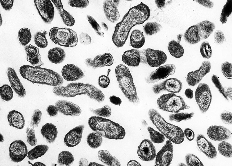
| [[Image:Coxiella burnetii.JPG|right|300px|C. burnetii, the Q fever causing agent]] | | [[Image:Coxiella burnetii.JPG|right|300px|C. burnetii, the Q fever causing agent]] | ||
|} | |} | ||
* Cattle, sheep, and goats are the primary reservoirs of C. burnetii. The infection has been noted in a wide variety of other animals, including other species of livestock and in domesticated pets. | |||
Cattle, sheep, and goats are the primary reservoirs of C. burnetii. The infection has been noted in a wide variety of other animals, including other species of livestock and in domesticated pets. | |||
* Coxiella burnetii does not usually cause clinical disease in these animals, although abortion in goats and sheep has been linked to C. burnetii infection. | * Coxiella burnetii does not usually cause clinical disease in these animals, although abortion in goats and sheep has been linked to C. burnetii infection. | ||
* Organisms are excreted in milk, urine, and feces of infected animals. Most importantly, during birthing, the organisms are shed in high numbers within the amniotic fluids and the placenta. | * Organisms are excreted in milk, urine, and feces of infected animals. Most importantly, during birthing, the organisms are shed in high numbers within the amniotic fluids and the placenta. | ||
Revision as of 21:40, 3 June 2017
Editor-In-Chief: C. Michael Gibson, M.S., M.D. [1]
|
Q fever Microchapters |
|
Diagnosis |
|---|
|
Treatment |
|
Case Studies |
|
Q fever pathophysiology On the Web |
|
American Roentgen Ray Society Images of Q fever pathophysiology |
|
Risk calculators and risk factors for Q fever pathophysiology |
Overview
Pathophysiology
 |
- Cattle, sheep, and goats are the primary reservoirs of C. burnetii. The infection has been noted in a wide variety of other animals, including other species of livestock and in domesticated pets.
- Coxiella burnetii does not usually cause clinical disease in these animals, although abortion in goats and sheep has been linked to C. burnetii infection.
- Organisms are excreted in milk, urine, and feces of infected animals. Most importantly, during birthing, the organisms are shed in high numbers within the amniotic fluids and the placenta.
- The organisms are resistant to heat, drying, and many common disinfectants. These features enable the bacteria to survive for long periods in the environment.
- Infection of humans usually occurs by inhalation of these organisms from air that contains airborne barnyard dust contaminated by dried placental material, birth fluids, and excreta of infected herd animals.
- Humans are often very susceptible to the disease, and very few organisms may be required to cause infection. Ingestion of contaminated milk, followed by regurgitation and inspiration of the contaminated food, is a less common mode of transmission.
- Other modes of transmission to humans, including tick bites and human to human transmission, are rare.