Pulmonary hypertension CT: Difference between revisions
Ralph Matar (talk | contribs) No edit summary |
m (Robot: Changing Category:Disease state to Category:Disease) |
||
| Line 37: | Line 37: | ||
[[Category:Cardiology]] | [[Category:Cardiology]] | ||
[[Category:Pulmonology]] | [[Category:Pulmonology]] | ||
[[Category:Disease | [[Category:Disease]] | ||
[[Category:Mature chapter]] | [[Category:Mature chapter]] | ||
Revision as of 22:37, 9 December 2011
|
Pulmonary Hypertension Microchapters |
|
Diagnosis |
|---|
|
Treatment |
|
Case Studies |
|
Pulmonary hypertension CT On the Web |
|
American Roentgen Ray Society Images of Pulmonary hypertension CT |
|
Risk calculators and risk factors for Pulmonary hypertension CT |
Editor-In-Chief: C. Michael Gibson, M.S., M.D. [1], Richard Channick, M.D.; Assistant Editor(s)-in-Chief: Ralph Matar, Lisa Prior, Ann Slater, R.N.
Pulmonary hypertension CT-scan[1]:
CT scanning is a valuable, noninvasive procedure for confirming the presence of pulmonary hypertension.
Different types of CT imaging have been used to rule out certain etiologies of pulmonary hypertension and to delineate the anatomy of the pulmonary vasculature:
1-Contrast-enhanced images may show intraluminal abnormalities in the arteries and veins, which are useful for confirming etiologies such as thromboembolic disease.
2-High-resolution CT (HRCT) scanning of the chest has a role in the evaluation of pulmonary hypertension in patients with suspected diffuse lung disease, like in patients with scleroderma, interstitial lung disease, and histiocytosis X.
3- Spiral CT scan in a patient with pulmonary hypertension can also reveal enlarged pulmonary arteries and an absence of thrombosis.
- The upper limit of normal for the diameter of the pulmonary artery is 28.6 mm. A value greater than 28.6 mm suggests increased pressures in the pulmonary system.
Multi Sliced CT
Images shown below are courtesy of RadsWiki and copylefted
-
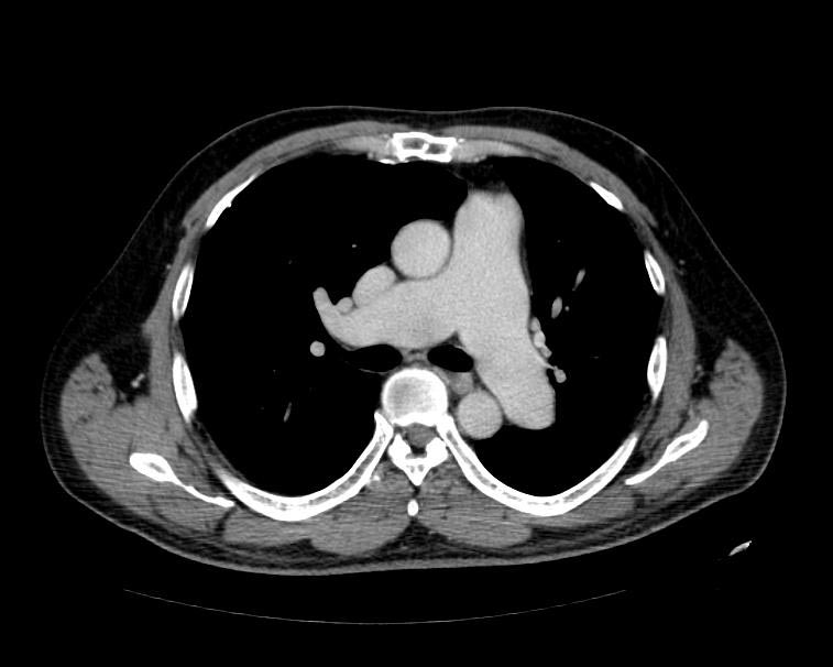
MSCT: Pulmonary hypertension. Note increase in diameter of pulmonary artery.
-
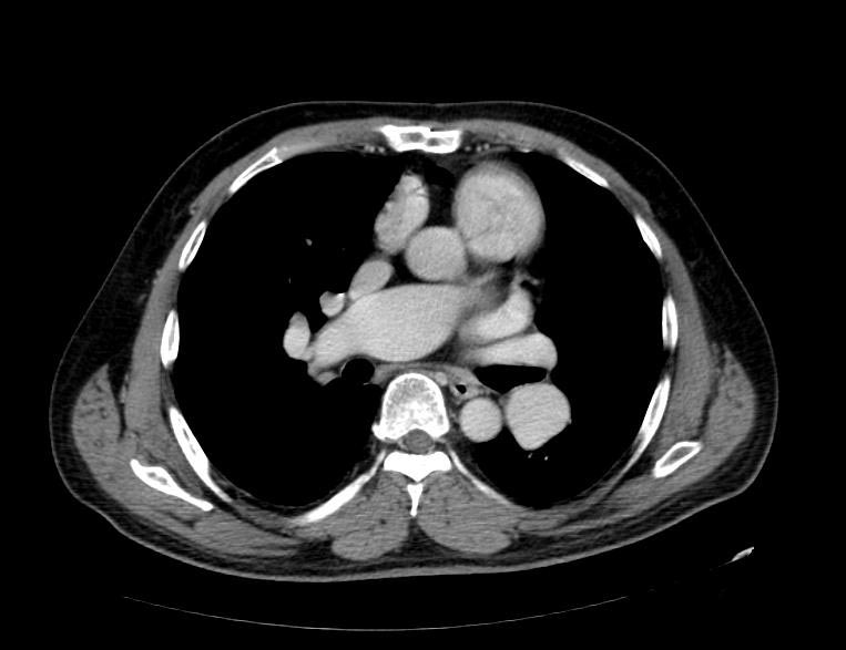
MSCT: Pulmonary hypertension. Note increase in diameter of pulmonary artery.


CT-scan of a patient with advanced Histiocytosis X associated with severe pulmonary hypertension[2]:
