Pulmonary hypertension CT: Difference between revisions
Rim Halaby (talk | contribs) No edit summary |
No edit summary |
||
| (6 intermediate revisions by 3 users not shown) | |||
| Line 1: | Line 1: | ||
__NOTOC__ | __NOTOC__ | ||
{{Pulmonary hypertension}} | {{Pulmonary hypertension}} | ||
{{CMG}}, Richard Channick, M.D.; '''Assistant Editor(s)-in-Chief:''' [[User:Ralph Matar|Ralph Matar]], [[User:Lisa Prior|Lisa Prior]], [[Ann Slater|Ann Slater, R.N.]] | {{CMG}}, Richard Channick, M.D.; '''Assistant Editor(s)-in-Chief:''' [[User:Ralph Matar|Ralph Matar]], [[User:Lisa Prior|Lisa Prior]], [[Ann Slater|Ann Slater, R.N.]]; {{Jose}} | ||
==Overview== | ==Overview== | ||
CT | A [[lung]] CT scan is helpful in the differential diagnosis of [[pulmonary hypertension]]. Different types of CT imaging have been used to rule out certain etiologies of pulmonary hypertension and to evaluate the anatomy of the pulmonary vasculature. | ||
Different types of CT imaging have been used to rule out certain etiologies of pulmonary hypertension and to | |||
==CT== | ==CT== | ||
*Contrast-enhanced images may show intraluminal abnormalities in the arteries and veins, which are useful for confirming etiologies such as [[thromboembolism|thromboembolic]] disease. | *Contrast-enhanced images may show intraluminal abnormalities in the arteries and veins, which are useful for confirming etiologies such as [[thromboembolism|thromboembolic]] disease. | ||
*[[High-resolution CT]] (HRCT) scanning of the chest has a role in the evaluation of pulmonary hypertension in patients with suspected diffuse lung disease, like in patients with [[scleroderma]], [[interstitial lung disease]], and [[histiocytosis X]]. | *[[High-resolution CT]] (HRCT) scanning of the chest has a role in the evaluation of pulmonary hypertension in patients with suspected diffuse lung disease, like in patients with [[scleroderma]], [[interstitial lung disease]], and [[histiocytosis X]]. | ||
*A [[spiral CT scan]] in a patient with pulmonary hypertension can also reveal enlarged pulmonary arteries and an absence of [[thrombosis]]. | |||
*[[ | **The normal upper limit for the diameter of the pulmonary artery is 28.6 mm. A value greater than 28.6 mm suggests increased pressure in the pulmonary system.<ref>Pulmonary Hypertension Imaging,Author: Davinder Jassal, MD, FACC, FRCPC; Chief Editor: Eugene C Lin, MD</ref> | ||
**The upper limit | * A CT scan is 100% specific for the diagnosis of pulmonary veno-occlusive disease (PVOD).<ref name="pmid15208112">{{cite journal| author=Resten A, Maitre S, Humbert M, Rabiller A, Sitbon O, Capron F et al.| title=Pulmonary hypertension: CT of the chest in pulmonary venoocclusive disease. | journal=AJR Am J Roentgenol | year= 2004 | volume= 183 | issue= 1 | pages= 65-70 | pmid=15208112 | doi=10.2214/ajr.183.1.1830065 | pmc= | url=http://www.ncbi.nlm.nih.gov/entrez/eutils/elink.fcgi?dbfrom=pubmed&tool=sumsearch.org/cite&retmode=ref&cmd=prlinks&id=15208112 }} </ref> The characteristics findings of PVOD on CT scan are:<ref name="pmid15208112">{{cite journal| author=Resten A, Maitre S, Humbert M, Rabiller A, Sitbon O, Capron F et al.| title=Pulmonary hypertension: CT of the chest in pulmonary venoocclusive disease. | journal=AJR Am J Roentgenol | year= 2004 | volume= 183 | issue= 1 | pages= 65-70 | pmid=15208112 | doi=10.2214/ajr.183.1.1830065 | pmc= | url=http://www.ncbi.nlm.nih.gov/entrez/eutils/elink.fcgi?dbfrom=pubmed&tool=sumsearch.org/cite&retmode=ref&cmd=prlinks&id=15208112 }} </ref> | ||
** Subpleural thickened septal lines | |||
** Centrilobular ground-glass opacities (vs panlobular opacities in idiopathic pulmonary arterial hypertension) | |||
** [[Adenopathy]] | |||
===Multi Sliced CT=== | ===Multi Sliced CT=== | ||
| Line 20: | Line 21: | ||
<div align="left"> | <div align="left"> | ||
<gallery heights="175" widths="175"> | <gallery heights="175" widths="175"> | ||
Image:Pulmonary-artery-hypertension-101.jpg| | Image:Pulmonary-artery-hypertension-101.jpg|Pulmonary hypertension. Note increase in diameter of pulmonary artery. | ||
Image:Pulmonary-artery-hypertension-102.jpg| | Image:Pulmonary-artery-hypertension-102.jpg|Pulmonary hypertension. Note increase in diameter of pulmonary artery. | ||
</gallery> | </gallery> | ||
</div> | </div> | ||
| Line 27: | Line 28: | ||
===CT-Scan of a Patient with Advanced Histiocytosis X Associated With Severe Pulmonary Hypertension<ref>Severe Pulmonary Hypertension in Histiocytosis X,Am. J. Respir. Crit. Care Med., Volume 161, Number 1, January 2000, 216-223</ref>=== | ===CT-Scan of a Patient with Advanced Histiocytosis X Associated With Severe Pulmonary Hypertension<ref>Severe Pulmonary Hypertension in Histiocytosis X,Am. J. Respir. Crit. Care Med., Volume 161, Number 1, January 2000, 216-223</ref>=== | ||
<gallery> | <gallery> | ||
Image:Ct-scan for a patient with advanced Histiocytosis X associated with severe pulmonary hypertension.jpeg| | Image:Ct-scan for a patient with advanced Histiocytosis X associated with severe pulmonary hypertension.jpeg|CT-scan for a patient with advanced histiocytosis X associated with severe pulmonary hypertension. | ||
</gallery> | </gallery> | ||
| Line 34: | Line 35: | ||
{{WH}} | {{WH}} | ||
{{WS}} | {{WS}} | ||
[[Category:Medicine]] | |||
[[Category:Cardiology]] | [[Category:Cardiology]] | ||
[[Category:Pulmonology]] | [[Category:Pulmonology]] | ||
[[Category: | [[Category:Emergency medicine]] | ||
[[Category: | [[Category:Up-To-Date]] | ||
[[Category:Radiology]] | |||
Latest revision as of 13:42, 9 June 2021
|
Pulmonary Hypertension Microchapters |
|
Diagnosis |
|---|
|
Treatment |
|
Case Studies |
|
Pulmonary hypertension CT On the Web |
|
American Roentgen Ray Society Images of Pulmonary hypertension CT |
|
Risk calculators and risk factors for Pulmonary hypertension CT |
Editor-In-Chief: C. Michael Gibson, M.S., M.D. [1], Richard Channick, M.D.; Assistant Editor(s)-in-Chief: Ralph Matar, Lisa Prior, Ann Slater, R.N.; José Eduardo Riceto Loyola Junior, M.D.[2]
Overview
A lung CT scan is helpful in the differential diagnosis of pulmonary hypertension. Different types of CT imaging have been used to rule out certain etiologies of pulmonary hypertension and to evaluate the anatomy of the pulmonary vasculature.
CT
- Contrast-enhanced images may show intraluminal abnormalities in the arteries and veins, which are useful for confirming etiologies such as thromboembolic disease.
- High-resolution CT (HRCT) scanning of the chest has a role in the evaluation of pulmonary hypertension in patients with suspected diffuse lung disease, like in patients with scleroderma, interstitial lung disease, and histiocytosis X.
- A spiral CT scan in a patient with pulmonary hypertension can also reveal enlarged pulmonary arteries and an absence of thrombosis.
- The normal upper limit for the diameter of the pulmonary artery is 28.6 mm. A value greater than 28.6 mm suggests increased pressure in the pulmonary system.[1]
- A CT scan is 100% specific for the diagnosis of pulmonary veno-occlusive disease (PVOD).[2] The characteristics findings of PVOD on CT scan are:[2]
- Subpleural thickened septal lines
- Centrilobular ground-glass opacities (vs panlobular opacities in idiopathic pulmonary arterial hypertension)
- Adenopathy
Multi Sliced CT
Images shown below are courtesy of RadsWiki and copylefted.
-
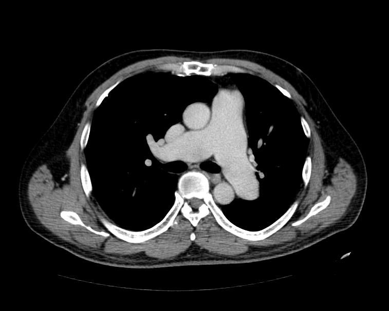
Pulmonary hypertension. Note increase in diameter of pulmonary artery.
-
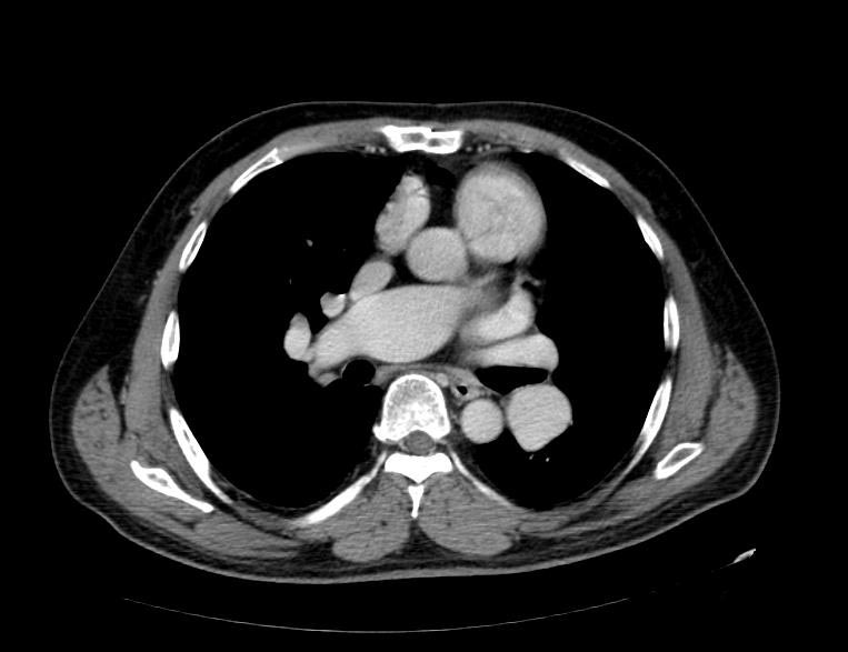
Pulmonary hypertension. Note increase in diameter of pulmonary artery.


CT-Scan of a Patient with Advanced Histiocytosis X Associated With Severe Pulmonary Hypertension[3]
-
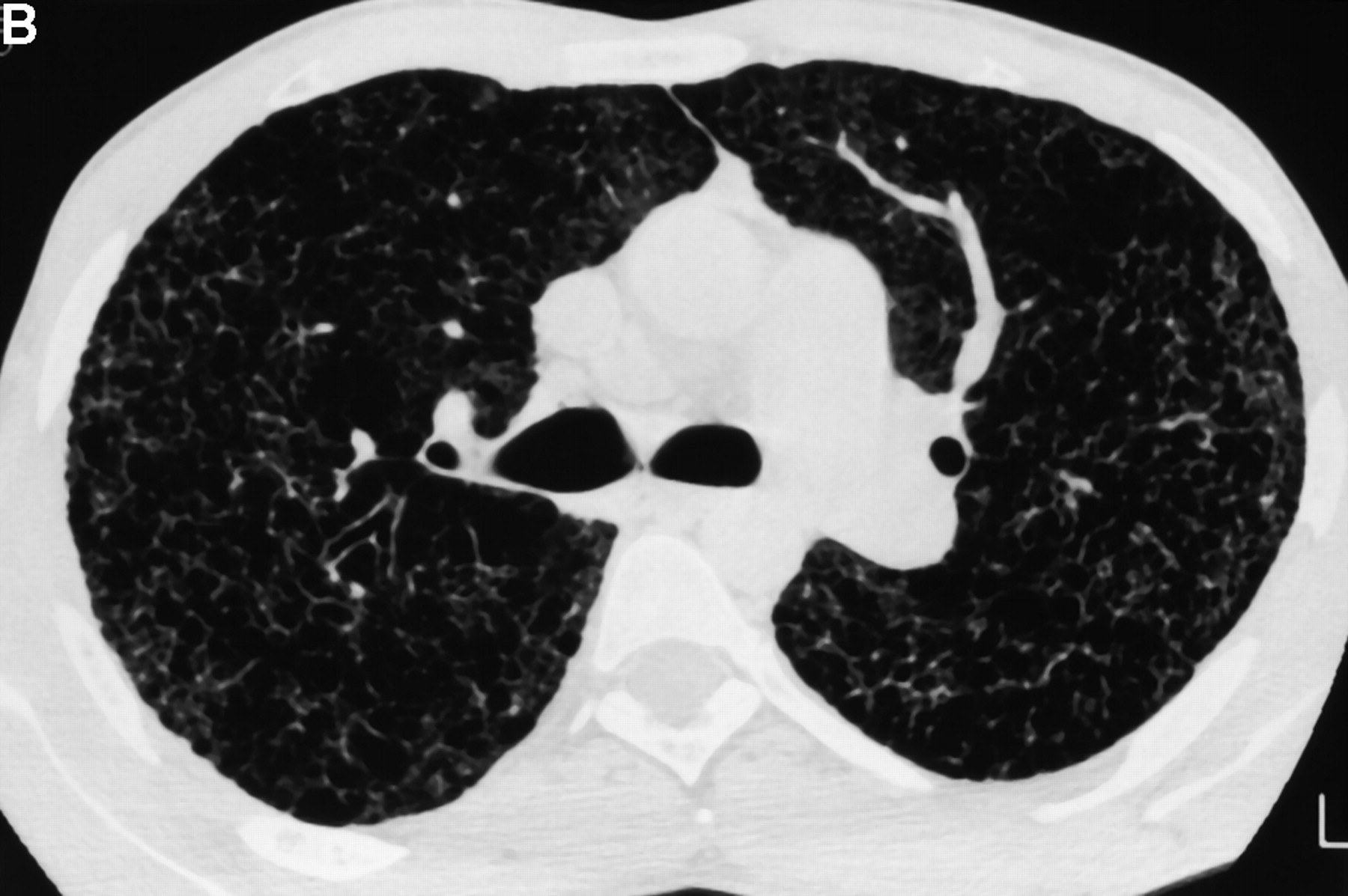
CT-scan for a patient with advanced histiocytosis X associated with severe pulmonary hypertension.

References
- ↑ Pulmonary Hypertension Imaging,Author: Davinder Jassal, MD, FACC, FRCPC; Chief Editor: Eugene C Lin, MD
- ↑ 2.0 2.1 Resten A, Maitre S, Humbert M, Rabiller A, Sitbon O, Capron F; et al. (2004). "Pulmonary hypertension: CT of the chest in pulmonary venoocclusive disease". AJR Am J Roentgenol. 183 (1): 65–70. doi:10.2214/ajr.183.1.1830065. PMID 15208112.
- ↑ Severe Pulmonary Hypertension in Histiocytosis X,Am. J. Respir. Crit. Care Med., Volume 161, Number 1, January 2000, 216-223