Peptic ulcer pathophysiology: Difference between revisions
mNo edit summary |
No edit summary |
||
| Line 8: | Line 8: | ||
==Pathophysiology== | ==Pathophysiology== | ||
A major causative factor (60% of gastric and 90% of duodenal ulcers) is chronic [[inflammation]] due to ''[[Helicobacter pylori]]'' that colonizes (''i.e.'' settles there after entering the body) the [[Pyloric antrum|antral]] [[mucosa]]. The immune system is unable to clear the infection, despite the appearance of antibodies. Thus, the [[bacterium]] can cause a chronic active [[gastritis]] (type B gastritis), resulting in a defect in the regulation of [[gastrin]] production by that part of the stomach, and gastrin secretion is increased. [[Gastrin]], in turn, stimulates the production of [[gastric acid]] by parietal cells. The acid erodes the [[mucosa]] and causes the ulcer. | A major causative factor (60% of gastric and 90% of duodenal ulcers) is chronic [[inflammation]] due to ''[[Helicobacter pylori]]'' that colonizes (''i.e.'' settles there after entering the body) the [[Pyloric antrum|antral]] [[mucosa]]. The immune system is unable to clear the infection, despite the appearance of antibodies. Thus, the [[bacterium]] can cause a chronic active [[gastritis]] (type B gastritis), resulting in a defect in the regulation of [[gastrin]] production by that part of the stomach, and [[gastrin]] secretion is increased. [[Gastrin]], in turn, stimulates the production of [[gastric acid]] by parietal cells. The acid erodes the [[mucosa]] and causes the ulcer. | ||
Another major cause is the use of [[NSAID]]s (see above). The gastric mucosa protects itself from [[gastric acid]] with a layer of mucus, the secretion of which is stimulated by certain prostaglandins. NSAIDs block the function of [[cyclooxygenase]] 1 (''cox-1''), which is essential for the production of these prostaglandins. Newer NSAIDs ([[celecoxib]], [[rofecoxib]]) only inhibit''cox-2'', which is less essential in the gastric mucosa, and roughly halve the risk of NSAID-related gastric ulceration. | Another major cause is the use of [[NSAID]]s (see above). The gastric mucosa protects itself from [[gastric acid]] with a layer of mucus, the secretion of which is stimulated by certain prostaglandins. NSAIDs block the function of [[cyclooxygenase]] 1 (''cox-1''), which is essential for the production of these prostaglandins. Newer NSAIDs ([[celecoxib]], [[rofecoxib]]) only inhibit''cox-2'', which is less essential in the gastric mucosa, and roughly halve the risk of NSAID-related gastric ulceration. | ||
| Line 33: | Line 33: | ||
===Microscopic Pathology=== | ===Microscopic Pathology=== | ||
A gastric peptic ulcer is a mucosal defect which penetrates the [[muscularis mucosae]] and muscularis propria, produced by acid-pepsin aggression. Ulcer margins are perpendicular and present chronic gastritis. During the active phase, the base of the ulcer shows 4 zones: inflammatory exudate, fibrinoid necrosis, granulation tissue and fibrous tissue. The fibrous base of the ulcer may contain vessels with thickened wall or with thrombosis.<ref name="pathologyatlas">{{cite web | url=http://www.pathologyatlas.ro/Peptic%20ulcer.html| title=ATLAS OF PATHOLOGY|accessdate=2007-08-26}}</ref> | A gastric peptic ulcer is a mucosal defect which penetrates the [[muscularis mucosae]] and muscularis propria, produced by acid-pepsin aggression. Ulcer margins are perpendicular and present chronic gastritis. During the active phase, the base of the ulcer shows 4 zones: [[inflammatory]] exudate, fibrinoid necrosis, granulation tissue and fibrous tissue. The fibrous base of the ulcer may contain vessels with thickened wall or with thrombosis.<ref name="pathologyatlas">{{cite web | url=http://www.pathologyatlas.ro/Peptic%20ulcer.html| title=ATLAS OF PATHOLOGY|accessdate=2007-08-26}}</ref> | ||
==References== | ==References== | ||
Revision as of 17:44, 12 October 2017
|
Peptic ulcer Microchapters |
|
Diagnosis |
|---|
|
Treatment |
|
Surgery |
|
Case Studies |
|
2017 ACG Guidelines for Peptic Ulcer Disease |
|
Guidelines for the Indications to Test for, and to Treat, H. pylori Infection |
|
Guidlines for factors that predict the successful eradication when treating H. pylori infection |
|
Guidelines to document H. pylori antimicrobial resistance in the North America |
|
Guidelines for evaluation and testing of H. pylori antibiotic resistance |
|
Guidelines for when to test for treatment success after H. pylori eradication therapy |
|
Guidelines for penicillin allergy in patients with H. pylori infection |
|
Peptic ulcer pathophysiology On the Web |
|
American Roentgen Ray Society Images of Peptic ulcer pathophysiology |
|
Risk calculators and risk factors for Peptic ulcer pathophysiology |
Editor-In-Chief: C. Michael Gibson, M.S., M.D. [1]
Please help WikiDoc by adding more content here. It's easy! Click here to learn about editing.
Overview
Pathophysiology
A major causative factor (60% of gastric and 90% of duodenal ulcers) is chronic inflammation due to Helicobacter pylori that colonizes (i.e. settles there after entering the body) the antral mucosa. The immune system is unable to clear the infection, despite the appearance of antibodies. Thus, the bacterium can cause a chronic active gastritis (type B gastritis), resulting in a defect in the regulation of gastrin production by that part of the stomach, and gastrin secretion is increased. Gastrin, in turn, stimulates the production of gastric acid by parietal cells. The acid erodes the mucosa and causes the ulcer.
Another major cause is the use of NSAIDs (see above). The gastric mucosa protects itself from gastric acid with a layer of mucus, the secretion of which is stimulated by certain prostaglandins. NSAIDs block the function of cyclooxygenase 1 (cox-1), which is essential for the production of these prostaglandins. Newer NSAIDs (celecoxib, rofecoxib) only inhibitcox-2, which is less essential in the gastric mucosa, and roughly halve the risk of NSAID-related gastric ulceration.
Tobacco smoking, blood group, spices and other factors that were suspected to cause ulcers until late in the 20th century, are actually of relatively minor importance in the development of peptic ulcers.[1]
Glucocorticoids lead to atrophy of all epithelial tissues. Their role in ulcerogenesis is relatively small.
There is debate as to whether Stress in the psychological sense can influence the development of peptic ulcers (see Stress and ulcers above). Burns and head trauma, however, can lead to "stress ulcers", and it is reported in many patients who are on mechanical ventilation.
Smoking leads to atherosclerosis and vascular spasms, causing vascular insufficiency and promoting the development of ulcers through ischemia.
Overuse of laxatives is also known to cause peptic ulcers.
A family history is often present in duodenal ulcers, especially when blood group O is also present. Inheritance appears to be unimportant in gastric ulcers.
Gastrinomas (Zollinger-Ellison syndrome), rare gastrin-secreting tumors, cause multiple and difficult to heal ulcers.

Gross Pathology
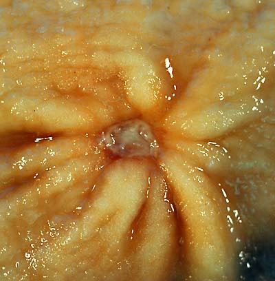
Gastric ulcers are most often localized on the lesser curvature of the stomach. The ulcer is a round to oval parietal defect ("hole"), 2 to 4 cm diameter, with a smooth base and perpendicular borders. These borders are not elevated or irregular as in the ulcerative form of gastric cancer. Surrounding mucosa may present radial folds, as a consequence of the parietal scarring.
Microscopic Pathology
A gastric peptic ulcer is a mucosal defect which penetrates the muscularis mucosae and muscularis propria, produced by acid-pepsin aggression. Ulcer margins are perpendicular and present chronic gastritis. During the active phase, the base of the ulcer shows 4 zones: inflammatory exudate, fibrinoid necrosis, granulation tissue and fibrous tissue. The fibrous base of the ulcer may contain vessels with thickened wall or with thrombosis.[2]
References
- ↑ For nearly 100 years, scientists and doctors thought that ulcers were caused by stress, spicy food, and alcohol. Treatment involved bed rest and a bland diet. Later, researchers added stomach acid to the list of causes and began treating ulcers with antacids.National Digestive Diseases Information Clearinghouse
- ↑ "ATLAS OF PATHOLOGY". Retrieved 2007-08-26.