Occipital vein
| Cardiology Network |
 Discuss Occipital vein further in the WikiDoc Cardiology Network |
| Adult Congenital |
|---|
| Biomarkers |
| Cardiac Rehabilitation |
| Congestive Heart Failure |
| CT Angiography |
| Echocardiography |
| Electrophysiology |
| Cardiology General |
| Genetics |
| Health Economics |
| Hypertension |
| Interventional Cardiology |
| MRI |
| Nuclear Cardiology |
| Peripheral Arterial Disease |
| Prevention |
| Public Policy |
| Pulmonary Embolism |
| Stable Angina |
| Valvular Heart Disease |
| Vascular Medicine |
Editor-In-Chief: C. Michael Gibson, M.S., M.D. [1]
The occipital vein begins in a plexus at the back part of the vertex of the skull.
From the plexus emerges a single vessel, which pierces the cranial attachment of the Trapezius and, dipping into the suboccipital triangle, joins the deep cervical and vertebral veins.
Occasionally it follows the course of the occipital artery and ends in the internal jugular; in other instances, it joins the posterior auricular vein and through it opens into the external jugular.
The parietal emissary vein connects it with the superior sagittal sinus; and as it passes across the mastoid portion of the temporal bone, it receives the mastoid emissary vein which connects it with the transverse sinus.
The occipital diploic vein sometimes joins it.
Additional images
-
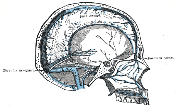
Sagittal section of the skull, showing the sinuses of the dura.
