Neonatal respiratory distress syndrome pathophysiology
|
Neonatal respiratory distress syndrome Microchapters |
|
Differentiating Neonatal respiratory distress syndrome from other Diseases |
|---|
|
Diagnosis |
|
Treatment |
|
Case Studies |
|
Neonatal respiratory distress syndrome pathophysiology On the Web |
|
American Roentgen Ray Society Images of Neonatal respiratory distress syndrome pathophysiology |
|
FDA on Neonatal respiratory distress syndrome pathophysiology |
|
CDC on Neonatal respiratory distress syndrome pathophysiology |
|
Neonatal respiratory distress syndrome pathophysiology in the news |
|
Blogs on Neonatal respiratory distress syndrome pathophysiology |
|
Directions to Hospitals Treating Neonatal respiratory distress syndrome |
|
Risk calculators and risk factors for Neonatal respiratory distress syndrome pathophysiology |
Editor-In-Chief: C. Michael Gibson, M.S., M.D. [1]
Pathophysiology
The lungs are developmentally deficient in a material called surfactant, which allows the alveoli to remain open throughout the normal cycle of inhalation and exhalation. Surfactant is a complex system of lipids, proteins and glycoproteins which are produced in specialized lung cells called Type II cells or Type II pneumocytes. The surfactant is packaged by the cell in structures called lamellar bodies, and extruded into the alveoli. The lamellar bodies then unfold into a complex lining of the alveoli. This layer reduces the surface tension of the fluid that lines the alveolar walls. During exhalation the walls of the alveoli come in contact and surface tension tends to cause them to stick together, preventing re-inflation. By reducing surface tension, surfactant allows the alveoli to re-expand with inspiration. Without adequate amounts of surfactant, the alveoli collapse and are very difficult to expand. Microscopically, a surfactant deficient lung is characterized by collapsed alveoli alternating with hyperaerated alveoli, vascular congestion and, in time, hyaline membranes. Hyaline membranes are composed of fibrin, cellular debris, red blood cells, rare neutrophils and macrophages. They appear as an eosinophilic, amorphous material, lining or filling the alveolar spaces and blocking gas exchange. As a result, blood passing through the lungs is unable to pick up oxgen and unload carbon dioxide from the alveolar spaces. Blood oxygen levels fall and carbon dioxide rises, resulting in rising blood acid levels and hypoxia. Structural immaturity, as manifest by low numbers of alveoli, also contributes to the disease process. Therapeutic oxygen and positive-pressure ventilation, while potentially life-saving, can also damage the lung. The diagnosis is made by the clinical picture and the chest xray, which demonstrates decreased lung volumes (bell-shaped chest), absence of the thymus (after about 6 hours), a small (0.5-1 mm), discrete, uniform infiltrate (sometimes described as a "ground glass" appearance) that involves all lobes of the lung, and air-bronchograms (ie the infiltrate will outline the larger airways passages which remain air-filled). In severe cases, this becomes exaggerated until the cardiac borders become inapparent (a 'white-out' appearance).
Gross Pathology
The characteristic pathology seen in babies who die from RDS was the source of the name "hyaline membrane disease". These waxy-appearing layers line the collapsed tiny air sacs ("alveoli") of the lung. In addition, the lungs show bleeding, over-distention of airways and damage to the lining cells.
The organs generally showed no abnormalities other than those of immaturity expected at this gestational age. There was moderate diffuse subarachnoid hemorrhage and a small amount of blood in the pleural and pericardial cavities.
-
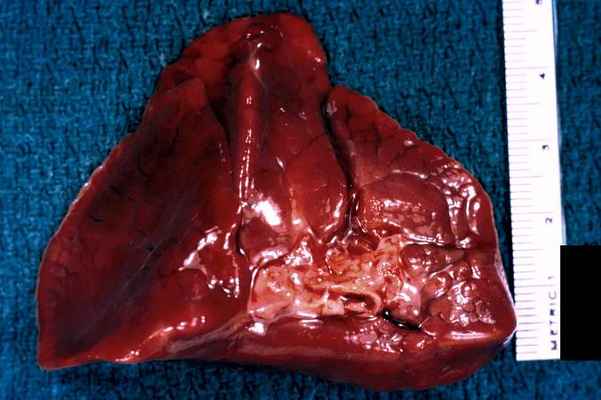
This is a gross photograph of lung demonstrating hyaline membrane disease (Infant respiratory distress syndrome) and atelectasis.

Microscopic Pathology
-
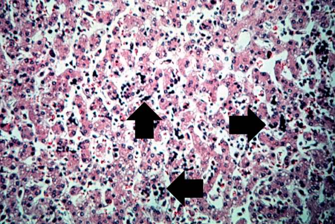
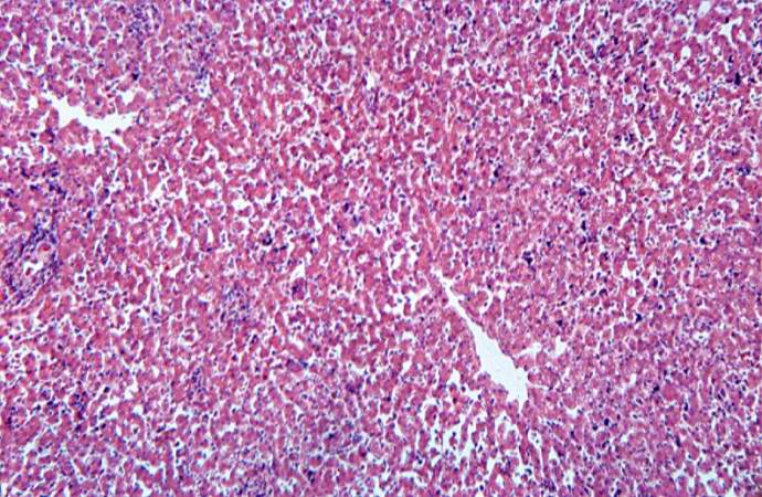
This high-power photomicrograph of liver shows more clearly the immature blood cell precursors (arrows) which represent extramedullary hematopoiesis of the liver. The liver is a normal site of fetal hematopoiesis and, for this stage of gestation, extramedullary hematopoiesis of the liver is normal.
-
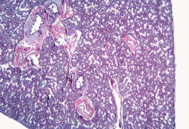
This low-power photomicrograph of lung demonstrates hypercellular pulmonary interstitium and small air spaces (as compared to adult lungs).
-
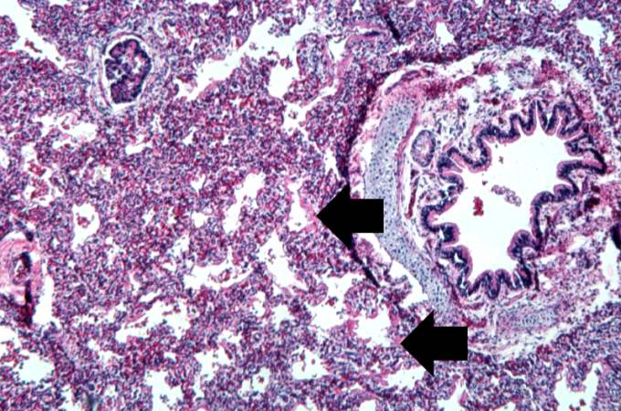
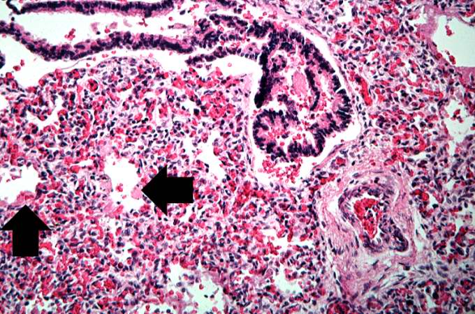
This is a medium-power photomicrograph showing a large bronchus with cartilage. Interstitial congestion with numerous red cells is apparent. Even at this magnification hyaline membranes (arrows) can be seen lining the alveoli.


-
This high-power photomicrograph shows an airway with adjacent lung tissue. Some alveoli have hyaline membranes (arrows). There is severe congestion of the interstitium throughout this section.
-
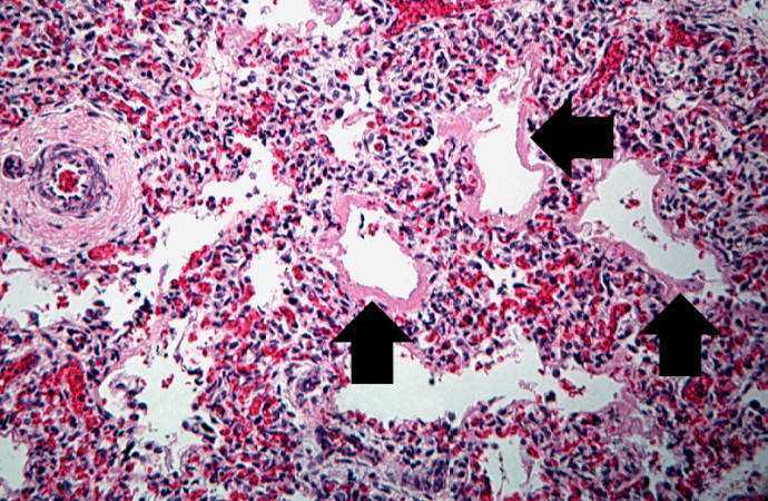
This medium-power photomicrograph shows the pink acellular homogeneous material lining the alveoli which comprises the hyaline membranes (arrows). The interstitium shows congestion, as in previous sections.
-
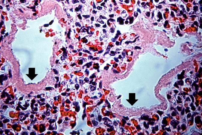
This higher-power photomicrograph shows more clearly the hyaline membranes (arrows) and the congestion in the interstitium.


-
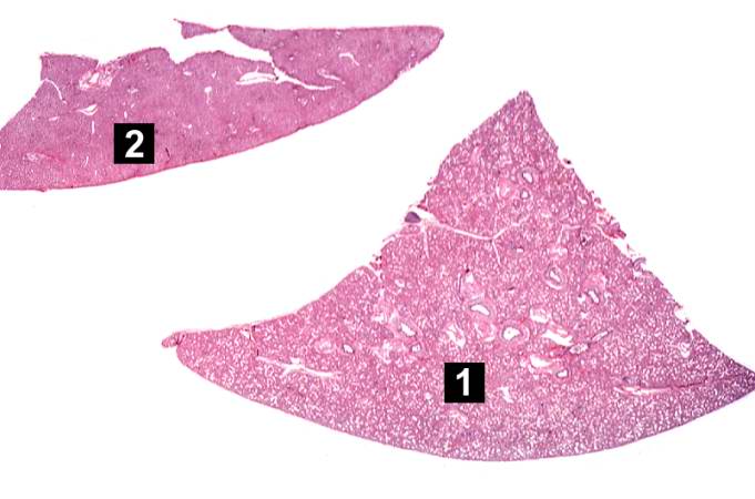
This is a low-power photomicrograph of a triangular-shaped section of lung (1) and an oblong section of liver (2). The lack of open air spaces in this neonatal lung indicates its immaturity.
-
This is a low-power photomicrograph of liver which contains dark blue-stained cells in the hepatic sinusoids. These are immature blood cell precursors and this represents extramedullary hematopoiesis of the liver.

