Nausea and vomiting pathophysiology
|
Nausea and vomiting Microchapters |
|
Diagnosis |
|---|
|
Treatment |
|
Case Studies |
|
Nausea and vomiting pathophysiology On the Web |
|
American Roentgen Ray Society Images of Nausea and vomiting pathophysiology |
|
Risk calculators and risk factors for Nausea and vomiting pathophysiology |
Editor-In-Chief: C. Michael Gibson, M.S., M.D. [1] Associate Editor(s)-in-Chief: Vishnu Vardhan Serla M.B.B.S. [2]
Overview
The physiology of nausea and vomiting encompass psychological states, the central nervous system, the autonomic nervous system, gastric dysrhythmias, and the endocrine system. Each individual has a threshold for nausea that changes minute by minute. Stimuli giving rise to nausea and vomiting originate from visceral, vestibular, and chemoreceptor trigger zone inputs which are mediated by serotonin/dopamine, histamine/acetylcholine, and serotonin/dopamine, respectively.
Pathophysiology
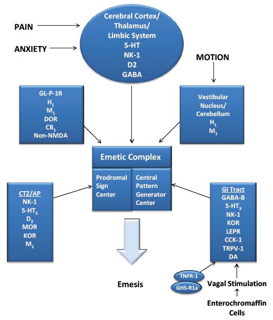
The physiology of nausea and vomiting encompass psychological states, the central nervous system, the autonomic nervous system, gastric dysrhythmias, and the endocrine system. Each individual has a threshold for nausea that changes minute by minute. At any given moment, the threshold depends on the interaction of certain inherent factors of the individual with the more changeable psychological states of anxiety, anticipation, expectation, and adaptation. Stimuli giving rise to nausea and vomiting originate from visceral, vestibular, and chemoreceptor trigger zone inputs which are mediated by serotonin/dopamine, histamine/acetylcholine, and serotonin/dopamine, respectively. These relationships between these areas are the basis of the pharmacotherapy.[1]
Vomiting Center
The vomiting center lies in the medulla oblongata and comprises the reticular formation and the nucleus of the tractus solitarius.
When there is stimulus, it activates motor pathways descend from this center and trigger vomiting.
These efferent pathways travel within the 5th, 7th, 9th, 10th, and 12th cranial nerves to the upper gastrointestinal tract, within vagal and sympathetic nerves to the lower tract, and within spinal nerves to the diaphragm and abdominal muscles.
The vomiting center can be activated directly by irritants or indirectly following input from gastrointestinal tract, cerebral cortex and thalamus, vestibular region, and chemoreceptor trigger zone (CRTZ). [2]
The CRTZ is closest in proximity, lying between the medulla and the floor of the fourth ventricle, and it is not protected by the blood-brain barrier.
The chemoreceptor trigger zone at the base of the fourth ventricle has numerous dopamine D2 receptors, serotonin 5-HT3 receptors, opioid receptors, acetylcholine receptors, and receptors for substance P. Stimulation of different receptors are involved in different pathways leading to emesis, in the final common pathway substance P appears to be involved.[3]
The vestibular system which sends information to the brain via cranial nerve VIII (vestibulocochlear nerve). It plays a major role in motion sickness and is rich in muscarinic receptors and histamine H1 receptors.
Cranial nerve X (vagus nerve), which is activated when the pharynx is irritated, leading to a gag reflex.
Vagal and enteric nervous system inputs that transmit information regarding the state of the gastrointestinal system. Irritation of the GI mucosa by chemotherapy, radiation, distention or acute infectious gastroenteritis activates the 5-HT3 receptors of these inputs.
The CNS mediates vomiting arising from psychiatric disorders and stress.
Vomiting:
The act of vomiting consists of 3 steps:[4]
- '''Nausea''' is an unpleasant and difficult to describe psychic experience. Physiologically,nausea is typically associated with decreased gastric motility and increased tone in the small intestine. Also, there is often reverse peristalsis in the proximal small intestine.
- '''Retching''' ("dry heaves") refers to spasmodic respiratory movements conducted with a closedglottis. While this is occurring, the antrum of the stomach contracts and the fundus and cardia relax.
- '''Emesis''' is when gastric and often small intestinal contents are propelled up to and out of the mouth.
and is caused by three types of outputs initiated by themedulla:Motor, parasympathetic nervous system (PNS) and sympathetic nervous system (SNS).
- Increased salivation to protect the enamel of teeth from stomach acids (excessive vomiting leads to caries). This is part of the PNS output.
- Reverse peristalsis, starting from the middle of the small intestine, sweeping up the contents of the digestive tract into the stomach, through the relaxed pyloric sphincter.
- A lowering of intrathoracic pressure (by inspiration against a closed glottis), coupled with an increase in abdominal pressure as the abdominal muscles contract, propels stomach contents into the esophagus without involvement of the reverse peristalsis. The lower esophageal sphincter relaxes.
- Vomiting also initiates a SNS response causing both sweating and increased heart rate.
The neurotransmitters that regulate vomiting are poorly understood, but inhibitors of dopamine, histamine and serotonin are all used to suppress vomiting, suggesting that these play a role in the initiation or maintenance of a vomiting cycle. Vasopressin and neurokinin may also be involved.
Content
As the stomach secretes acid, vomit contains a high concentration of hydronium ions and is thus strongly acidic. Recent food intake will be reflected in the gastric vomit.
The content of the vomitus (vomit) may be of medical interest.
Fresh blood in the vomit is called hematemesis ("blood vomiting").
Old blood bears resemblance to coffee grounds (as the iron in the blood is oxidized), and is termed "coffee ground vomiting".
Bile can enter the vomit during subsequent heaves due to duodenal contraction if the vomiting is severe.
Fecal vomiting is often a consequence of intestinal obstruction, and is treated as a warning sign of this potentially serious problem ("signum mali ominis"); such vomiting is sometimes called "miserere". If food has recently been consumed, then partly digested food may show up in the vomit.
References
- ↑ Singh P, Yoon SS, Kuo B (January 2016). "Nausea: a review of pathophysiology and therapeutics". Therap Adv Gastroenterol. 9 (1): 98–112. doi:10.1177/1756283X15618131. PMC 4699282. PMID 26770271.
- ↑ Hornby PJ. Central neurocircuitry associated with emesis. Am J Med 2001;111:106S-12S. PMID 11749934.
- ↑ Rhodes VA (December 1990). "Nausea, vomiting, and retching". Nurs Clin North Am. 25 (4): 885–900. PMID 2235641.
