Myelofibrosis: Difference between revisions
(→MRI) |
(→MRI) |
||
| Line 41: | Line 41: | ||
<gallery> | <gallery> | ||
Image:Myelofibrosis_MRI_001.jpg | Image:Myelofibrosis_MRI_001.jpg|Myelofibrosis | ||
Image:Myelofibrosis_MRI_002.jpg | Image:Myelofibrosis_MRI_002.jpg|Myelofibrosis | ||
</gallery> | </gallery> | ||
<gallery> | <gallery> | ||
Image:Myelofibrosis_MRI_003.jpg | Image:Myelofibrosis_MRI_003.jpg|Myelofibrosis | ||
Image:Myelofibrosis_MRI_004.jpg | Image:Myelofibrosis_MRI_004.jpg|Myelofibrosis | ||
</gallery> | </gallery> | ||
Revision as of 14:14, 19 January 2009
Template:DiseaseDisorder infobox Template:Search infobox Editor-In-Chief: C. Michael Gibson, M.S., M.D. [1]
Please Take Over This Page and Apply to be Editor-In-Chief for this topic: There can be one or more than one Editor-In-Chief. You may also apply to be an Associate Editor-In-Chief of one of the subtopics below. Please mail us [2] to indicate your interest in serving either as an Editor-In-Chief of the entire topic or as an Associate Editor-In-Chief for a subtopic. Please be sure to attach your CV and or biographical sketch.
Overview
Myelofibrosis with myeloid metaplasia, also known as agnogenic myeloid metaplasia, chronic idiopathic myelofibrosis, and primary myelofibrosis,[1] was first described in 1879 and is currently classified as a myeloproliferative disease caused by the growth and proliferation of an abnormal bone marrow stem cell, resulting in the replacement of the bone marrow with fibrous connective tissue. An eponym for the disease is Heuck-Assmann disease, or Assmann's Disease.
Presentation
The bone marrow is replaced by collagen fibrosis, impairing the patient's ability to generate new blood cells resulting in a progressive anemia. It is usually reactive following other myeloproliferative disorders, such as polycythemia rubra vera or essential thrombocytosis. Extramedullary hematopoeisis occurs as the haemopoetic cells migrate away from the bone marrow, to the liver and spleen. Patients often have hepatosplenomegaly and poikilocytosis. The mean survival is 5 years and causes of death include infection, bleeding, organ failure, portal hypertension, and leukemic transformation.
Diagnosis
Diagnosis is based upon:
- Normochromic normocytic anaemia
- Red cell poikilocytosis on blood film (tear drop RBCs)
- JAK 2 mutation on Val 617 Phe locus in 50%
- Raised lactate dehydrogenase
- Raised neutrophil alkaline phosphatase score
- Bone marrow biopsy may show increased cellularity and fibrosis
Diagnostic Findings
MRI
-
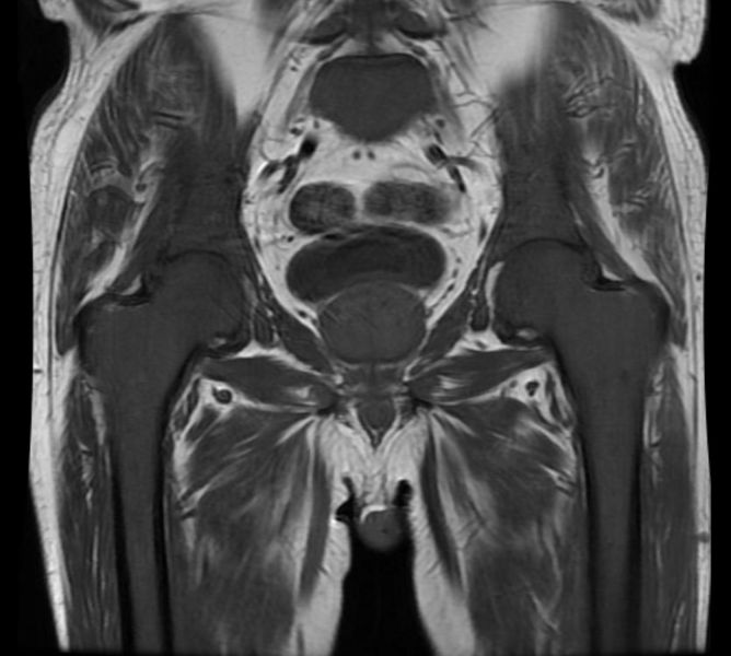
Myelofibrosis
-

Myelofibrosis


-

Myelofibrosis
-

Myelofibrosis


References
- ↑ Older terms include "myelofibrosis with myeloid metaplasia" and "agnogenic myeloid metaplasia". The World Health Organization utilizes the name "chronic idiopathic myelofibrosis", while the International Working Group on Myelofibrosis Research and Treatment calls the disease "primary myelofibrosis".