Mycosis fungoides physical examination
|
Cutaneous T cell lymphoma Microchapters |
Editor-In-Chief: C. Michael Gibson, M.S., M.D. [1]; Associate Editor(s)-in-Chief: Sogand Goudarzi, MD [2]
Overview
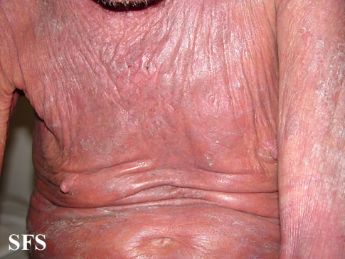
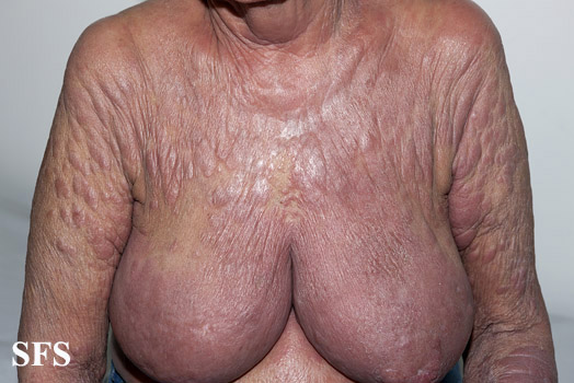
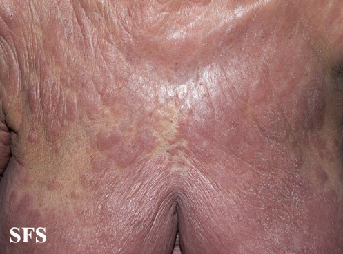
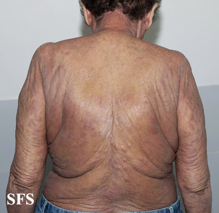
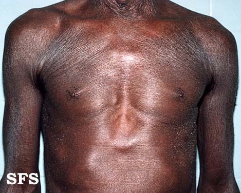
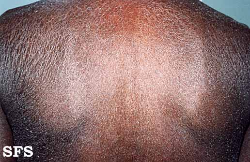
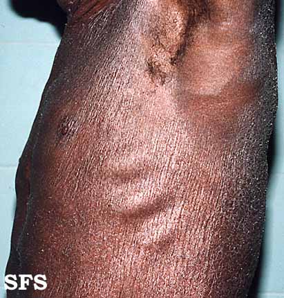
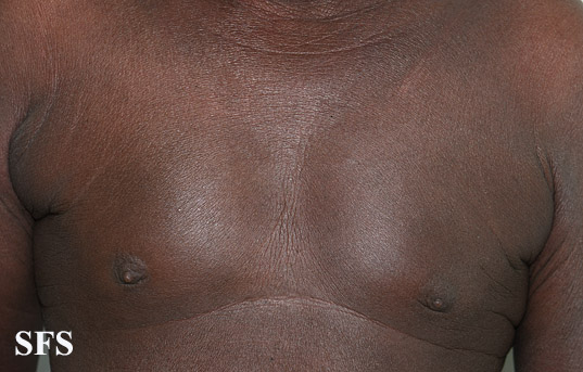
Common physical examination findings of cutaneous T cell lymphoma include fever, rash, pruritus, ulcer, chest tenderness, abdomen tenderness, bone tenderness, peripheral lymphadenopathy, and central lymphadenopathy.
Vital Signs
Skin
- Skin examination of patients with mycosis fungoides should be done generally.
- Skin lesions and lymphadenopathy should be observed and monitor their changes.
HEENT
- HEENT examination of patients with mycosis fungoides is usually normal.
Neck
- Neck examination of patients with mycosis fungoides is usually normal.
Lungs
- Pulmonary examination of patients with mycosis fungoides is usually normal.
- Pulmonary function tests and comprehensive metabolic panel (including liver and kidney function) should be monitored during treatment with methotrexate.
Heart
- Cardiovascular examination of patients with mycosis fungoides is usually normal.
Abdomen
- Abdominal examination of patients with mycosis fungoides is usually normal.
Back
- Back examination of patients with mycosis fungoides is usually normal.
Genitourinary
- Genitourinary examination of patients with mycosis fungoides is usually normal.
Neuromuscular
- Neuromuscular examination of patients with mycosis fungoides is usually normal
Extremities
- Extremities examination of patients with mycosis fungoides is usually normal.
- Clubbing
- Cyanosis
- Pitting/non-pitting edema of the upper/lower extremities
- Muscle atrophy
- Fasciculations in the upper/lower extremities
References
Template:WH Template:WS ==Physical Examination[1]==
Vital Signs
HEENT
Chest Exam
- Thoracic masses suggestive of central lymphadenopathy
- Chest tenderness
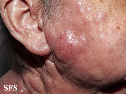
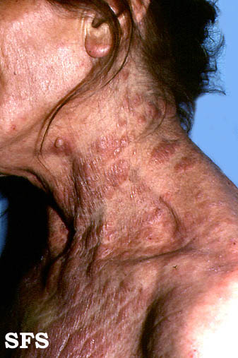
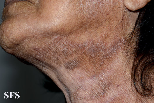
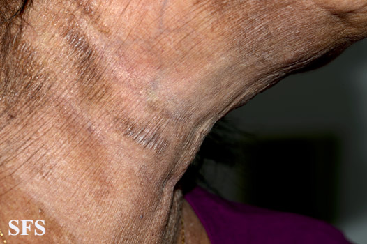
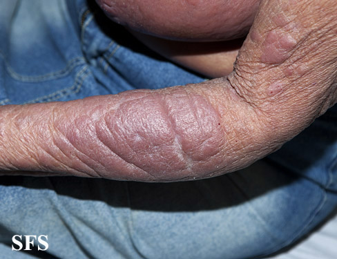
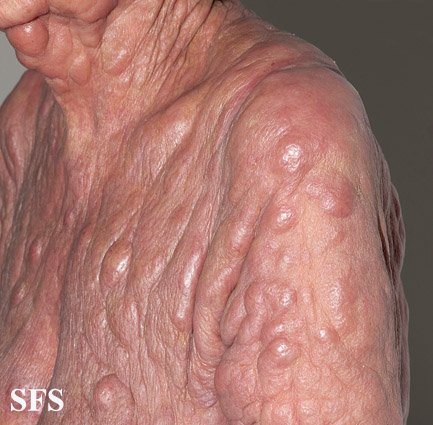
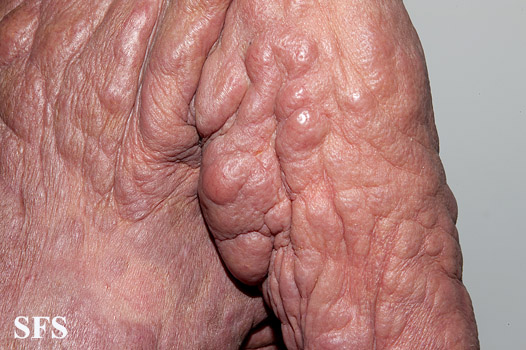
Skin
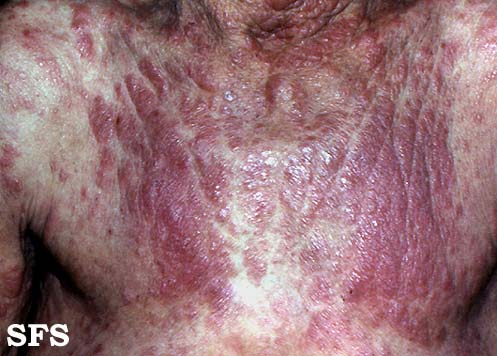
- Generalized erythroderma
- Ulcer
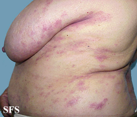
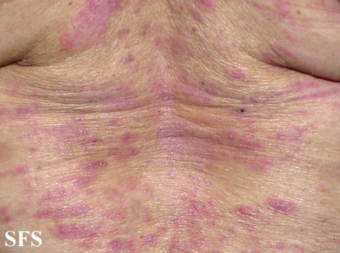
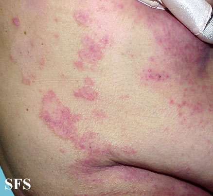
- Rash
- Pruritus
Abdomen
- Abdominal masses suggestive of central lymphadenopathy
- Abdomen tenderness
Extremities
- Peripheral lymphadenopathy
- Bone tenderness
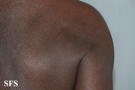
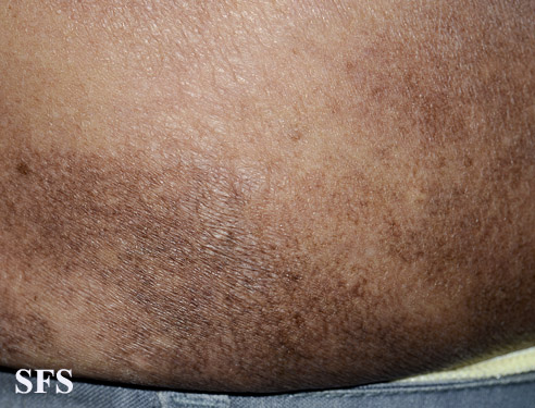
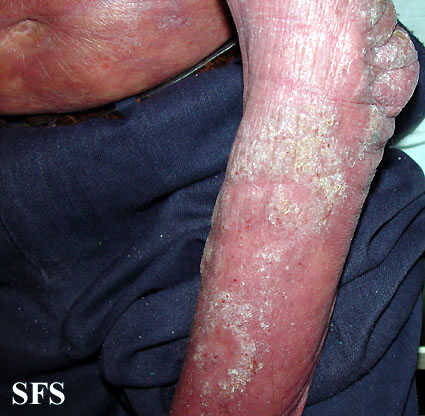
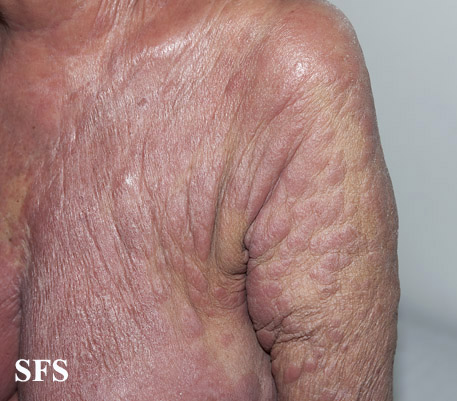
| Name | Description |
|---|---|
| Premycotic (pretumor) phase |
|
| Patch phase |
|
| Plaque phase |
|
| Tumor phase |
|
References
- ↑ 1.0 1.1 Cutaneous T cell lymphoma. Surveillance, Epidemiology, and End Results . http://seer.cancer.gov/seertools/hemelymph/51f6cf56e3e27c3994bd52f7/ Accessed on January 19, 2016
- ↑ Cutaneous T cell lymphoma. Canadian Cancer Society. http://www.cancer.ca/en/cancer-information/cancer-type/non-hodgkin-lymphoma/non-hodgkin-lymphoma/types-of-nhl/cutaneous-t-cell-lymphoma/?region=on Accessed on January 19, 2016
