Mycosis fungoides pathophysiology
|
Cutaneous T cell lymphoma Microchapters |
Editor-In-Chief: C. Michael Gibson, M.S., M.D. [1];Associate Editor(s)-in-Chief: Sogand Goudarzi, MD [2]
Overview
Cutaneous T cell lymphoma arises from T-cell lymphocytes, which are normally involved in the cell mediated immune response. On microscopic histopathological analysis, atypical lymphoid cells, polymorphous inflammatory infiltrate in the dermis, and lymphocytes with cerebroid nuclei are characteristic findings of mycosis fungoides.
Pathophysiology
- Cutaneous T cell lymphoma arises from T-cell lymphocytes, which are normally involved in the cell mediated immune response.[1]
- The tumor cells originate from memory T cells or skin homing CD4+ T cells expressing cutaneous lymphocyte antigen (CLA) and chemokine receptors CCR4 and CCR7.[2]
- It is understood that cutaneous t cell lymphoma (maycosis fungoides, Sezary sydrome ) is the result of malignant T cell that derived from a mature CD41 CD45RO1 memory T cells.[3]
- Malignant T cell express adhesion molecules such as CCR4 and CLA.[3]
- Malignant T cell in Sezary syndrome (Sezary cells) have a different phenotype, they express CCR7 and L-selectin 4.[3][4]
- Immunohistochemistry shows expression T-cell antigens such as CD7, CD5, CD26, or CD2 and CD3.[3]
- [Pathogen name] is usually transmitted via the [transmission route] route to the human host.* Following transmission/ingestion, the [pathogen] uses the [entry site] to invade the [cell name] cell.
- [Disease or malignancy name] arises from [cell name]s, which are [cell type] cells that are normally involved in [function of cells].
- The progression to [disease name] usually involves the [molecular pathway].
- The pathophysiology of [disease/malignancy] depends on the histological subtype.
- Sezary syndrome and mycosis fungoides are T-cell lymphomas that primary manifest as multiple cutaneous lesions.
- Mycosis Fungoides is the most common type of cutaneous T cell lymphoma.
- Sézary's cells are T-cells that have pathological quantities of mucopolysaccharides.
- Sézary's disease is sometimes considered a late stage of mycosis fungoides.
Gross Pathology
- On gross pathology, irregular, erythematous, circular patches are characteristic findings of cutaneous T cell lymphoma.
-
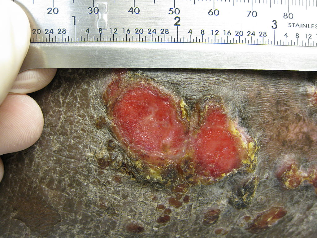
Plaque of mycosis fungoides
-
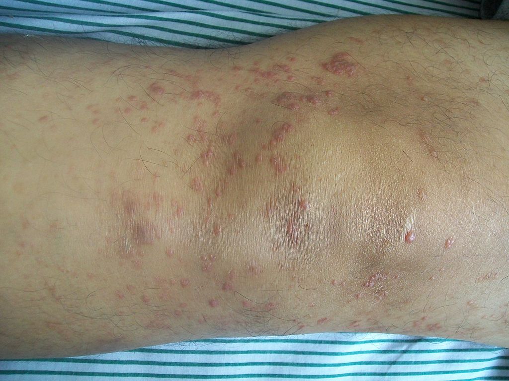
Mycosis fungoides knee


Microscopic Pathology
- Mycosis fungoides pathological course may be divided into three main stages:
- Premycotic stage
- Mycotic stage
- Tumoros stage
- The premycotic stage
- Non-diagnostic and represented by chronic nonspecific dermatisis associated with psoriasiform changes in epidermis
- The mycotic stage
- Shows a polymorphous inflammatory infiltrate in the dermis that contains small numbers of frankly atypical lymphoid cells
- These cells may line up individually along the epidermal basal layer
- The presence of spongiosis is highly suggestive of mycosis fungoides
- Tumoros stage
- Expansion of the dermis due to dense infiltration by medium sized lymphocytes that are typically characterized by a cerebroid nuclei.
-
Sézary's disease
-
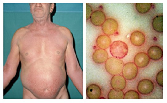
61-year-old man presented in 1972 with unrelenting pruritus of six months’ duration. On the right is his peripheral blood film stained with Periodic Acid-Schiff (PAS) showing a neoplastic T cell (Sézary cell).
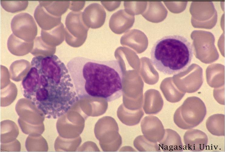
-
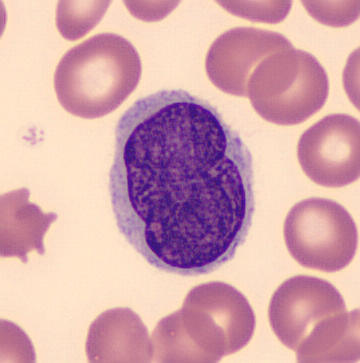
Pleomorphic abnormal T cell with the characteristic cerebriform nuclei (Peripheral blood - MGG stain)
-
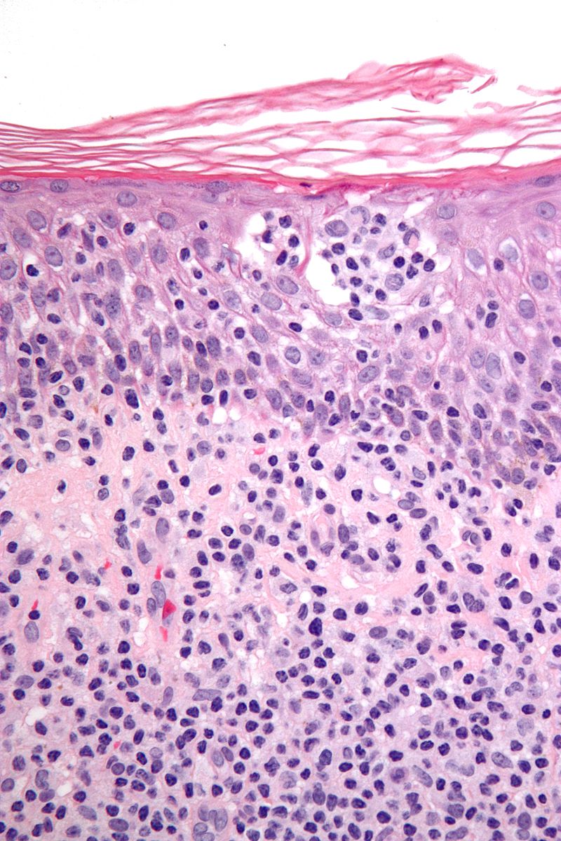
Features: Nests of lymphocytes in the epidermis; "Pautrier microabscesses". Single lymphocytes in epidermis; "lymphocyte exocytosis". Short linear arrays of lymphocytes along the basal layer of the epidermis; "epidermotropism".


Genetics
- Development of cutaneous T cell lymphoma is the result of multiple genetic mutations.[5][6][7][8][9]
OR
- Genes involved in the pathogenesis of cutaneous T cell lymphoma include:
- Other deletion such as ZEB1, ARID1A, DNMT3A, CDKN2A, FAS, ATM, CTCF, STAT5B, PRKCQ and somatic mutations (NFKB2, CD28, RHOA) observed in gene sequencing.[12]
- The development of cutaneous T cell lymphoma is the result of multiple genetic mutations such as:[13][14][15][16]
- There is not a classic chromosomal translocation in cutaeous T cell lymphoma( MF and SS ) significant
chromosomal instability has been noted. Losses on 1p, 10q, 13q, and 17p and gains of 4, 17q, and 18 have been identified [17]
- deletions and translocations in different chromosomes or chromosomal segments
- Chromosomal amplification of JunB at 19p12 observed in mycosis fungoides and Sezary syndrome.[17]
References
- ↑ Wilcox RA (January 2016). "Cutaneous T-cell lymphoma: 2016 update on diagnosis, risk-stratification, and management". Am. J. Hematol. 91 (1): 151–65. doi:10.1002/ajh.24233. PMC 4715621. PMID 26607183.
- ↑ Fuhlbrigge RC, Kieffer JD, Armerding D, Kupper TS (October 1997). "Cutaneous lymphocyte antigen is a specialized form of PSGL-1 expressed on skin-homing T cells". Nature. 389 (6654): 978–81. doi:10.1038/40166. PMID 9353122.
- ↑ 3.0 3.1 3.2 3.3 Foss, Francine M.; Girardi, Michael (2017). "Mycosis Fungoides and Sezary Syndrome". Hematology/Oncology Clinics of North America. 31 (2): 297–315. doi:10.1016/j.hoc.2016.11.008. ISSN 0889-8588.
- ↑ Campbell, J. J.; Clark, R. A.; Watanabe, R.; Kupper, T. S. (2010). "Sezary syndrome and mycosis fungoides arise from distinct T-cell subsets: a biologic rationale for their distinct clinical behaviors". Blood. 116 (5): 767–771. doi:10.1182/blood-2009-11-251926. ISSN 0006-4971.
- ↑ Shin, J.; Monti, S.; Aires, D. J.; Duvic, M.; Golub, T.; Jones, D. A.; Kupper, T. S. (2007). "Lesional gene expression profiling in cutaneous T-cell lymphoma reveals natural clusters associated with disease outcome". Blood. 110 (8): 3015–3027. doi:10.1182/blood-2006-12-061507. ISSN 0006-4971.
- ↑ Wong, Henry K.; Mishra, Anjali; Hake, Timothy; Porcu, Pierluigi (2011). "Evolving Insights in the Pathogenesis and Therapy of Cutaneous T-cell lymphoma (Mycosis Fungoides and Sezary Syndrome)". British Journal of Haematology. 155 (2): 150–166. doi:10.1111/j.1365-2141.2011.08852.x. ISSN 0007-1048.
- ↑ Wilcox, Ryan A. (2011). "Cutaneous T-cell lymphoma: 2011 update on diagnosis, risk-stratification, and management". American Journal of Hematology. 86 (11): 928–948. doi:10.1002/ajh.22139. ISSN 0361-8609.
- ↑ Whittaker, Sean (2006). "Biological Insights into the Pathogenesis of Cutaneous T-Cell Lymphomas (CTCL)". Seminars in Oncology. 33: 3–6. doi:10.1053/j.seminoncol.2005.12.015. ISSN 0093-7754.
- ↑ Henry K. Wong (2013). "Novel biomarkers, dysregulated epigenetics, and therapy in cutaneous T-cell lymphoma". Discovery medicine. 16 (87): 71–78. PMID 23998443. Unknown parameter
|month=ignored (help) - ↑ Karenko, Leena; Hahtola, Sonja; Päivinen, Suvi; Karhu, Ritva; Syrjä, Sanna; Kähkönen, Marketta; Nedoszytko, Boguslaw; Kytölä, Soili; Zhou, Ying; Blazevic, Vesna; Pesonen, Maria; Nevala, Hanna; Nupponen, Nina; Sihto, Harri; Krebs, Inge; Poustka, Annemarie; Roszkiewicz, Jadwiga; Saksela, Kalle; Peterson, Pärt; Visakorpi, Tapio; Ranki, Annamari (2005). "Primary Cutaneous T-Cell Lymphomas Show a Deletion or Translocation AffectingNAV3, the HumanUNC-53Homologue". Cancer Research. 65 (18): 8101–8110. doi:10.1158/0008-5472.CAN-04-0366. ISSN 0008-5472.
- ↑ Lin, Yea-Lih; Pasero, Philippe (2012). "Interference Between DNA Replication and Transcription as a Cause of Genomic Instability". Current Genomics. 13 (1): 65–73. doi:10.2174/138920212799034767. ISSN 1389-2029.
- ↑ Choi, Jaehyuk; Goh, Gerald; Walradt, Trent; Hong, Bok S; Bunick, Christopher G; Chen, Kan; Bjornson, Robert D; Maman, Yaakov; Wang, Tiffany; Tordoff, Jesse; Carlson, Kacie; Overton, John D; Liu, Kristina J; Lewis, Julia M; Devine, Lesley; Barbarotta, Lisa; Foss, Francine M; Subtil, Antonio; Vonderheid, Eric C; Edelson, Richard L; Schatz, David G; Boggon, Titus J; Girardi, Michael; Lifton, Richard P (2015). "Genomic landscape of cutaneous T cell lymphoma". Nature Genetics. 47 (9): 1011–1019. doi:10.1038/ng.3356. ISSN 1061-4036.
- ↑ Leena Karenko, Sonja Hahtola, Suvi Paivinen, Ritva Karhu, Sanna Syrja, Marketta Kahkonen, Boguslaw Nedoszytko, Soili Kytola, Ying Zhou, Vesna Blazevic, Maria Pesonen, Hanna Nevala, Nina Nupponen, Harri Sihto, Inge Krebs, Annemarie Poustka, Jadwiga Roszkiewicz, Kalle Saksela, Part Peterson, Tapio Visakorpi & Annamari Ranki (2005). "Primary cutaneous T-cell lymphomas show a deletion or translocation affecting NAV3, the human UNC-53 homologue". Cancer research. 65 (18): 8101–8110. doi:10.1158/0008-5472.CAN-04-0366. PMID 16166283. Unknown parameter
|month=ignored (help) - ↑ Yaohua Zhang, Yang Wang, Richard Yu, Yuanshen Huang, Mingwan Su, Cheng Xiao, Magdalena Martinka, Jan P. Dutz, Xuejun Zhang, Zhizhong Zheng & Youwen Zhou (2012). "Molecular markers of early-stage mycosis fungoides". The Journal of investigative dermatology. 132 (6): 1698–1706. doi:10.1038/jid.2012.13. PMID 22377759. Unknown parameter
|month=ignored (help) - ↑ Alexander Ungewickell, Aparna Bhaduri, Eon Rios, Jason Reuter, Carolyn S. Lee, Angela Mah, Ashley Zehnder, Robert Ohgami, Shashikant Kulkarni, Randall Armstrong, Wen-Kai Weng, Dita Gratzinger, Mahkam Tavallaee, Alain Rook, Michael Snyder, Youn Kim & Paul A. Khavari (2015). "Genomic analysis of mycosis fungoides and Sezary syndrome identifies recurrent alterations in TNFR2". Nature genetics. 47 (9): 1056–1060. doi:10.1038/ng.3370. PMID 26258847. Unknown parameter
|month=ignored (help) - ↑ Jaehyuk Choi, Gerald Goh, Trent Walradt, Bok S. Hong, Christopher G. Bunick, Kan Chen, Robert D. Bjornson, Yaakov Maman, Tiffany Wang, Jesse Tordoff, Kacie Carlson, John D. Overton, Kristina J. Liu, Julia M. Lewis, Lesley Devine, Lisa Barbarotta, Francine M. Foss, Antonio Subtil, Eric C. Vonderheid, Richard L. Edelson, David G. Schatz, Titus J. Boggon, Michael Girardi & Richard P. Lifton (2015). "Genomic landscape of cutaneous T cell lymphoma". Nature genetics. 47 (9): 1011–1019. doi:10.1038/ng.3356. PMID 26192916. Unknown parameter
|month=ignored (help) - ↑ 17.0 17.1 Mao, Xin; Orchard, Guy; Lillington, Debra M.; Russell-Jones, Robin; Young, Bryan D.; Whittaker, Sean (2003). "Genetic alterations in primary cutaneous CD30+ anaplastic large cell lymphoma". Genes, Chromosomes and Cancer. 37 (2): 176–185. doi:10.1002/gcc.10184. ISSN 1045-2257.