Lung mass imaging
|
Lung Mass Microchapters |
|
Diagnosis |
|---|
|
Treatment |
|
Lung mass imaging On the Web |
|
American Roentgen Ray Society Images of Lung mass imaging |
Editor-In-Chief: C. Michael Gibson, M.S., M.D. [1]Associate Editor(s)-in-Chief: Maria Fernanda Villarreal, M.D. [2]
Overview
Computed tomography is the method of choice for the evaluation of lung masses. The evaluation of lung masses will depend on several characteristics, such as: calcification, margins, location, distribution, and attenuation. Further evaluation of lung masses, should include other diagnostic studies, such as: bronchoscopy, sputum cytology, or mediastinoscopy. Other imaging study useful for the malignancy evaluation of lung masses is PET scanning, which may be helpful for the detection of occult disease and malignancy assessment.[1]
Imaging
- The evaluation of lung masses will depend on the following characteristics:
Calcification
- Calcification patterns are commonly seen in granulomatous disease and hamartomas
- Calcification patterns are normally a sign of benignancy
- Characteristic benign calcification patterns of pulmonary nodule, include:
- Diffuse
- Central
- Laminated
- Popcorn
Size
- Any area of pulmonary opacification that measures more than 30 mm
Location
- Locations of lung mass, include:
- Pleural
- Endobronchial
- Parenchymal
Margins
- Different types of margins for lung mass, include:
- Lobulated or scalloped margins
- Intermediate malignancy probability
- Smooth margins
- Associated with benignancy
Attenuation
- Different types of attenuation for lung mass, include:
- Solid
- Malignancy rate of only 7%
- Calcified
- Partly solid
- Malignancy rate of 63%
- Ground glass
- Malignancy rate of 18%
Plain Radiograph
- On plain radiograph, characteristic findings of lung mass, include:[2]
- Rounded or spiculated mass
- Bulky hilum (representing the tumor and local nodal involvement)
- Lobar collapse
- Cavitation may be seen as an air-fluid level
- Pleural effusion
CT
- Computed tomography is the method of choice for the evaluation of lung masses
- On CT scan, characteristic findings of lung mass, include:
- Single pulmonary nodule or mass
- Localized area of parenchymal consolidation
- Bubble-like areas of low attenuation within the mass are a characteristic finding
- Hilar and mediastinal lymphadenopathy is uncommon
- Persistent peripheral consolidation
MRI
- On MRI, there are no characteristic findings of lung mass[3]
- MRI may be helpful for the assessment of mediastinal lymph nodes and diagnose distant metastasis (in malignant lung masses)
Gallery
Plain Radiograph
-
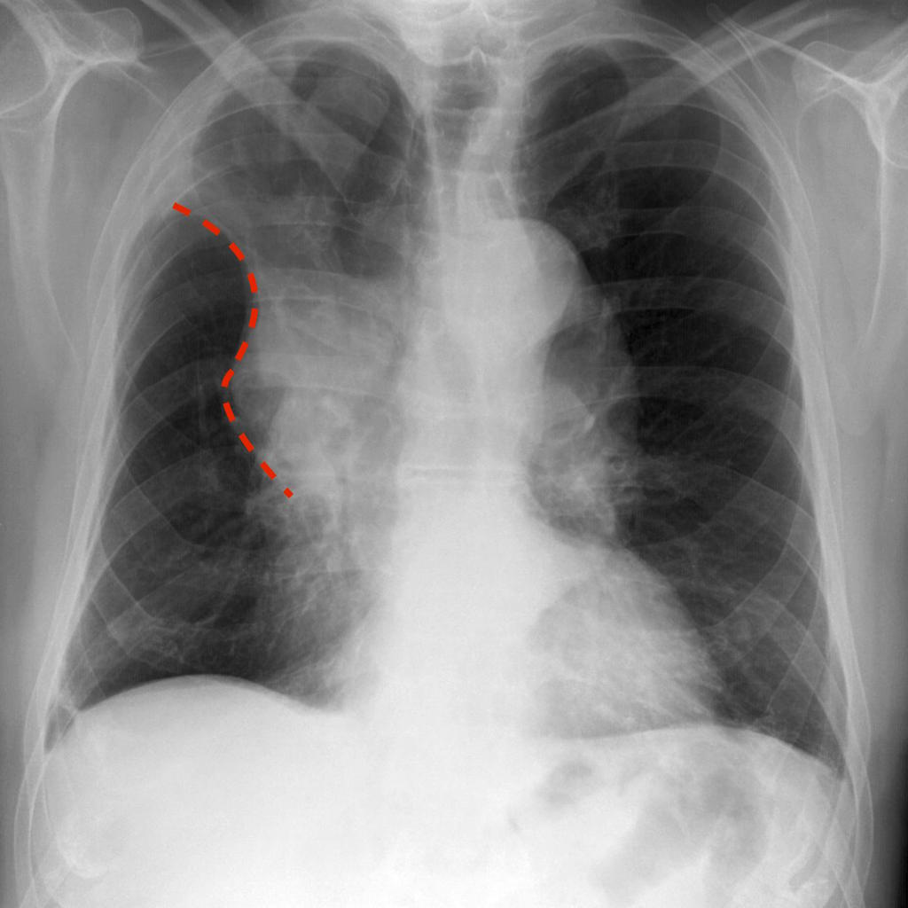
Golden "S" Sign (or reverse "S" sign of Golden) : right upper lobar collapse (the right upper lobe appearing dense and shifting medially and upwards, with a central mass expanding the hilum
-
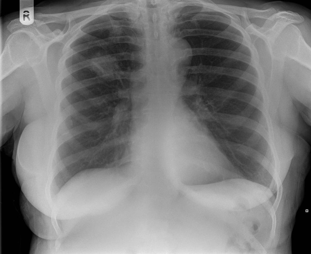
Squameous cell lung cancer: lung cavitating mass left upper lobe adjacent to the oblique fissure. The prominent air-fluid level is best seen on the lateral radiograph
-
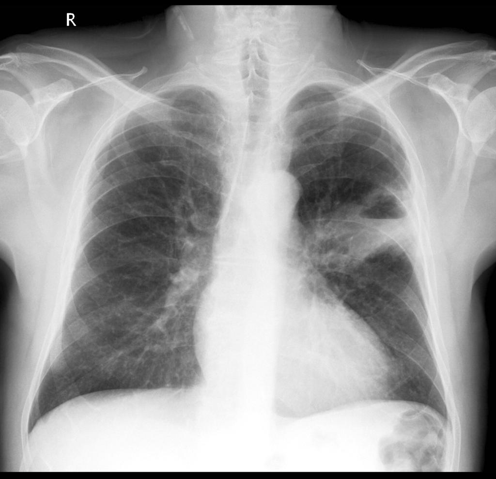
Luftsichel sign: curvilinear opacity at the left apex represents compensatory hyperinflation of the left lower lobe
-
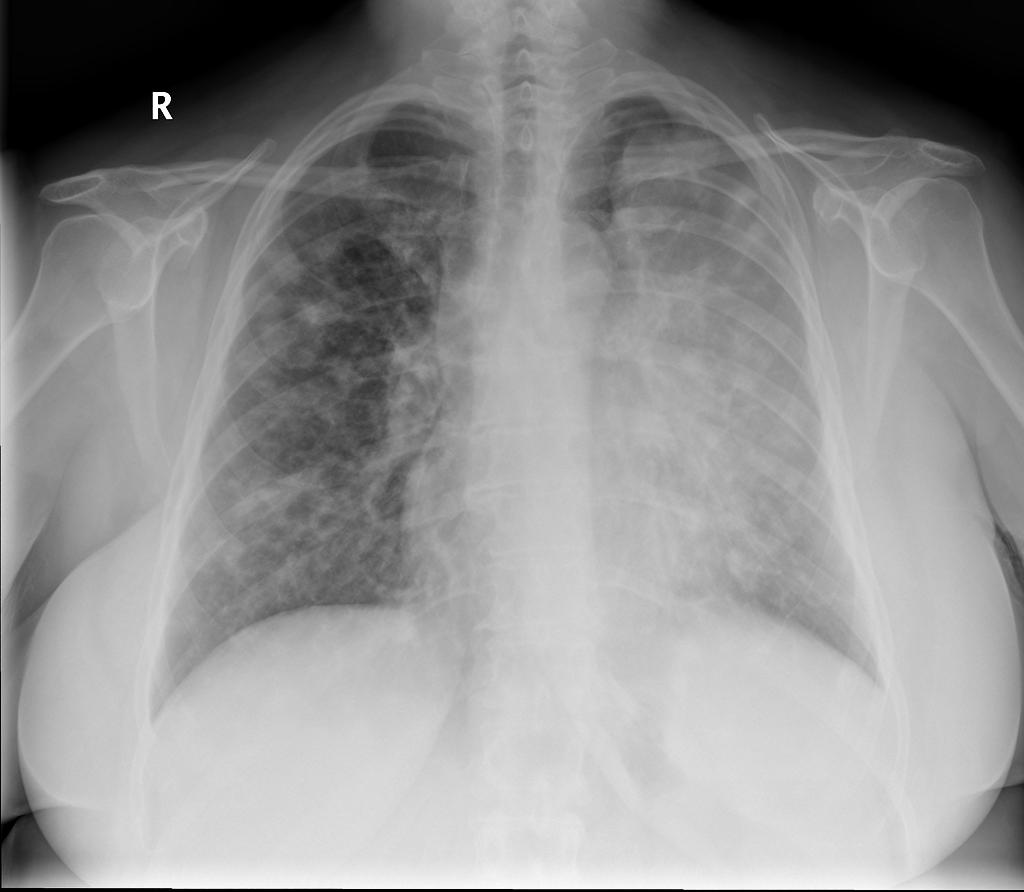
Coin lesion sign: round or oval, well-circumscribed lesion, compatible with primary lung cancer


CT
Malignant Lung Mass
-
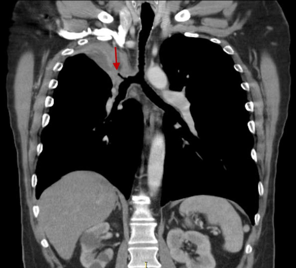
Bronchogenic lung carcincoma: upper lobe collapse
-
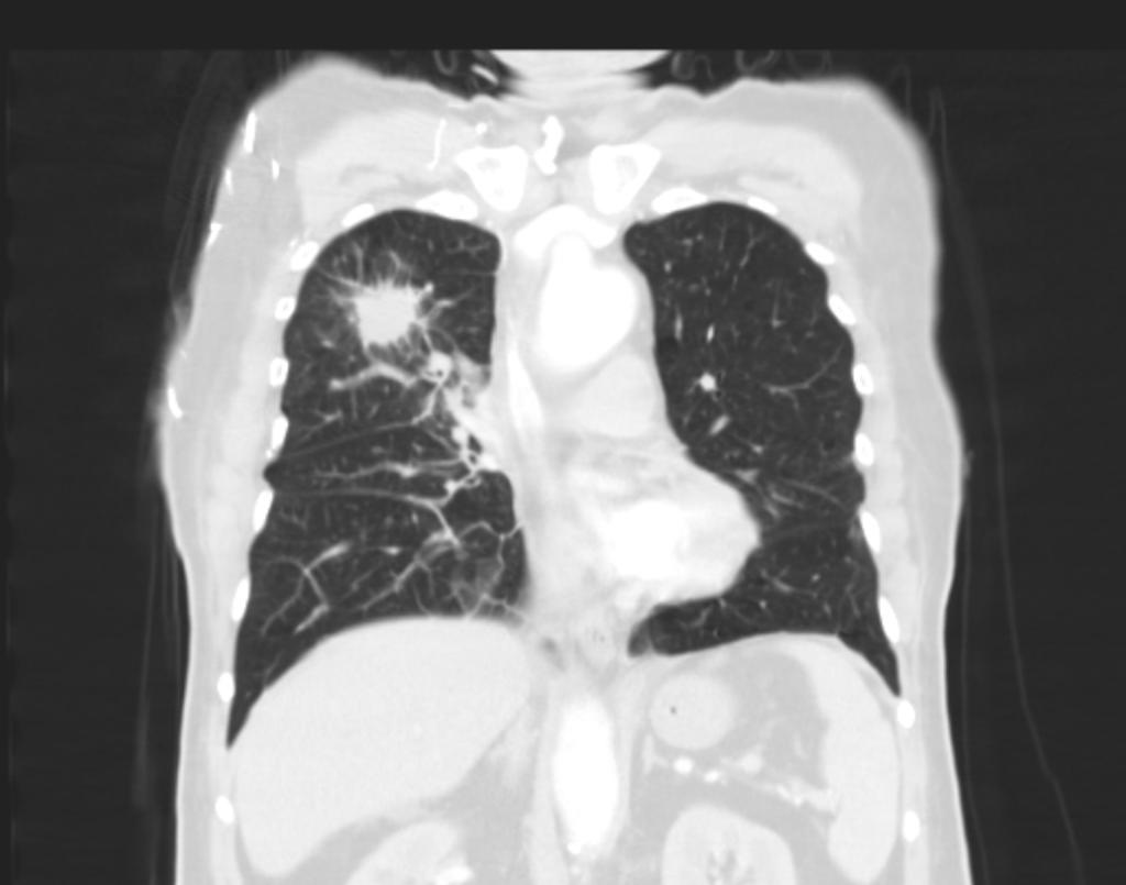
Bronchogenic lung carcincoma: upper lobe with lymphangitic spread
-
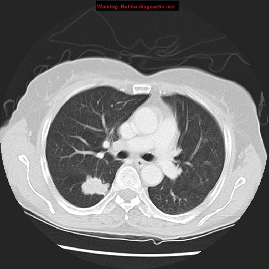
Adenocarcinoma of the lung: ground-glass attenuation corresponds to a lepidic growth pattern and the solid component correspond to invasive patterns.
-
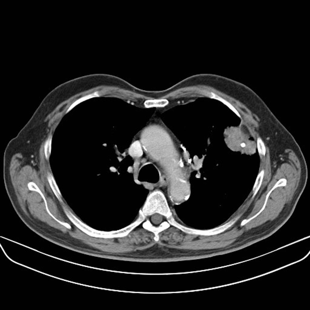
Squamous cell lung carcinoma: Peripheral squamous cell lung carcinoma may be seen as a solid nodule/mass with or without an irregular border. The irregular margin can be attributed to a desmoplastic reaction or infiltrative growth


Benign Lung Mass
-
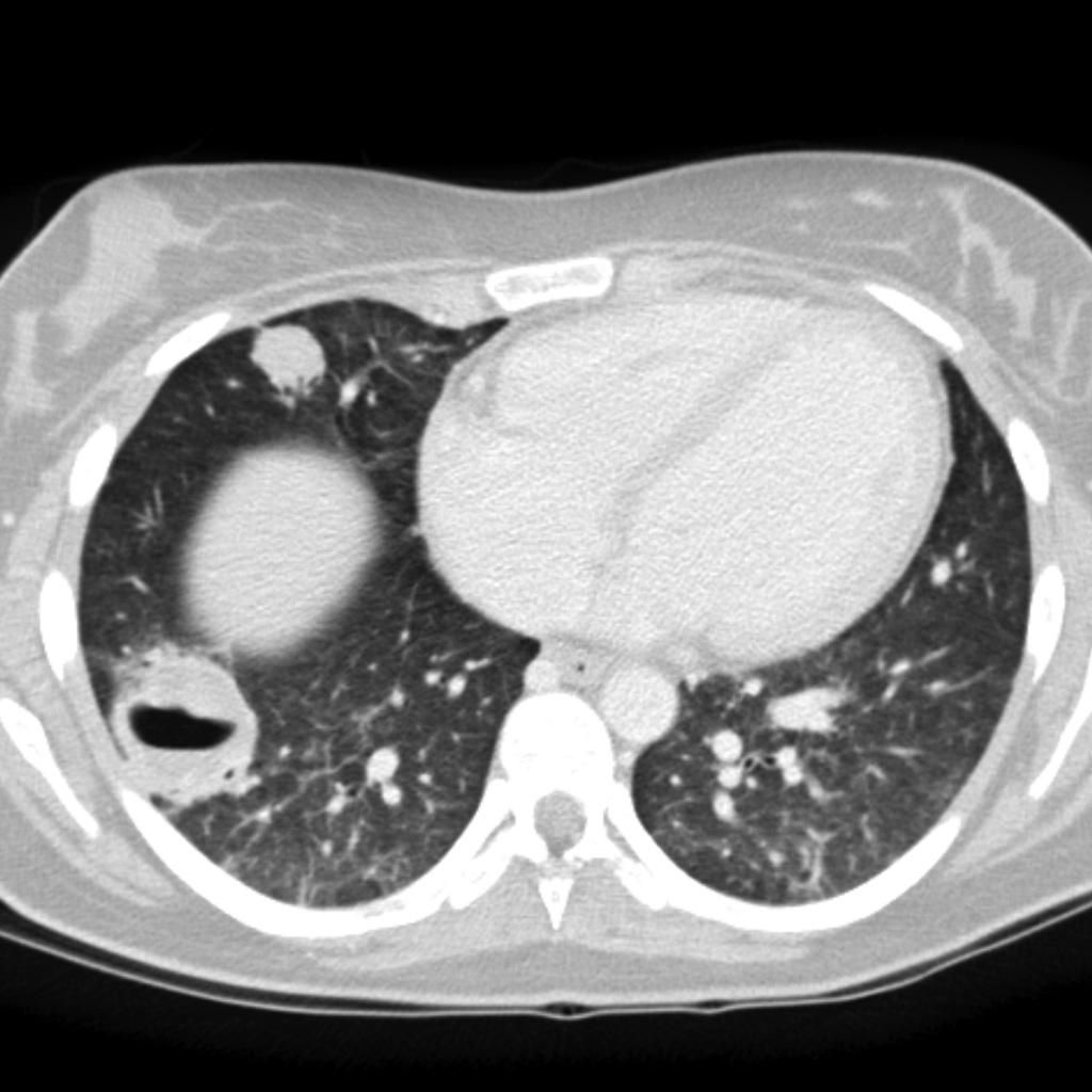
Wegener granulomatosis

References
- ↑ Albert RH, Russell JJ (2009). "Evaluation of the solitary pulmonary nodule". Am Fam Physician. 80 (8): 827–31. PMID 19835344.
- ↑ Kundel HL (1981). "Predictive value and threshold detectability of lung tumors". Radiology. 139 (1): 25–9. doi:10.1148/radiology.139.1.7208937. PMID 7208937.
- ↑ Hochhegger B, Marchiori E, Sedlaczek O, Irion K, Heussel CP, Ley S, Ley-Zaporozhan J, Soares Souza A, Kauczor HU (2011). "MRI in lung cancer: a pictorial essay". Br J Radiol. 84 (1003): 661–8. doi:10.1259/bjr/24661484. PMC 3473490. PMID 21697415.