Lower Limb: Difference between revisions
Irfan Dotani (talk | contribs) |
Matt Pijoan (talk | contribs) No edit summary |
||
| (24 intermediate revisions by 2 users not shown) | |||
| Line 1: | Line 1: | ||
{{CMG}}; | {{CMG}}; {{AE}} {{I.D.}} | ||
{| class="wikitable" | {| class="wikitable" | ||
| Line 23: | Line 22: | ||
| Hip | | Hip | ||
| Ilium | | | ||
*[[Ilium]] | |||
*[[Ischium]] | |||
*[[Pubic Bone]] | |||
*[[Acetabulum]] | |||
*[[Foramen obturatum]] | |||
| [[Piriformis muscle|Piriformis]] | | | ||
*[[Piriformis muscle|Piriformis]] | |||
*[[Superior gemellus]] | |||
*[[Inferior gemellus]] | |||
*[[Tensor fasciae latae]] | |||
*[[Sartorius]] | |||
*[[Gluteus medius]] | |||
*[[Gluteus minimus]] | |||
| Iliofemoral ligament | | | ||
*[[Iliofemoral ligament]] | |||
*[[Pubofemoral ligament]] | |||
*[[Hip|Ischiofemoral ligament]] | |||
*[[Hip joint capsule]] | |||
| Gluteal artery | | | ||
*[[Gluteal artery]] | |||
*[[Pudendal artery]] | |||
*[[Perforating arteries]] | |||
*[[Femoral artery]] | |||
*[[Obturator artery]] | |||
| Great saphenous vein | | | ||
*[[Great saphenous vein]] | |||
*[[Femoral vein]] | |||
| Saphenous nerve | | | ||
*[[Saphenous nerve]] | |||
*[[Obturator nerve]] | |||
*[[Femoral nerve]] | |||
*[[Clunial nerve]] | |||
*[[Sciatic nerve]] | |||
*[[Cutaneous nerve]] | |||
*[[Gluteal nerve]] | |||
*[[Pudendal nerve]] | |||
|- | |- | ||
| Line 39: | Line 69: | ||
| Knee | | Knee | ||
| Femur | | | ||
*[[Femur]] | |||
*[[Tibia]] | |||
*[[Patella]] | |||
| Quadriceps femoris muscle | | | ||
*[[Quadriceps femoris muscle]] | |||
*[[Hamstring]] | |||
*[[Gastrocnemius muscle]] | |||
*[[Vastus mediali]] | |||
*[[Vastus lateralis muscle]] | |||
*[[Popliteus muscle]] | |||
*[[Soleus muscle]] | |||
*[[Articularis genus muscle]] | |||
| Anterior cruciate ligament (ACL) | | | ||
*[[Anterior cruciate ligament]](ACL) | |||
*[[Posterior cruciate ligament]](PCL) | |||
*[[Medial collateral ligament]](MCL) | |||
*[[Lateral collateral ligament]](LCL) | |||
*[[patellofemoral joint]] | |||
*[[tibiofemoral joint]] | |||
| Genicular artery | | | ||
*[[Genicular artery]] | |||
*[[Popliteal artery]] | |||
*[[Tibial artery]] | |||
| Varicose veins | | | ||
*[[Varicose veins]] | |||
*[[Femoral veins]] | |||
| Sciatic nerve | | | ||
*[[Sciatic nerve]] | |||
*[[Tibial nerve]] | |||
*[[Peroneal nerve]] | |||
|- | |- | ||
| Line 55: | Line 110: | ||
| Ankle | | Ankle | ||
| Fibula | | | ||
*[[Fibula]] | |||
*[[Tibula]] | |||
*[[Talus]] | |||
*[[Medial malleolus]] | |||
*[[Lateral malleolus]] | |||
| Anterior tibial | | | ||
*[[Anterior tibial]] | |||
*[[Posterior tibial]] | |||
*[[Peroneal tibial] | |||
*[[Extensors]] | |||
*[[Flexors]] | |||
| Tibiofibular ligament | | | ||
*[[Tibiofibular ligament]] | |||
*[[Deltoid ligament]] | |||
*[[Tibiofibular Syndesmosis joint]] | |||
*[[Motrise Joint]] | |||
*[[Plantar fascia]] | |||
| Anterior tibial artery | | | ||
*[[Anterior tibial artery]] | |||
*[[Peroneal (fibular) artery]] | |||
*[[Anterior medial malieolar artery]] | |||
*[[Plantar artery]] | |||
*[[Communicating branch]] | |||
| Popliteal vein | | | ||
*[[Popliteal vein]] | |||
*[[saphenous vein]] | |||
*[[Femoral vein]] | |||
*[[Tributaries of LSV]] | |||
| Peroneal vein | | | ||
*[[Peroneal vein]] | |||
*[[Sural nerve]] | |||
*[[Tibial nerve]] | |||
*[[Fibular nerve]] | |||
|- | |- | ||
| Line 71: | Line 154: | ||
| Thigh | | Thigh | ||
| Femur | | | ||
*[[Femur]] | |||
*[[Tibia]] | |||
*[[Fibula]] | |||
| Quadriceps femoris muscle | | | ||
*[[Quadriceps femoris muscle]] | |||
*[[Hamstring]] | |||
*[[Biceps femoris muscle]] | |||
*[[Vastus medialis]] | |||
*[[Adductor longus muscle]] | |||
*[[Vastus lateralis muscle]] | |||
*[[Sartorius muscle]] | |||
*[[Semitendinosus muscle]] | |||
*[[Semimembranosus muscle]] | |||
*[[Gracilis muscle]] | |||
*[[Adductor magnus muscle]] | |||
*[[Pectineus muscle]] | |||
*[[Adductor brevis muscle]] | |||
*[[Illiopsoas]] | |||
*[[Illiacus muscle]] | |||
*[[Tensor fasciae latae muscle]] | |||
*[[External obturator muscle]] | |||
*[[Quadratus femoris muscle]] | |||
*[[Articularis genus muscle]] | |||
| Hip joint capsule | | | ||
*[[Hip joint capsule]] | |||
*[[Iliofemoral ligament]] | |||
*[[Pubofemoral ligament]] | |||
*[[Ischiofemoral ligament]] | |||
| Gluteal artery | | | ||
*[[ Gluteal artery]] | |||
*[[Pudendal artery]] | |||
*[[Perforating arteries]] | |||
*[[Femoral artery]] | |||
*[[Obturator artery]] | |||
| Great saphenous vein | | | ||
*[[Great saphenous vein]] | |||
*[[Femoral vein]] | |||
| Saphenous nerve | | | ||
*[[Saphenous nerve]] | |||
*[[Obturator nerve]] | |||
*[[Femoral nerve]] | |||
*[[Clunial nerve]] | |||
*[[Sciatic nerve]] | |||
*[[Cutaneous nerve]] | |||
*[[Gluteal nerve]] | |||
*[[Pudendal nerve]] | |||
|- | |- | ||
| Line 87: | Line 211: | ||
| Foot | | Foot | ||
| Phalanges | | | ||
*[[Phalanges]] | |||
*[[Metatarsals]] | |||
*[[Cuneiform bones]] | |||
*[[Cuboid bone]] | |||
*[[Navicular bone]] | |||
| Abductor hallucis muscle | | | ||
*[[Abductor hallucis muscle]] | |||
*[[Extensor digitorum brevis muscle]] | |||
*[[Flexor digitorum brevis muscle]] | |||
*[[Tibialis anterior muscle]] | |||
*[[Extensor hallucis longus muscle]] | |||
*[[Flexor hallucis Brevis muscle]] | |||
*[[Plantar interossei muscles]] | |||
*[[Quadratus plantar muscle]] | |||
*[[Abductor digiti minimi muscle of foot]] | |||
*[[Lumbricals of the hand]] | |||
*[[Dorsal interossei of the foot]] | |||
*[[Extensor hallucis brevis muscle]] | |||
| Inferior (Distal) Tibiofibular Joint | | | ||
*[[Inferior (Distal) Tibiofibular Joint]] | |||
*[[Talocalcaneal Joint]] | |||
*[[Talocalcaneonavicular Joint]] | |||
*[[Calcaneocuboid Joint]] | |||
*[[Naviculocuneiform Joint]] | |||
*[[Cuboideonavicular Joint]] | |||
*[[Intercuneiform And Cuneocuboid Joints]] | |||
*[[Tarsometatarsal Joints]] | |||
*[[Intermetatarsal Joints]] | |||
*[[Metatarsophalangeal Joints]] | |||
*[[Interphalangeal Joints]] | |||
*[[Cuboideonavicular ligament]] | |||
*[[Intercuneiform ligament]] | |||
*[[Metatarsal ligament]] | |||
| Dorsalis pedis artery | | | ||
*[[Dorsalis pedis artery]] | |||
*[[Posterior tibial artery]] | |||
*[[Anterior tibial artery]] | |||
*[[Arcuate artery of the foot]] | |||
*[[Medial plantar artery]] | |||
*[[Plantar arc]] | |||
*[[Deep plantar artery]] | |||
*[[Plantar metatarsal arteries]] | |||
*[[Medial tarsal arteries]] | |||
*[[Proper plantar digital arteries]] | |||
| Superficial dorsal vein | | | ||
*[[Superficial dorsal vein]] | |||
*[[Lateral plantar vein]] | |||
*[[Saphenous vein]] | |||
*[[Posterior tibial vein]] | |||
| Lateral plantar nerve | | | ||
*[[Lateral plantar nerve]] | |||
*[[Tibial nerve]] | |||
*[[Medial plantar nerve]] | |||
*[[Plantar digital nerves]] | |||
|} | |} | ||
| Line 111: | Line 284: | ||
|Ilium | |Ilium | ||
|The ilium forms the sacroiliac joint with the sacrum along its medial side and forms the superior end of the hip joint at the acetabulum. The sacroiliac joint is a planar joint that allows a slight degree of gliding between the pelvis and the spinal column. The hip joint is a ball-and-socket joint that permits the thigh to have a free range of motion. | |The ilium forms the sacroiliac joint with the sacrum along its medial side and forms the superior end of the hip joint at the acetabulum. The sacroiliac joint is a planar joint that allows a slight degree of gliding between the pelvis and the spinal column. The hip joint is a ball-and-socket joint that permits the thigh to have a free range of motion. | ||
| | |[[File:Pelvis_diagram.png|300px|thumb|By Je at uwo at English Wikipedia (Transferred from en.wikipedia to Commons.) [Public domain], via Wikimedia Commons]] | ||
|- | |- | ||
|Ischium | |Ischium | ||
|Forms the lower and back part of the hip bone. | |Forms the lower and back part of the hip bone. | ||
| | |[[File:500px-Gray341.png|300px|thumb|By Henry Vandyke Carter - Henry Gray (1918) Anatomy of the Human Body (See "Book" section below)Bartleby.com: Gray's Anatomy, Plate 341, Public Domain, https://commons.wikimedia.org/w/index.php?curid=108259]] | ||
|- | |- | ||
|Pubic Bone | |Pubic Bone | ||
|The ventral and anterior of the three parts that come together to create the pelvic bone. | |The ventral and anterior of the three parts that come together to create the pelvic bone. | ||
| | |[[File:Bassin osseux.jpg|300px|thumb|By Auteur: d.renardOriginal uploader was D.renard at fr.wikipedia - Dessin personnel avec légendesOriginally from fr.wikipedia; description page is/was here., Copyrighted free use, https://commons.wikimedia.org/w/index.php?curid=1370283]] | ||
|- | |- | ||
|Acetabulum | |Acetabulum | ||
|A cup-shaped opening on each side of the pelvic girdle formed where the ischium, ilium, and pubis all meet, and into which the head of the femur inserts. | |A cup-shaped opening on each side of the pelvic girdle formed where the ischium, ilium, and pubis all meet, and into which the head of the femur inserts. | ||
| | |[[File:Pelvic_girdle_illustration.svg.png|300px|thumb|By Original: U.S. National Cancer Institute; Vectorization: Fred the Oyster; German translation kopiersperre/Rothwild (Own work based on: Illu pelvic girdle.jpg) [CC BY-SA 4.0 (https://creativecommons.org/licenses/by-sa/4.0)], via Wikimedia Commons]] | ||
|- | |- | ||
|Foramen obturatum | |Foramen obturatum | ||
|The large opening created by the ischium and pubis bones of the pelvis through which nerves and blood vessels pass. | |The large opening created by the ischium and pubis bones of the pelvis through which nerves and blood vessels pass. | ||
| | |[[File:Pelvis_diagram.png|300px|thumb|By Je at uwo at English Wikipedia (Transferred from en.wikipedia to Commons.) [Public domain], via Wikimedia Commons]] | ||
|} | |} | ||
| Line 139: | Line 312: | ||
|Femur | |Femur | ||
|Supports the weight of the body and allowing motion of the leg. The femur articulates proximally with the acetabulum of the pelvis forming the hip joint, and distally with the tibia and patella to form the knee joint. | |Supports the weight of the body and allowing motion of the leg. The femur articulates proximally with the acetabulum of the pelvis forming the hip joint, and distally with the tibia and patella to form the knee joint. | ||
| | |[[File:Femur_head.png|300px|thumb|See page for author [Public domain or Public domain], via Wikimedia Commons]] | ||
|- | |- | ||
|Tibia | |Tibia | ||
|It forms the knee joint with the femur and the ankle joint with the fibula and tarsus. | |It forms the knee joint with the femur and the ankle joint with the fibula and tarsus. | ||
| | |[[File:250px-Tibia - frontal view.png|300px|thumb|Henry Vandyke Carter [Public domain], via Wikimedia Commons]] | ||
|- | |- | ||
|Patella | |Patella | ||
|The patella increases the leverage that the quadriceps tendon can exert on the femur by increasing the angle at which it acts. Also acts as protection for the muscles underneath the patella. | |The patella increases the leverage that the quadriceps tendon can exert on the femur by increasing the angle at which it acts. Also acts as protection for the muscles underneath the patella. | ||
| | |[[File:Illu_lower_extremity.jpg|240px|thumb|Public Domain, https://commons.wikimedia.org/w/index.php?curid=789643]] | ||
|} | |} | ||
| Line 159: | Line 332: | ||
|Fibula | |Fibula | ||
|Long, thin and lateral bone of the lower leg. It runs parallel to the tibia, or shin bone, and plays a significant role in stabilizing the ankle and supporting the muscles of the lower leg. | |Long, thin and lateral bone of the lower leg. It runs parallel to the tibia, or shin bone, and plays a significant role in stabilizing the ankle and supporting the muscles of the lower leg. | ||
| | |[[File:500px-Fibula - anterior view.png|300px|thumb|By Anatomography - en:Anatomography (setting page of this image), CC BY-SA 2.1 jp, https://commons.wikimedia.org/w/index.php?curid=24719980]] | ||
|- | |- | ||
|Tibula | |Tibula | ||
|Known as the shinbone and is the second largest bone in the body. Helps with weight-bearing and stabilization. | |Known as the shinbone and is the second largest bone in the body. Helps with weight-bearing and stabilization. | ||
| | |[[File:Braus_1921_293.png|300px|thumb|By Braus, Hermann [Public domain], via Wikimedia Commons]] | ||
|- | |- | ||
|Talus | |Talus | ||
|The talus bone forms the primary connection between the lower leg and foot and is vital for mobility. In fact, the structure of the talus bone is so unique it can form the connection between numerous other bones such as the tibia, fibula, calcaneus (heel) and navicular or tarsal bones found in the foot. | |The talus bone forms the primary connection between the lower leg and foot and is vital for mobility. In fact, the structure of the talus bone is so unique it can form the connection between numerous other bones such as the tibia, fibula, calcaneus (heel) and navicular or tarsal bones found in the foot. | ||
| | |[[File:Gray273.png|240px|thumb|Henry Vandyke Carter [Public domain], via Wikimedia Commons]] | ||
|- | |- | ||
|Medial malleolus | |Medial malleolus | ||
|The medial malleolus is the prominence on the inner side of the ankle, formed by the lower end of the tibia. | |The medial malleolus is the prominence on the inner side of the ankle, formed by the lower end of the tibia. | ||
| | |[[File:Gray357.png|300px|thumb|By Henry Vandyke Carter - Henry Gray (1918) Anatomy of the Human Body (See "Book" section below)Bartleby.com: Gray's Anatomy, Plate 357, Public Domain, https://commons.wikimedia.org/w/index.php?curid=566495]] | ||
|- | |- | ||
|Lateral malleolus | |Lateral malleolus | ||
|The lateral malleolus is the prominence on the outer side of the ankle, formed by the lower end of the tibia. | |The lateral malleolus is the prominence on the outer side of the ankle, formed by the lower end of the tibia. | ||
| | |[[File:Gray1239.png|300px|thumb|Henry Vandyke Carter [Public domain], via Wikimedia Commons]] | ||
|} | |} | ||
| Line 187: | Line 360: | ||
|Femur | |Femur | ||
|Supports the weight of the body and allowing motion of the leg. The femur articulates proximally with the acetabulum of the pelvis forming the hip joint, and distally with the tibia and patella to form the knee joint. | |Supports the weight of the body and allowing motion of the leg. The femur articulates proximally with the acetabulum of the pelvis forming the hip joint, and distally with the tibia and patella to form the knee joint. | ||
| | |[[File:fibulaimage.jpg|300px|thumb|By Henry Vandyke Carter - Henry Gray (1918) Anatomy of the Human Body (See "Book" section below)Bartleby.com: Gray's Anatomy, Plate 244, Public Domain, https://commons.wikimedia.org/w/index.php?curid=30613]] | ||
|- | |- | ||
|Tibia | |Tibia | ||
|It forms the knee joint with the femur and the ankle joint with the fibula and tarsus. | |It forms the knee joint with the femur and the ankle joint with the fibula and tarsus. | ||
| | |[[File:tibiapresent.jpg|300px|thumb|By Braus, Hermann - Anatomie des Menschen: ein Lehrbuch für Studierende und Ärzte, Public Domain, https://commons.wikimedia.org/w/index.php?curid=29934112]] | ||
|- | |- | ||
|Fibula | |Fibula | ||
|Long, thin and lateral bone of the lower leg. It runs parallel to the tibia, or shin bone, and plays a significant role in stabilizing the ankle and supporting the muscles of the lower leg. | |Long, thin and lateral bone of the lower leg. It runs parallel to the tibia, or shin bone, and plays a significant role in stabilizing the ankle and supporting the muscles of the lower leg. | ||
| | |[[File:download-1.jpg|300px|thumb|By Anatomography - en:Anatomography (setting page of this image), CC BY-SA 2.1 jp, https://commons.wikimedia.org/w/index.php?curid=24719980]] | ||
|} | |} | ||
| Line 207: | Line 380: | ||
|Phalanges | |Phalanges | ||
|The phalanges of the foot help us balance, walk, and run. | |The phalanges of the foot help us balance, walk, and run. | ||
| | |[[File:Images.jpg|300px|thumb|By Mariana Ruiz Villarreal (LadyofHats); retouches by Nyks - Own work. Image renamed from Image:Human hand bones simple-edit1-2.svg, Public Domain, https://commons.wikimedia.org/w/index.php?curid=3949051]] | ||
|- | |- | ||
|Metatarsals | |Metatarsals | ||
|Metatarsals are convex in shape (arch upward), are long bones, and give the foot its arch. They work with connective tissues, ligaments, and tendons to provide movement in the foot | |Metatarsals are convex in shape (arch upward), are long bones, and give the foot its arch. They work with connective tissues, ligaments, and tendons to provide movement in the foot | ||
| | |[[File:metatarsalsimages.jpg|300px|thumb|By BodyParts3D is made by DBCLS. - Polygon data is from BodyParts3D, CC BY-SA 2.1 jp, https://commons.wikimedia.org/w/index.php?curid=28131678]] | ||
|- | |- | ||
|Cuneiform bones | |Cuneiform bones | ||
|This bone is cube-shaped and connects the foot and the ankle. It also provides stability to the foot. | |This bone is cube-shaped and connects the foot and the ankle. It also provides stability to the foot. | ||
| | |[[File:500px-Gray268.png|300px|thumb|By Henry Vandyke Carter - Henry Gray (1918) Anatomy of the Human Body (See "Book" section below)Bartleby.com: Gray's Anatomy, Plate 268, Public Domain, https://commons.wikimedia.org/w/index.php?curid=792353]] | ||
|- | |- | ||
|Cuboid bone | |Cuboid bone | ||
|The cuboid bone is one of the seven tarsal bones located on the lateral (outer) side of the foot. This bone is cube-shaped and connects the foot and the ankle. It also provides stability to the foot. | |The cuboid bone is one of the seven tarsal bones located on the lateral (outer) side of the foot. This bone is cube-shaped and connects the foot and the ankle. It also provides stability to the foot. | ||
| | |[[File:cuboidbonedownload.jpg|300px|thumb|By Henry Vandyke Carter - Henry Gray (1918) Anatomy of the Human Body (See "Book" section below)Bartleby.com: Gray's Anatomy, Plate 274, Public Domain, https://commons.wikimedia.org/w/index.php?curid=792356]] | ||
|- | |- | ||
|Navicular bone | |Navicular bone | ||
|The navicular is a boat-shaped bone located in the top inner side of the foot, just above the transverse. It helps connect the talus, or anklebone, to the cuneiform bones of the foot | |The navicular is a boat-shaped bone located in the top inner side of the foot, just above the transverse. It helps connect the talus, or anklebone, to the cuneiform bones of the foot | ||
| | |[[File:navicularboneimage.jpg|300px|thumb|By Anatomist90 - Own work, CC BY-SA 3.0, https://commons.wikimedia.org/w/index.php?curid=17431658]] | ||
|} | |} | ||
| Line 239: | Line 412: | ||
|[[Piriformis muscle|Piriformis]] | |[[Piriformis muscle|Piriformis]] | ||
| | | | ||
* Part of | * Part of the lateral rotators of the hip, along with the quadratus femoris, gemellus inferior, gemellus superior, obturator externus, and obturator internus | ||
* The piriformis laterally rotates the femur with hip extension and abducts the femur with hip flexion | * The piriformis laterally rotates the femur with hip extension and abducts the femur with hip flexion | ||
|Arise: | |Arise: | ||
| Line 255: | Line 428: | ||
* Superior gluteal | * Superior gluteal | ||
* Internal pudendal arteries | * Internal pudendal arteries | ||
| | |[[File:Sobo 1909 298.png|300px|thumb|By Henry Vandyke Carter - Henry Gray (1918) Anatomy of the Human Body (See "Book" section below)Bartleby.com: Gray's Anatomy, Plate 341, Public Domain, https://commons.wikimedia.org/w/index.php?curid=108259]] | ||
|- | |- | ||
| | | | ||
| Line 273: | Line 446: | ||
Arterial supply: | Arterial supply: | ||
*Inferior gluteal artery | *Inferior gluteal artery | ||
| | |[[File:superiorgemellus.jpg|300px|thumb|Henry Vandyke Carter [Public domain], via Wikimedia Commons]] | ||
|- | |- | ||
| | | | ||
| Line 290: | Line 463: | ||
Arterial supply: | Arterial supply: | ||
*Inferior gluteal artery | *Inferior gluteal artery | ||
| | |[[File:inferiorgemellus.jpg|300px|thumb|Henry Vandyke Carter [Public domain], via Wikimedia Commons]] | ||
|- | |- | ||
| | | | ||
| Line 308: | Line 481: | ||
Arterial supply: | Arterial supply: | ||
*Superior gluteal and lateral circumflex femoral artery. | *Superior gluteal and lateral circumflex femoral artery. | ||
| | |[[File:TFL.png|300px|thumb|By Henry Vandyke Carter - Henry Gray (1918) Anatomy of the Human Body (See "Book" section below)Bartleby.com: Gray's Anatomy, Plate 341, Public Domain, https://commons.wikimedia.org/w/index.php?curid=108259]] | ||
|- | |- | ||
| | | | ||
| Line 325: | Line 498: | ||
Arterial supply: | Arterial supply: | ||
*Muscular branches of the femoral artery. | *Muscular branches of the femoral artery. | ||
| | |[[File:Sartorius.jpg|300px|thumb|Henry Vandyke Carter [Public domain], via Wikimedia Commons]] | ||
|- | |- | ||
| | | | ||
| Line 342: | Line 515: | ||
Arterial supply: | Arterial supply: | ||
*Superior gluteal artery | *Superior gluteal artery | ||
| | |[[File:Gluteusmedius.jpg|300px|thumb|By Henry Vandyke Carter - Henry Gray (1918) Anatomy of the Human Body (See "Book" section below)Bartleby.com: Gray's Anatomy, Plate 341, Public Domain, https://commons.wikimedia.org/w/index.php?curid=108259]] | ||
|- | |- | ||
| | | | ||
| Line 359: | Line 532: | ||
Arterial supply: | Arterial supply: | ||
*Superior gluteal artery | *Superior gluteal artery | ||
| | |[[File:Gluteusminimus.jpg|300px|thumb|By Original: U.S. National Cancer Institute; Vectorization: Fred the Oyster; German translation kopiersperre/Rothwild (Own work based on: Illu pelvic girdle.jpg) [CC BY-SA 4.0 (https://creativecommons.org/licenses/by-sa/4.0)], via Wikimedia Commons]] | ||
|- | |- | ||
| | | | ||
| Line 376: | Line 549: | ||
Arterial supply: | Arterial supply: | ||
*Inferior and superior gluteal arteries and the first perforating branch of the profunda femoris artery. | *Inferior and superior gluteal arteries and the first perforating branch of the profunda femoris artery. | ||
| | |[[File:Gluteusmaximus.jpg|300px|thumb|By Original: U.S. National Cancer Institute; Vectorization: Fred the Oyster; German translation kopiersperre/Rothwild (Own work based on: Illu pelvic girdle.jpg) [CC BY-SA 4.0 (https://creativecommons.org/licenses/by-sa/4.0)], via Wikimedia Commons]] | ||
|} | |} | ||
| Line 403: | Line 576: | ||
Arterial supply: | Arterial supply: | ||
*Medial circumflex femoral artery, inferior gluteal artery, 1st - 4th perforating arteries, obturator artery, and some superior muscular branches of popliteal artery. | *Medial circumflex femoral artery, inferior gluteal artery, 1st - 4th perforating arteries, obturator artery, and some superior muscular branches of popliteal artery. | ||
| | |[[File:Quadratusfemoris.jpg|300px|thumb|By Original: U.S. National Cancer Institute; Vectorization: Fred the Oyster; German translation kopiersperre/Rothwild (Own work based on: Illu pelvic girdle.jpg) [CC BY-SA 4.0 (https://creativecommons.org/licenses/by-sa/4.0)], via Wikimedia Commons]] | ||
|- | |- | ||
| | | | ||
| Line 420: | Line 593: | ||
Arterial supply: | Arterial supply: | ||
*Each head supplied by a sural branch of the popliteal artery. | *Each head supplied by a sural branch of the popliteal artery. | ||
| | |[[File:Gastrocnemius.png|300px|thumb|Henry Vandyke Carter [Public domain], via Wikimedia Commons]] | ||
|- | |- | ||
| | | | ||
| Line 437: | Line 610: | ||
Arterial supply: | Arterial supply: | ||
*Femoral artery, profunda femoris artery, and superior medial genicular branch of popliteal artery. | *Femoral artery, profunda femoris artery, and superior medial genicular branch of popliteal artery. | ||
| | |[[File:vastus-medialis.jpg|300px|thumb|Henry Vandyke Carter [Public domain], via Wikimedia Commons]] | ||
|- | |- | ||
| | | | ||
| Line 454: | Line 627: | ||
Arterial supply: | Arterial supply: | ||
*Lateral circumflex femoral artery. | *Lateral circumflex femoral artery. | ||
| | |[[File:vastuslateralis.jpg|300px|thumb|Henry Vandyke Carter [Public domain], via Wikimedia Commons]] | ||
|- | |- | ||
| | | | ||
| Line 471: | Line 644: | ||
Arterial supply: | Arterial supply: | ||
*Medial inferior genicular branch of the popliteal artery and muscular branch of the posterior tibial artery. | *Medial inferior genicular branch of the popliteal artery and muscular branch of the posterior tibial artery. | ||
| | |[[File:popliteus.jpg|300px|thumb|Henry Vandyke Carter [Public domain], via Wikimedia Commons]] | ||
|- | |- | ||
| | | | ||
| Line 488: | Line 661: | ||
Arterial supply: | Arterial supply: | ||
*Posterior tibial, peroneal, and sural arteries. | *Posterior tibial, peroneal, and sural arteries. | ||
| | |[[File:soleus.jpg|300px|thumb|By Henry Vandyke Carter - Henry Gray (1918) Anatomy of the Human Body (See "Book" section below)Bartleby.com: Gray's Anatomy, Plate 341, Public Domain, https://commons.wikimedia.org/w/index.php?curid=108259]] | ||
|} | |} | ||
| Line 515: | Line 688: | ||
Arterial supply: | Arterial supply: | ||
*Anterior tibial artery | *Anterior tibial artery | ||
| | |[[File:tibialisanterior.jpg|300px|thumb|By Original: U.S. National Cancer Institute; Vectorization: Fred the Oyster; German translation kopiersperre/Rothwild (Own work based on: Illu pelvic girdle.jpg) [CC BY-SA 4.0 (https://creativecommons.org/licenses/by-sa/4.0)], via Wikimedia Commons]] | ||
|- | |- | ||
| | | | ||
| Line 532: | Line 705: | ||
Arterial supply: | Arterial supply: | ||
*Muscular branches of sural, peroneal and posterior tibial arteries. | *Muscular branches of sural, peroneal and posterior tibial arteries. | ||
| | |[[File:Tibialisposterior.jpg|300px|thumb|By Original: U.S. National Cancer Institute; Vectorization: Fred the Oyster; German translation kopiersperre/Rothwild (Own work based on: Illu pelvic girdle.jpg) [CC BY-SA 4.0 (https://creativecommons.org/licenses/by-sa/4.0)], via Wikimedia Commons]] | ||
|- | |- | ||
| | | | ||
| Line 549: | Line 722: | ||
Arterial supply: | Arterial supply: | ||
*Anterior tibial artery | *Anterior tibial artery | ||
| | |[[File:extensorlongus.jpg|300px|thumb|By Original: U.S. National Cancer Institute; Vectorization: Fred the Oyster; German translation kopiersperre/Rothwild (Own work based on: Illu pelvic girdle.jpg) [CC BY-SA 4.0 (https://creativecommons.org/licenses/by-sa/4.0)], via Wikimedia Commons]] | ||
|- | |- | ||
| | | | ||
| Line 566: | Line 739: | ||
Arterial supply: | Arterial supply: | ||
*Anterior tibial artery | *Anterior tibial artery | ||
| | |[[File:extensorhlongus.jpg|300px|thumb|By Original: U.S. National Cancer Institute; Vectorization: Fred the Oyster; German translation kopiersperre/Rothwild (Own work based on: Illu pelvic girdle.jpg) [CC BY-SA 4.0 (https://creativecommons.org/licenses/by-sa/4.0)], via Wikimedia Commons]] | ||
|- | |- | ||
| | | | ||
| Line 583: | Line 756: | ||
Arterial supply: | Arterial supply: | ||
*Muscular branch of the posterior tibial artery. | *Muscular branch of the posterior tibial artery. | ||
| | |[[File:flexordlongus.jpg|300px|thumb|Henry Vandyke Carter [Public domain], via Wikimedia Commons]] | ||
|- | |- | ||
| | | | ||
| Line 600: | Line 773: | ||
Arterial supply: | Arterial supply: | ||
*Muscular branch of peroneal and posterior tibial artery. | *Muscular branch of peroneal and posterior tibial artery. | ||
| | |[[File:flexorhlongus.jpg|300px|thumb|By Henry Vandyke Carter - Henry Gray (1918) Anatomy of the Human Body (See "Book" section below)Bartleby.com: Gray's Anatomy, Plate 341, Public Domain, https://commons.wikimedia.org/w/index.php?curid=108259]] | ||
|} | |} | ||
| Line 627: | Line 800: | ||
Arterial supply: | Arterial supply: | ||
*Obturator artery and medial circumflex femoral artery. | *Obturator artery and medial circumflex femoral artery. | ||
| | |[[File:adductorbrevis.jpg|300px|thumb|By Henry Vandyke Carter - Henry Gray (1918) Anatomy of the Human Body (See "Book" section below)Bartleby.com: Gray's Anatomy, Plate 341, Public Domain, https://commons.wikimedia.org/w/index.php?curid=108259]] | ||
|- | |- | ||
| | | | ||
| Line 644: | Line 817: | ||
Arterial supply: | Arterial supply: | ||
*Obturator artery and medial circumflex femoral artery. | *Obturator artery and medial circumflex femoral artery. | ||
| | |[[File:adductorlongus.jpg|300px|thumb|Henry Vandyke Carter [Public domain], via Wikimedia Commons]] | ||
|- | |- | ||
| | | | ||
| Line 661: | Line 834: | ||
Arterial supply: | Arterial supply: | ||
*Obturator artery, medial circumflex femoral artery, and muscular branches of profunda femoris artery. | *Obturator artery, medial circumflex femoral artery, and muscular branches of profunda femoris artery. | ||
| | |[[File:gracilis.jpg|300px|thumb|Henry Vandyke Carter [Public domain], via Wikimedia Commons]] | ||
|- | |- | ||
| | | | ||
| Line 678: | Line 851: | ||
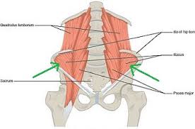
Arterial supply: | Arterial supply: | ||
*Lumbar branch of iliopsoas branch of the internal iliac artery. | *Lumbar branch of iliopsoas branch of the internal iliac artery. | ||
| | |[[File:iliacus.jpg|300px|thumb|By Henry Vandyke Carter - Henry Gray (1918) Anatomy of the Human Body (See "Book" section below)Bartleby.com: Gray's Anatomy, Plate 341, Public Domain, https://commons.wikimedia.org/w/index.php?curid=108259]] | ||
|- | |- | ||
| | | | ||
| Line 695: | Line 868: | ||
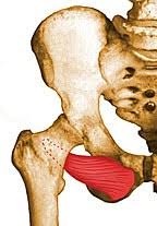
Arterial supply: | Arterial supply: | ||
*Obturator and medial circumflex femoral arteries. | *Obturator and medial circumflex femoral arteries. | ||
| | |[[File:obturatorexternus.jpg|300px|thumb|By Henry Vandyke Carter - Henry Gray (1918) Anatomy of the Human Body (See "Book" section below)Bartleby.com: Gray's Anatomy, Plate 341, Public Domain, https://commons.wikimedia.org/w/index.php?curid=108259]] | ||
|- | |- | ||
| | | | ||
| Line 712: | Line 885: | ||
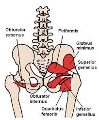
Arterial supply: | Arterial supply: | ||
*Internal pudendal and superior and inferior gluteal arteries. | *Internal pudendal and superior and inferior gluteal arteries. | ||
| | |[[File:obturatorinternus.jpg|300px|thumb|By Original: U.S. National Cancer Institute; Vectorization: Fred the Oyster; German translation kopiersperre/Rothwild (Own work based on: Illu pelvic girdle.jpg) [CC BY-SA 4.0 (https://creativecommons.org/licenses/by-sa/4.0)], via Wikimedia Commons]] | ||
|- | |- | ||
| | | | ||
| Line 729: | Line 902: | ||
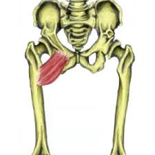
Arterial supply: | Arterial supply: | ||
*Internal pudendal and superior and inferior gluteal arteries. | *Internal pudendal and superior and inferior gluteal arteries. | ||
| | |[[File:pectineus.jpg|300px|thumb|By Original: U.S. National Cancer Institute; Vectorization: Fred the Oyster; German translation kopiersperre/Rothwild (Own work based on: Illu pelvic girdle.jpg) [CC BY-SA 4.0 (https://creativecommons.org/licenses/by-sa/4.0)], via Wikimedia Commons]] | ||
|- | |- | ||
| | | | ||
| Line 746: | Line 919: | ||
Arterial supply: | Arterial supply: | ||
*Superior and inferior gluteal and internal pudendal arteries. | *Superior and inferior gluteal and internal pudendal arteries. | ||
| | |[[File:piriformis.jpg|300px|thumb|By Original: U.S. National Cancer Institute; Vectorization: Fred the Oyster; German translation kopiersperre/Rothwild (Own work based on: Illu pelvic girdle.jpg) [CC BY-SA 4.0 (https://creativecommons.org/licenses/by-sa/4.0)], via Wikimedia Commons]] | ||
|- | |- | ||
| | | | ||
| Line 763: | Line 936: | ||
Arterial supply: | Arterial supply: | ||
*Lumbar branch of iliopsoas branch of the internal iliac artery. | *Lumbar branch of iliopsoas branch of the internal iliac artery. | ||
| | |[[File:psoas.jpg|300px]] | ||
|- | |- | ||
| | | | ||
| Line 780: | Line 953: | ||
Arterial supply: | Arterial supply: | ||
*Perforating branches of profunda femoris artery, inferior gluteal artery, and the superior muscular branches of the popliteal artery. | *Perforating branches of profunda femoris artery, inferior gluteal artery, and the superior muscular branches of the popliteal artery. | ||
| | |[[File:semimebranosus.jpg|300px|thumb|By Original: U.S. National Cancer Institute; Vectorization: Fred the Oyster; German translation kopiersperre/Rothwild (Own work based on: Illu pelvic girdle.jpg) [CC BY-SA 4.0 (https://creativecommons.org/licenses/by-sa/4.0)], via Wikimedia Commons]] | ||
|- | |- | ||
| | | | ||
|Semitendinosus | |Semitendinosus | ||
| | | | ||
* | *Extends the thigh and flexes the knee, and also rotates the tibia medially, especially when the knee is flexed. | ||
| | | | ||
Insertion: | Insertion: | ||
* | *Superior aspect of a medial portion of the tibial shaft. | ||
Arise: | Arise: | ||
* | *From common tendon with long head of biceps femoris from the superior medial quadrant of the posterior portion of the ischial tuberosity. | ||
| | | | ||
Innervation: | Innervation: | ||
* | *Tibial nerve. | ||
| | | | ||
Arterial supply: | Arterial supply: | ||
* | *Perforating branches of profunda femoris artery, inferior gluteal artery, and the superior muscular branches of the popliteal artery. | ||
| | |[[File:semitendinosus.jpg|300px|thumb|By Henry Vandyke Carter - Henry Gray (1918) Anatomy of the Human Body (See "Book" section below)Bartleby.com: Gray's Anatomy, Plate 341, Public Domain, https://commons.wikimedia.org/w/index.php?curid=108259]] | ||
|} | |} | ||
Latest revision as of 15:00, 31 July 2018
Editor-In-Chief: C. Michael Gibson, M.S., M.D. [1]; Associate Editor(s)-in-Chief: Irfan Dotani
Lower limb bony structures
| Parts | Function | Image | |
|---|---|---|---|
| Hip | Ilium | The ilium forms the sacroiliac joint with the sacrum along its medial side and forms the superior end of the hip joint at the acetabulum. The sacroiliac joint is a planar joint that allows a slight degree of gliding between the pelvis and the spinal column. The hip joint is a ball-and-socket joint that permits the thigh to have a free range of motion. | 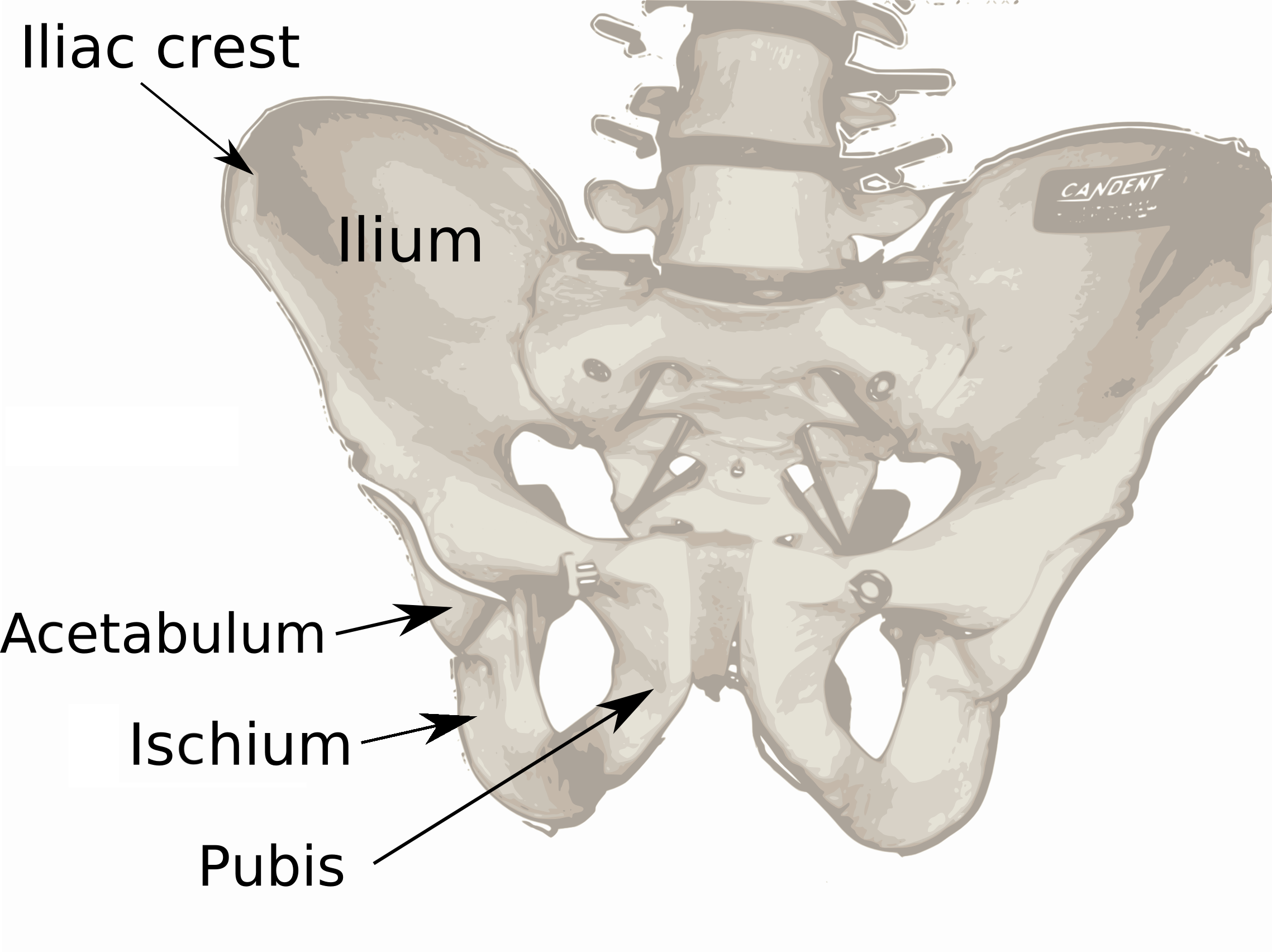 |
| Ischium | Forms the lower and back part of the hip bone. | 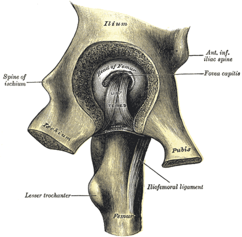 | |
| Pubic Bone | The ventral and anterior of the three parts that come together to create the pelvic bone. | 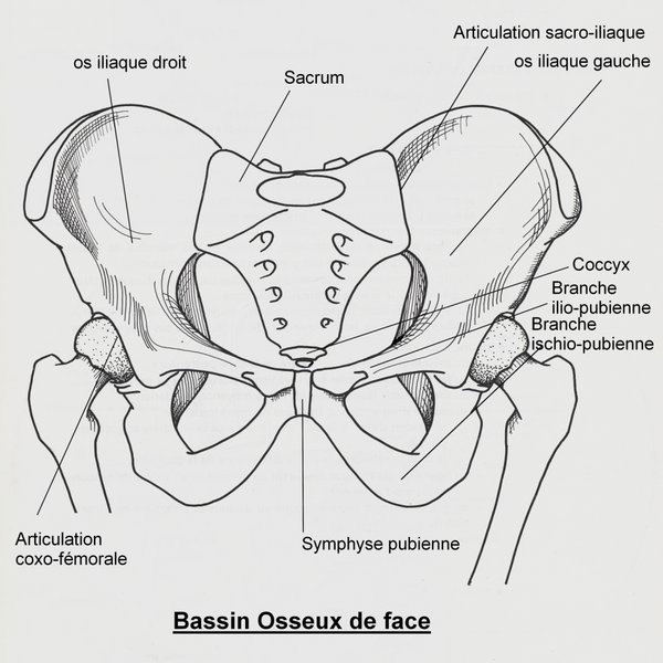 | |
| Acetabulum | A cup-shaped opening on each side of the pelvic girdle formed where the ischium, ilium, and pubis all meet, and into which the head of the femur inserts. | 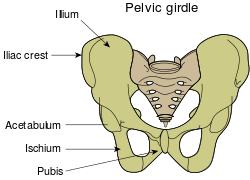 | |
| Foramen obturatum | The large opening created by the ischium and pubis bones of the pelvis through which nerves and blood vessels pass. |  |
| Parts | Function | Image | |
|---|---|---|---|
| Knee | Femur | Supports the weight of the body and allowing motion of the leg. The femur articulates proximally with the acetabulum of the pelvis forming the hip joint, and distally with the tibia and patella to form the knee joint. | 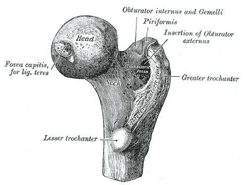 |
| Tibia | It forms the knee joint with the femur and the ankle joint with the fibula and tarsus. | 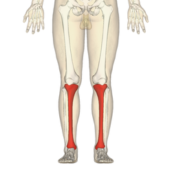 | |
| Patella | The patella increases the leverage that the quadriceps tendon can exert on the femur by increasing the angle at which it acts. Also acts as protection for the muscles underneath the patella. | 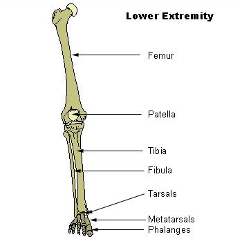 |
| Parts | Function | Image | |
|---|---|---|---|
| Ankle | Fibula | Long, thin and lateral bone of the lower leg. It runs parallel to the tibia, or shin bone, and plays a significant role in stabilizing the ankle and supporting the muscles of the lower leg. | 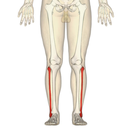 |
| Tibula | Known as the shinbone and is the second largest bone in the body. Helps with weight-bearing and stabilization. | 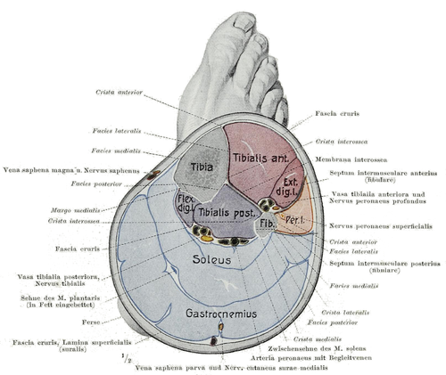 | |
| Talus | The talus bone forms the primary connection between the lower leg and foot and is vital for mobility. In fact, the structure of the talus bone is so unique it can form the connection between numerous other bones such as the tibia, fibula, calcaneus (heel) and navicular or tarsal bones found in the foot. | 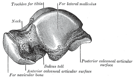 | |
| Medial malleolus | The medial malleolus is the prominence on the inner side of the ankle, formed by the lower end of the tibia. |  | |
| Lateral malleolus | The lateral malleolus is the prominence on the outer side of the ankle, formed by the lower end of the tibia. | 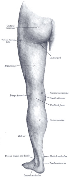 |
| Parts | Function | Image | |
|---|---|---|---|
| Thigh | Femur | Supports the weight of the body and allowing motion of the leg. The femur articulates proximally with the acetabulum of the pelvis forming the hip joint, and distally with the tibia and patella to form the knee joint. | |
| Tibia | It forms the knee joint with the femur and the ankle joint with the fibula and tarsus. | ||
| Fibula | Long, thin and lateral bone of the lower leg. It runs parallel to the tibia, or shin bone, and plays a significant role in stabilizing the ankle and supporting the muscles of the lower leg. |  |
| Parts | Function | Image | |
|---|---|---|---|
| Foot | Phalanges | The phalanges of the foot help us balance, walk, and run. | 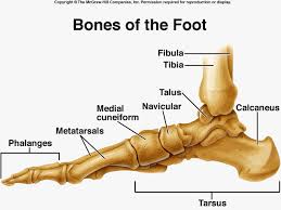 |
| Metatarsals | Metatarsals are convex in shape (arch upward), are long bones, and give the foot its arch. They work with connective tissues, ligaments, and tendons to provide movement in the foot | 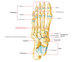 | |
| Cuneiform bones | This bone is cube-shaped and connects the foot and the ankle. It also provides stability to the foot. | 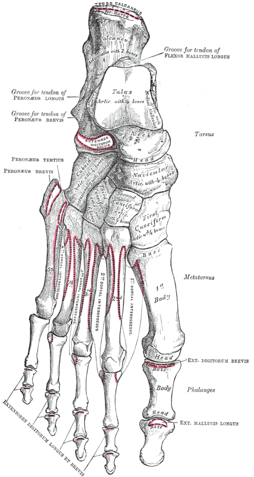 | |
| Cuboid bone | The cuboid bone is one of the seven tarsal bones located on the lateral (outer) side of the foot. This bone is cube-shaped and connects the foot and the ankle. It also provides stability to the foot. | 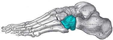 | |
| Navicular bone | The navicular is a boat-shaped bone located in the top inner side of the foot, just above the transverse. It helps connect the talus, or anklebone, to the cuneiform bones of the foot | 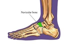 |
Lower limb muscles
| Muscle | Function | Insertion/Arise | Innervation | Blood supply | Image | |
|---|---|---|---|---|---|---|
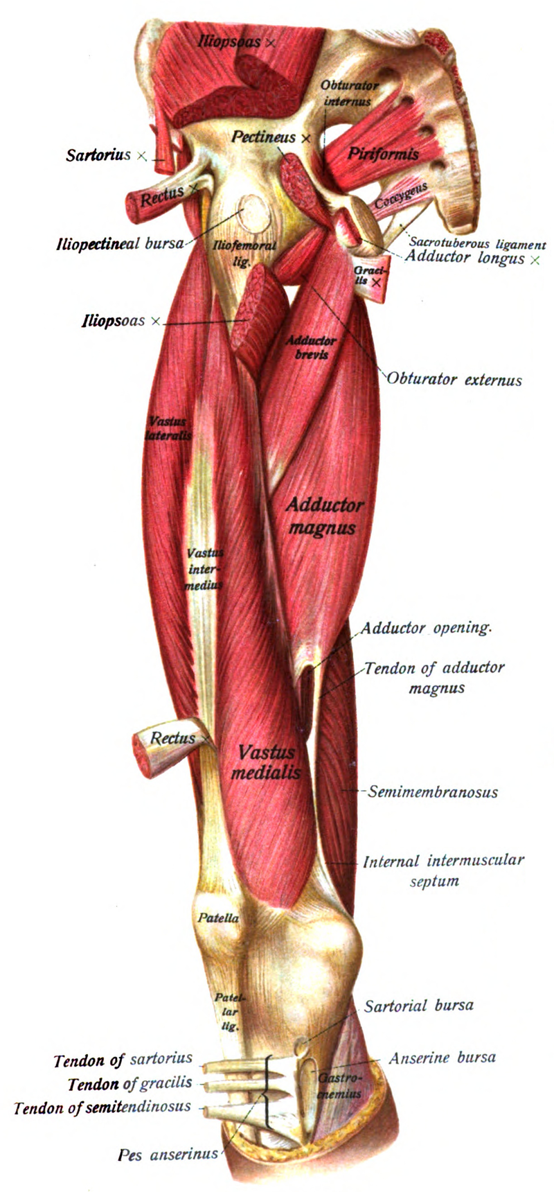
| Hip | Piriformis |
|
Arise:
Insertion:
|
Piriformis nerve:
|
Branches of the internal iliac artery:
|
 |
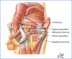
| Superior gemellus |
|
Insertion:
Arise:
|
Innervation:
|
Arterial supply:
|
 | |
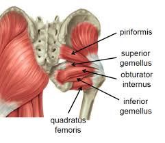
| Inferior gemellus |
|
Insertion:
Arise:
|
Innervation:
|
Arterial supply:
|
 | |
| Tensor fasciae latae |
|
Insertion:
Arise:
|
Innervation:
|
Arterial supply:
|
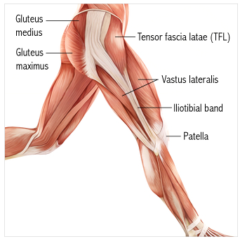 | |
| Sartorius |
|
Insertion:
Arise:
|
Innervation:
|
Arterial supply:
|
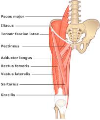 | |
| Gluteus medius |
|
Insertion:
Arise:
|
Innervation:
|
Arterial supply:
|
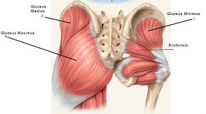 | |
| Gluteus minimus |
|
Insertion:
Arise:
|
Innervation:
|
Arterial supply:
|
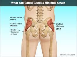 | |
| Gluteus maximus |
|
Insertion:
Arise:
|
Innervation:
|
Arterial supply:
|
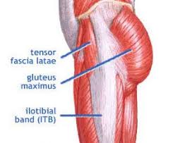 |
| Muscle | Function | Insertion/Arise | Innervation | Blood supply | Image | |
|---|---|---|---|---|---|---|
| Knee | Quadratus Femoris |
|
Insertion:
Arise:
|
Innervation:
|
Arterial supply:
|
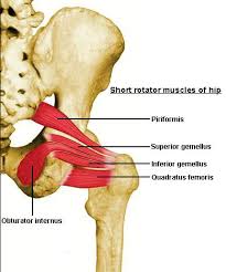 |
| Gastrocnemius muscle |
|
Insertion:
Arise:
|
Innervation:
|
Arterial supply:
|
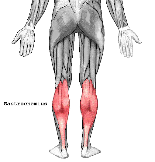 | |
| Vastus medialis |
|
Insertion:
Arise:
|
Innervation:
|
Arterial supply:
|
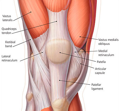 | |
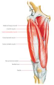
| Vastus lateralis muscle |
|
Insertion:
Arise:
|
Innervation:
|
Arterial supply:
|
 | |
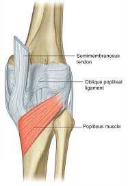
| Popliteus |
|
Insertion:
Arise:
|
Innervation:
|
Arterial supply:
|
 | |
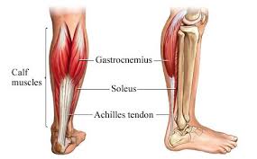
| Soleus |
|
Insertion:
Arise:
|
Innervation:
|
Arterial supply:
|
 |
| Muscle | Function | Insertion/Arise | Innervation | Blood supply | Image | |
|---|---|---|---|---|---|---|
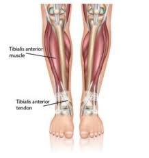
| Ankle | Anterior tibial |
|
Insertion:
Arise:
|
Innervation:
|
Arterial supply:
|
 |
| Posterior tibial |
|
Insertion:
Arise:
|
Innervation:
|
Arterial supply:
|
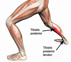 | |
| Extensor Digitorum Longus |
|
Insertion:
Arise:
|
Innervation:
|
Arterial supply:
|
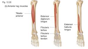 | |
| Extensor Hallucis Longus |
|
Insertion:
Arise:
|
Innervation:
|
Arterial supply:
|
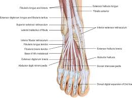 | |
| Flexor Digitorum Longus |
|
Insertion:
Arise:
|
Innervation:
|
Arterial supply:
|
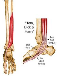 | |
| Flexor Hallucis Longus |
|
Insertion:
Arise:
|
Innervation:
|
Arterial supply:
|
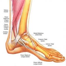 |
| Muscle | Function | Insertion/Arise | Innervation | Blood supply | Image | |
|---|---|---|---|---|---|---|
| Thigh | Adductor Brevis |
|
Insertion:
Arise:
|
Innervation:
|
Arterial supply:
|
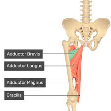 |
| Adductor Longus |
|
Insertion:
Arise:
|
Innervation:
|
Arterial supply:
|
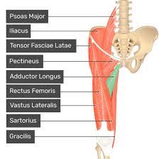 | |
| Gracilis |
|
Insertion:
Arise:
|
Innervation:
|
Arterial supply:
|
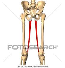 | |
| Iliacus |
|
Insertion:
Arise:
|
Innervation:
|
Arterial supply:
|
 | |
| Obturator Externus |
|
Insertion:
Arise:
|
Innervation:
|
Arterial supply:
|
 | |
| Obturator Internus |
|
Insertion:
Arise:
|
Innervation:
|
Arterial supply:
|
 | |
| Pectineus |
|
Insertion:
Arise:
|
Innervation:
|
Arterial supply:
|
 | |
| Piriformis |
|
Insertion:
Arise:
|
Innervation:
|
Arterial supply:
|
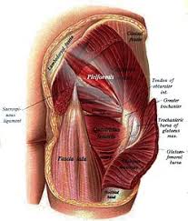 | |
| Psoas |
|
Insertion:
Arise:
|
Innervation:
|
Arterial supply:
|
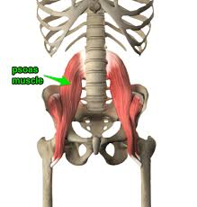
| |
| Semimembranosus |
|
Insertion:
Arise:
|
Innervation:
|
Arterial supply:
|
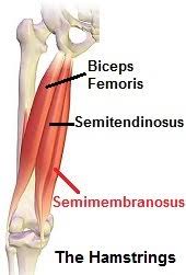 | |
| Semitendinosus |
|
Insertion:
Arise:
|
Innervation:
|
Arterial supply:
|
 |