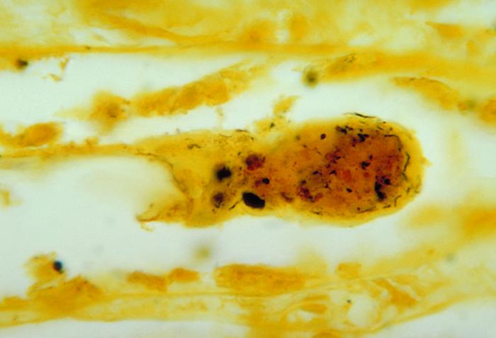
Leptospirosis laboratory findings: Difference between revisions
No edit summary |
|||
| Line 21: | Line 21: | ||
* Culture the bacteria from blood, urine or tissues | * Culture the bacteria from blood, urine or tissues | ||
* Other methods such as PCR, Immunostaining etc. | * Other methods such as PCR, Immunostaining etc. | ||
===Blood Tests=== | |||
Blood tests in leptospirosis include:<ref name="pmid13819407">{{cite journal| author=EDWARDS GA, DOMM BM| title=Human leptospirosis. | journal=Medicine (Baltimore) | year= 1960 | volume= 39 | issue= | pages= 117-56 | pmid=13819407 | doi= | pmc= | url=https://www.ncbi.nlm.nih.gov/entrez/eutils/elink.fcgi?dbfrom=pubmed&tool=sumsearch.org/cite&retmode=ref&cmd=prlinks&id=13819407 }} </ref> | Blood tests in leptospirosis include:<ref name="pmid13819407">{{cite journal| author=EDWARDS GA, DOMM BM| title=Human leptospirosis. | journal=Medicine (Baltimore) | year= 1960 | volume= 39 | issue= | pages= 117-56 | pmid=13819407 | doi= | pmc= | url=https://www.ncbi.nlm.nih.gov/entrez/eutils/elink.fcgi?dbfrom=pubmed&tool=sumsearch.org/cite&retmode=ref&cmd=prlinks&id=13819407 }} </ref> | ||
* '''CBC:''' Shows pancytopenia or peripheral leukocytosis with a left shift | * '''CBC:''' Shows pancytopenia or peripheral leukocytosis with a left shift | ||
| Line 28: | Line 28: | ||
* Elevated plasma creatinine | * Elevated plasma creatinine | ||
===Urinalysis=== | |||
* Proteinuria | * Proteinuria | ||
* Pyuria | * Pyuria | ||
* Microscopic hematuria | * Microscopic hematuria | ||
* Hyaline and granular casts | * Hyaline and granular casts | ||
===CSF Analysis === | |||
CSF findings are common in first or second week of illness.<ref name="pmid14902167">{{cite journal| author=BEESON PB, HANKEY DD| title=Leptospiral meningitis. | journal=AMA Arch Intern Med | year= 1952 | volume= 89 | issue= 4 | pages= 575-83 | pmid=14902167 | doi= | pmc= | url=https://www.ncbi.nlm.nih.gov/entrez/eutils/elink.fcgi?dbfrom=pubmed&tool=sumsearch.org/cite&retmode=ref&cmd=prlinks&id=14902167 }} </ref> | CSF findings are common in first or second week of illness.<ref name="pmid14902167">{{cite journal| author=BEESON PB, HANKEY DD| title=Leptospiral meningitis. | journal=AMA Arch Intern Med | year= 1952 | volume= 89 | issue= 4 | pages= 575-83 | pmid=14902167 | doi= | pmc= | url=https://www.ncbi.nlm.nih.gov/entrez/eutils/elink.fcgi?dbfrom=pubmed&tool=sumsearch.org/cite&retmode=ref&cmd=prlinks&id=14902167 }} </ref> | ||
* Opening pressure: normal or slightly elevated | * Opening pressure: normal or slightly elevated | ||
| Line 41: | Line 41: | ||
* Xanthochromasia is seen in severe Icteric leptospirosis<ref name="pmid20263193">{{cite journal| author=CARGILL WH, BEESON PB| title=The value of spinal fluid examination as a diagnostic procedure in Weil's disease. | journal=Ann Intern Med | year= 1947 | volume= 27 | issue= 3 | pages= 396-400 | pmid=20263193 | doi= | pmc= | url=https://www.ncbi.nlm.nih.gov/entrez/eutils/elink.fcgi?dbfrom=pubmed&tool=sumsearch.org/cite&retmode=ref&cmd=prlinks&id=20263193 }} </ref> | * Xanthochromasia is seen in severe Icteric leptospirosis<ref name="pmid20263193">{{cite journal| author=CARGILL WH, BEESON PB| title=The value of spinal fluid examination as a diagnostic procedure in Weil's disease. | journal=Ann Intern Med | year= 1947 | volume= 27 | issue= 3 | pages= 396-400 | pmid=20263193 | doi= | pmc= | url=https://www.ncbi.nlm.nih.gov/entrez/eutils/elink.fcgi?dbfrom=pubmed&tool=sumsearch.org/cite&retmode=ref&cmd=prlinks&id=20263193 }} </ref> | ||
===Microscopy=== | |||
* Dark field microscopy: Inorder to detect under dark field microscopy 10<sup>4</sup> leptospires/ml are necessary for one cell per field. | * Dark field microscopy: Inorder to detect under dark field microscopy 10<sup>4</sup> leptospires/ml are necessary for one cell per field. | ||
** Specimen: Blood, urine, CSF | ** Specimen: Blood, urine, CSF | ||
** Disadvantages: Test is insensitive and lacks specificity | ** Disadvantages: Test is insensitive and lacks specificity | ||
* Other microscopic techniques: Immunofluoroscence, Light microscopy | * Other microscopic techniques: Immunofluoroscence, Light microscopy | ||
===Antigen detection tests=== | |||
Leptospiral antigens can be identified from different specimen such as blood, urine with higher sensitivity than dark field microspy. Techniques include: | Leptospiral antigens can be identified from different specimen such as blood, urine with higher sensitivity than dark field microspy. Techniques include: | ||
* Radioimmunoassay (RIA) | * Radioimmunoassay (RIA) | ||
* Enzyme-linked immunosorbent assay (ELISA) | * Enzyme-linked immunosorbent assay (ELISA) | ||
===Culture=== | |||
'''Specimen for culture''' | '''Specimen for culture''' | ||
* First week of illness: Blood, CSF | * First week of illness: Blood, CSF | ||
* Second week onwards: Urine<ref name="pmid7989538">{{cite journal| author=Bal AE, Gravekamp C, Hartskeerl RA, De Meza-Brewster J, Korver H, Terpstra WJ| title=Detection of leptospires in urine by PCR for early diagnosis of leptospirosis. | journal=J Clin Microbiol | year= 1994 | volume= 32 | issue= 8 | pages= 1894-8 | pmid=7989538 | doi= | pmc=263898 | url=https://www.ncbi.nlm.nih.gov/entrez/eutils/elink.fcgi?dbfrom=pubmed&tool=sumsearch.org/cite&retmode=ref&cmd=prlinks&id=7989538 }} </ref> | * Second week onwards: Urine<ref name="pmid7989538">{{cite journal| author=Bal AE, Gravekamp C, Hartskeerl RA, De Meza-Brewster J, Korver H, Terpstra WJ| title=Detection of leptospires in urine by PCR for early diagnosis of leptospirosis. | journal=J Clin Microbiol | year= 1994 | volume= 32 | issue= 8 | pages= 1894-8 | pmid=7989538 | doi= | pmc=263898 | url=https://www.ncbi.nlm.nih.gov/entrez/eutils/elink.fcgi?dbfrom=pubmed&tool=sumsearch.org/cite&retmode=ref&cmd=prlinks&id=7989538 }} </ref> | ||
| Line 58: | Line 58: | ||
Leptospira can be cultured in Ellinghausen-McCullough-Johnson-Harris medium, which is incubated at 28 to 30ºC.<ref name="pmid3754265">{{cite journal| author=Rule PL, Alexander AD| title=Gellan gum as a substitute for agar in leptospiral media. | journal=J Clin Microbiol | year= 1986 | volume= 23 | issue= 3 | pages= 500-4 | pmid=3754265 | doi= | pmc=268682 | url=https://www.ncbi.nlm.nih.gov/entrez/eutils/elink.fcgi?dbfrom=pubmed&tool=sumsearch.org/cite&retmode=ref&cmd=prlinks&id=3754265 }} </ref> The median time to positivity is three weeks with a maximum of 3 months. This makes culture techniques useless for diagnostic purposes, but is commonly used in research. | Leptospira can be cultured in Ellinghausen-McCullough-Johnson-Harris medium, which is incubated at 28 to 30ºC.<ref name="pmid3754265">{{cite journal| author=Rule PL, Alexander AD| title=Gellan gum as a substitute for agar in leptospiral media. | journal=J Clin Microbiol | year= 1986 | volume= 23 | issue= 3 | pages= 500-4 | pmid=3754265 | doi= | pmc=268682 | url=https://www.ncbi.nlm.nih.gov/entrez/eutils/elink.fcgi?dbfrom=pubmed&tool=sumsearch.org/cite&retmode=ref&cmd=prlinks&id=3754265 }} </ref> The median time to positivity is three weeks with a maximum of 3 months. This makes culture techniques useless for diagnostic purposes, but is commonly used in research. | ||
===Serological Tests=== | |||
Serological test are useful to detect leptospira-specific IgM antibodies in the early acute phase of illness, especially after first week of clinical symptoms. Antibodies production start 5-7 days after the onset of the initial presentation. Serological test in the 1st week will give false negative results. | Serological test are useful to detect leptospira-specific IgM antibodies in the early acute phase of illness, especially after first week of clinical symptoms. Antibodies production start 5-7 days after the onset of the initial presentation. Serological test in the 1st week will give false negative results. | ||
<br>'''Screening tests''' | <br>'''Screening tests''' | ||
* Enzyme-linked immunosorbent assay (ELISA): sensitivity of 90% and specificity of 94%.<ref name="pmid11271784">{{cite journal| author=Zochowski WJ, Palmer MF, Coleman TJ| title=An evaluation of three commercial kits for use as screening methods for the detection of leptospiral antibodies in the UK. | journal=J Clin Pathol | year= 2001 | volume= 54 | issue= 1 | pages= 25-30 | pmid=11271784 | doi= | pmc=1731274 | url=https://www.ncbi.nlm.nih.gov/entrez/eutils/elink.fcgi?dbfrom=pubmed&tool=sumsearch.org/cite&retmode=ref&cmd=prlinks&id=11271784 }} </ref> Positive test results shows high IgM titre in a single serum sample or a 4-fold rise in titre in a paired tests is consistent with current or recent infection. | * Enzyme-linked immunosorbent assay (ELISA): sensitivity of 90% and specificity of 94%.<ref name="pmid11271784">{{cite journal| author=Zochowski WJ, Palmer MF, Coleman TJ| title=An evaluation of three commercial kits for use as screening methods for the detection of leptospiral antibodies in the UK. | journal=J Clin Pathol | year= 2001 | volume= 54 | issue= 1 | pages= 25-30 | pmid=11271784 | doi= | pmc=1731274 | url=https://www.ncbi.nlm.nih.gov/entrez/eutils/elink.fcgi?dbfrom=pubmed&tool=sumsearch.org/cite&retmode=ref&cmd=prlinks&id=11271784 }} </ref> Positive test results shows high IgM titre in a single serum sample or a 4-fold rise in titre in a paired tests is consistent with current or recent infection. | ||
'''Confirmatory tests''' | '''Confirmatory tests''' | ||
* Microscopic agglutination test (MAT): Specificity of 94%. All positive screening tests should be confirmed by the MAT. Agglutinating antibodies can detects both IgM and IgG classes and are detectable from about Days 7 to 10 after onset of symptoms. | * Microscopic agglutination test (MAT): Specificity of 94%. All positive screening tests should be confirmed by the MAT. Agglutinating antibodies can detects both IgM and IgG classes and are detectable from about Days 7 to 10 after onset of symptoms. | ||
Revision as of 22:18, 4 March 2017

|
Leptospirosis Microchapters |
|
Diagnosis |
|---|
|
Treatment |
|
Case Studies |
|
Leptospirosis laboratory findings On the Web |
|
American Roentgen Ray Society Images of Leptospirosis laboratory findings |
|
Risk calculators and risk factors for Leptospirosis laboratory findings |
Editor-In-Chief: C. Michael Gibson, M.S., M.D. [1];Associate Editor(s)-in-Chief: Venkata Sivakrishna Kumar Pulivarthi M.B.B.S [2]
Overview
The diagnosis of leptospirosis is based upon clinical suspicion and lab diagnosis, so lab tests should be considered in a patient with a history of contact with potentially infected animals, soil or surface waters contaminated by animal urine.[1] Leptospires can be found in blood and CSF for the first 7 to 10 days and then in the urine. Hence, in the early diagnosis specimen of choice should be blood or CSF for culture. From the second week onwards serological tests are useful in the diagnosis.
Laboratory findings
Laboratory Findings
As the clinical manifestations of the disease are non specific, the clinical diagnosis is difficult. The laboratory investigations for leptospirosis should be considered in patient with an abrupt onset of fever, chills, conjunctival suffusion, headache, myalgia and jaundice with history of occupational exposure to infected animals or contaminated with animal urine.[2] Laboratory criteria for the diagnosis of leptospirosis are presence of one or more of the following:[1]
- Culture positivity
- Antibody titre of ≥1 in 320 by Microscopic Agglutination test (MAT) in a single serum sample
- Seroconversion in paired sera collected in the acute and convalescent phase established by ELISA IgM and/or MAT methods
- Evidence of leptospira antigen by molecular methods.
Laboratory investigations useful in the diagnosis of leptospirosis include:
- Identification of leptospires in tissues using antibodies labelled with fluorescent markers
- Antibody detection by serological studies
- Culture the bacteria from blood, urine or tissues
- Other methods such as PCR, Immunostaining etc.
Blood Tests
Blood tests in leptospirosis include:[3]
- CBC: Shows pancytopenia or peripheral leukocytosis with a left shift
- Elevated ESR
- Liver functional tests: Mild elevation in aminotransferases, bilirubin, and alkaline phosphatase.[4]
- Elevated plasma creatinine
Urinalysis
- Proteinuria
- Pyuria
- Microscopic hematuria
- Hyaline and granular casts
CSF Analysis
CSF findings are common in first or second week of illness.[5]
- Opening pressure: normal or slightly elevated
- Cells: Lymphocyte predominance[5]
- Protein: Normal to elevated[3]
- Glucose: Normal
- Xanthochromasia is seen in severe Icteric leptospirosis[6]
Microscopy
- Dark field microscopy: Inorder to detect under dark field microscopy 104 leptospires/ml are necessary for one cell per field.
- Specimen: Blood, urine, CSF
- Disadvantages: Test is insensitive and lacks specificity
- Other microscopic techniques: Immunofluoroscence, Light microscopy
Antigen detection tests
Leptospiral antigens can be identified from different specimen such as blood, urine with higher sensitivity than dark field microspy. Techniques include:
- Radioimmunoassay (RIA)
- Enzyme-linked immunosorbent assay (ELISA)
Culture
Specimen for culture
- First week of illness: Blood, CSF
- Second week onwards: Urine[7]
Leptospira can be cultured in Ellinghausen-McCullough-Johnson-Harris medium, which is incubated at 28 to 30ºC.[8] The median time to positivity is three weeks with a maximum of 3 months. This makes culture techniques useless for diagnostic purposes, but is commonly used in research.
Serological Tests
Serological test are useful to detect leptospira-specific IgM antibodies in the early acute phase of illness, especially after first week of clinical symptoms. Antibodies production start 5-7 days after the onset of the initial presentation. Serological test in the 1st week will give false negative results.
Screening tests
- Enzyme-linked immunosorbent assay (ELISA): sensitivity of 90% and specificity of 94%.[9] Positive test results shows high IgM titre in a single serum sample or a 4-fold rise in titre in a paired tests is consistent with current or recent infection.
Confirmatory tests
- Microscopic agglutination test (MAT): Specificity of 94%. All positive screening tests should be confirmed by the MAT. Agglutinating antibodies can detects both IgM and IgG classes and are detectable from about Days 7 to 10 after onset of symptoms.
References
- ↑ 1.0 1.1 Forbes AE, Zochowski WJ, Dubrey SW, Sivaprakasam V (2012). "Leptospirosis and Weil's disease in the UK". QJM. 105 (12): 1151–62. doi:10.1093/qjmed/hcs145. PMID 22843698.
- ↑ LastName, FirstName (2003). Human leptospirosis : guidance for diagnosis, surveillance and control. Geneva: World Health Organization. ISBN 9241545895.
- ↑ 3.0 3.1 EDWARDS GA, DOMM BM (1960). "Human leptospirosis". Medicine (Baltimore). 39: 117–56. PMID 13819407.
- ↑ Bharti AR, Nally JE, Ricaldi JN, Matthias MA, Diaz MM, Lovett MA; et al. (2003). "Leptospirosis: a zoonotic disease of global importance". Lancet Infect Dis. 3 (12): 757–71. PMID 14652202.
- ↑ 5.0 5.1 BEESON PB, HANKEY DD (1952). "Leptospiral meningitis". AMA Arch Intern Med. 89 (4): 575–83. PMID 14902167.
- ↑ CARGILL WH, BEESON PB (1947). "The value of spinal fluid examination as a diagnostic procedure in Weil's disease". Ann Intern Med. 27 (3): 396–400. PMID 20263193.
- ↑ Bal AE, Gravekamp C, Hartskeerl RA, De Meza-Brewster J, Korver H, Terpstra WJ (1994). "Detection of leptospires in urine by PCR for early diagnosis of leptospirosis". J Clin Microbiol. 32 (8): 1894–8. PMC 263898. PMID 7989538.
- ↑ Rule PL, Alexander AD (1986). "Gellan gum as a substitute for agar in leptospiral media". J Clin Microbiol. 23 (3): 500–4. PMC 268682. PMID 3754265.
- ↑ Zochowski WJ, Palmer MF, Coleman TJ (2001). "An evaluation of three commercial kits for use as screening methods for the detection of leptospiral antibodies in the UK". J Clin Pathol. 54 (1): 25–30. PMC 1731274. PMID 11271784.