Intracranial pressure: Difference between revisions
| Line 24: | Line 24: | ||
*In 1950s, therapeutic hypothermia (goal core temperature of 32-34C) was first introduced as a treatment for brain injury. | *In 1950s, therapeutic hypothermia (goal core temperature of 32-34C) was first introduced as a treatment for brain injury. | ||
*In early 1800s, the Monro-Kellie hypothesis and the CSF physiology was first introduced by Alexander Monro and George Kellie. | |||
*Hans Queckenstedt's was the first person to use lumbar needle for ICP monitoring. | |||
* | * | ||
Revision as of 04:08, 27 August 2020
| Intracranial pressure | |
 | |
|---|---|
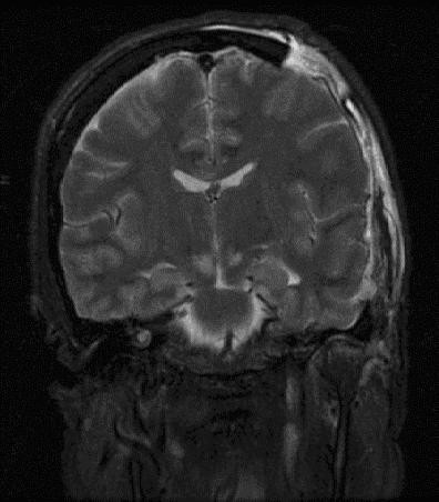
| Severely high ICP can cause herniation. |
Editor-In-Chief: C. Michael Gibson, M.S., M.D. [1] Associate Editor(s)-in-Chief: Luke Rusowicz-Orazem, B.S., Sabeeh Islam, MBBS[2]
Overview
Intracranial pressure, (ICP), is the pressure exerted by three structures inside the cranium; brain parenchyma, CSF and blood. The norma ICP is 10-15 mmHg and is usually maintained by equilibrium of the intracranial contents. Intracranial hypertension ( IH), is elevation of the pressure in the cranium. It typically occurs when the ICP is >20 mmHg.
Historical Perspective
- In 1950s, therapeutic hypothermia (goal core temperature of 32-34C) was first introduced as a treatment for brain injury.
- In early 1800s, the Monro-Kellie hypothesis and the CSF physiology was first introduced by Alexander Monro and George Kellie.
- Hans Queckenstedt's was the first person to use lumbar needle for ICP monitoring.
Classification[edit | edit source]
- Elevated intracranial pressure or Intracranial hypertension may be classified into two subtypes/groups:
- Acute
- Chronic
- Intracranial hypertension may also be classified as various stages:
- Stage 1: Minimal increases in ICP due to compensatory mechanisms
- Stage 2:
- Any change in volume greater than 100–120 mL
- Exhaustion of compensatory mechanisms
- Compromise of neuronal oxygenation and systemic arteriolar vasoconstriction to increase MAP and CP
- Stage 3:
- Sustained increased ICP
- Dramatic changes in ICP with small changes in volume
- The ICP approaches the MAP,
Increased ICP:
Intracranial pressure, (ICP), is the pressure exerted by three structures inside the cranium; brain parenchyma, CSF and blood. The norma ICP is 10-15 mmHg and is usually maintained by equilibrium of the intracranial contents.
Intracranial Hypertension:
Intracranial hypertension ( IH), is elevation of the pressure in the cranium. It typically occurs when the ICP is >20 mmHg.
Pathophysiology
Intracranial components and their proportions:
- Brain parenchyma volume: 1400 ml (80%)
- CSF volume: 10 ml (10%)
- Blood volume: 10 ml (10%)
The Monro-Kellie Hypothesis:
- The Monro-Kellie hypothesis explains the relationship between the contents of the cranium and intracranial pressure. It explains the underlying pathophysiology of elevated intracranial pressure or intracranial hypertension
- In normal physiological state, intracranial contents (the brain tissue, the blood, and the cerebrospinal fluid) maintain an equilibrium state and keep the ICP within normal range by acting as compensatory mechanisms for small volume changes
- Compensatory mechanisms are being exhausted by large volume changes, eventually causing significantly elevated intracranial pressures and potential herniation
Intracranial compliance:
- There is an inverse relationship between intracranial components and the compliance.
- Generally the normal compliance is maintained by compensatory mechanisms such as
- Increased CSF reabsorption via thecal sac
- Increased venoconstriction to decrease cerebral venous flow
- Decreased cerebral venous flow via increased extracranial drainage
Cerebral Blood Flow (Ohm's Law):
- Cerebral blood flow is generally assessed by subtracting jugular venous pressure from carotid arterial pressure and dividing by cerebrovascular resistance, as follows:
- CBF = (CAP - JVP) ÷ CVR
- Cerebral perfusion is assessed by cerebral perfusion pressure (CPP). CPP is calculated by subtracting ICP from mean arterial pressure, as follows:
- CPP = MAP - ICP
- In normal physiological states, ICP and CPP is maintained by autoregulation.
Several pathophysiologic mechanisms are thought to be involved in the pathogenesis of Increased Intracaranial pressure (ICP) or Intracranial hypertension (ICH). All mechanisms eventually lead to brain injury from brain stem compression and decreased cerebral blood supply or ischemia. These mechanisms are as follows:
- Mass effect
- It can occur secondary to brain tumor, contusions, subdural or epidural hematoma, or abscess
- Cerebral edema or Generalized brain swelling
- It can occur secondary to ischemic-anoxia states, hypertensive encephalopathy, pseudotumor cerebri, hypercarbia, and hepatocerebral syndrome.
- These conditions tend to decrease the cerebral perfusion pressure but with minimal tissue shifts.
- Increase in venous pressure
- Secondary to venous sinus thrombosis, heart failure, neck surgery or obstruction of superior mediastinal or jugular veins.
- Obstruction to CSF flow
- Secondary to hydrocephalus, extensive meningeal disease (e.g., infectious, carcinomatous, granulomatous, or hemorrhagic), or obstruction in cerebral convexities and superior sagittal sinus (decreased absorption).
- Increased CSF production
- Meningitis, subarachnoid hemorrhage, or choroid plexus tumor.
- Increased cerebral blood flow (CBF)
- Increased CBF is generally seen in conditions associated with hypercapnia and hypoxia
- Drugs
- Idiopathic
- Pseudotumor cerebri
- Mass effect
Causes
Common Causes
- Aneurysm
- Arnold-chiari malformation
- Behçet's disease
- Brain tumor
- Cerebral edema
- Cerebral venous sinus thrombosis
- Choroid plexus tumor
- Chronic kidney disease
- Colloid cyst of third ventricle
- Contusions
- Crouzon craniofacial dysostosis
- Cushing's syndrome
- Dural arteriovenous fistula
- Encephalitis
- Epidural haemorrhage
- Epidural hematoma
- Erdheim-chester disease
- Excess cerebrospinal fluid
- Head trauma
- Hydrocephalus
- Hypertensive brain hemorrhage
- Hypertensive encephalopathy
- Idiopathic intracranial hypertension
- Insulin like growth factor 1
- Intracranial granuloma
- Intracranial haemorrhage
- Intraventricular hemorrhage
- Meningioma
- Meningitis
- Meningoencephalitis
- Multiple hamartoma syndrome
- Obstruction of superior mediastinal veins
- Obstruction of jugular veins
- Status epilepticus
- Stroke
- Subarachnoid haemorrhage
- Subdural haemorrhage
- Subdural hematoma
- Vasculitis
- Venous sinus thrombosis
Differential Diagnosis of Increased Intracranial Pressure (ICP)
- Increased Intracaranial pressure (ICP) or Intracranial hypertension (ICH) must be differentiated from other diseases that cause headache, nausea, vomiting and neurologic deficits such as tumor, abscess or space occupying lesion, venous sinus thrombosis, neck surgery, Obstructive hydrocephalus, meningitis, subarachnoid hemorrhage, choroid plexus papilloma, and Malignant systemic hypertension.
Epidemiology and Demographics
- The prevalence of intracranial hypertension is approximately 1.0 per 100,000 individuals worldwide.
Gender
- Idiopathic ICH is more prevalent among women of childbearing age.
Risk Factors
- Common risk factors in the development of Increased Intracaranial pressure (ICP) or Intracranial hypertension (ICH) include underlying pathologies such as; mass lesions, abscesses, and hematomas.
- Other risk factors include
- Obesity
- Chronic hypertension
- Women of childbearing age
Natural History, Complications and Prognosis
- Early clinical features include nausea, vomiting, and confusion.
- If left untreated, patients may progress to have severe neurologic consequences such as brain herniation, brain death, respiratory depression, brain infections, coma and death.
- Common complications of intracranial hypertension include brain herniation and neurologic deficits.
Diagnosis
Diagnostic Criteria
- The diagnosis of Increased Intracaranial pressure (ICP) or Intracranial hypertension (ICH) is made when ICP is >20 mmHg.
History and Symptoms
- Symptoms of elevated intracranial pressure may include the following:
- Headache
- Nausea
- Vomiting
- Hyperventilation (due to injury to brain stem or tegmentum is damaged.[1]
- Changes in your behavior
- Weakness or problems with moving or talking
- Lack of energy or sleepiness
- Seizure
Physical Examination
- Physical examination may be remarkable for
- Ocular palsies (abducens palsy)
- Periorbital bruising
- Altered level of consciousness
- Papilledema
- Pupillary dilatation
- Cushing's triad ( Elevated systolic blood pressure, a widened pulse pressure, bradycardia, and an abnormal respiratory pattern.
- Cheyne-Stokes respiration
- Bulging of fontanels in infants
Laboratory Findings
- There are no specific laboratory findings associated with Increased Intracaranial pressure (ICP) or Intracranial hypertension (ICH).
Electrocardiogram
- There are no ECG findings associated with Increased Intracaranial pressure (ICP) or Intracranial hypertension (ICH).
X-ray
- There are no x-ray findings associated with Increased Intracaranial pressure (ICP) or Intracranial hypertension (ICH).
CT scan
- CT scan may be helpful in the diagnosis of Increased Intracaranial pressure (ICP) or Intracranial hypertension (ICH).
- Findings on CT scan suggestive of Increased Intracaranial pressure (ICP) or Intracranial hypertension (ICH) include presence of mass lesions, midline shift or hemorrhage
- CT scan is particularly helpful for people with acute rise in ICP
MRI
- MR venography (MRV) is preferred over MRI for the diagnosis of cerebral venous thrombosis
- MRI has a greater sensitivity to detect subtle intracranial masses (eg, gliomatosis cerebri) and meningeal-based pathologies and should be done if no contraindications (eg, pacemakers, metallic clips in head, metallic foreign bodies) present
Other Diagnostic Studies
Other diagnostic studies for Increased Intracaranial pressure (ICP) or Intracranial hypertension (ICH) include invasive and non-invasive ICP monitoring, particularly preferred in patients with no CT or MRI findings, at risk of developing increased ICP, and comatosed.
- Invasive ICP monitoring usually involves 4 anatomic sites
- Intraventricular
- Intraparenchymal
- Subarachnoid
- Epidural
- Noninvasive devices still need further large randomized trials to prove their clinical efficacy. They are not used in clinical practice but are still under investigation and include
- Transcranial Doppler (TCD)
- Tissue resonance analysis (TRA)
- Ocular sonography
- Intraocular pressure
- Tympanic membrane displacement
Treatment
Medical Therapy
- The management of intracranial hypertension is generally directed towards treating the cause/etiology of the raised intracranial pressure.
- Intracranial hypertension is considered a medical emergency and the management includes emergent resuscitative as well as specific treatment.
Resuscitation:
General principles for resuscitation include
- Maintain oxygen
- Head elevation
- Hyperventilation to achieve a PaCO2 of 26-30 mmHg
- Osmotic diuresis with intravenous mannitol and Lasix
- Appropriate sedation, if patient requires intubation. Propofol is considered to be the preferred agent.
- Therapeutic hypothermia to achieve a low metabolic state
- Appropriate choice of fluids to achieve euvolemic state. Avoid hypotonic agents
- Allow permissive hypertension. Treat hypertension only when CPP >120 mmHg and ICP >20 mmHg
- Seizure prophylaxis with anticonvulsant therapy.
Other therapies for intracranial hypertension:
- Osmotic diuresis can be achieved by hypertonic saline bolus or mannitol. Hypertonic saline is usually considered to be more effective compared to mannitol for acute ICP reduction.
Mannitol can be given as a bolus of 1 g/kg when prepared as 20% solution. The dose is usually repeated every 6-8 hours. It should be used cautiously in patients with renal insufficiency. Intravenous Lasix (0.5 to 1 mg/kg) is usually given with mannitol.
- Glucocorticoids are usually preferred when the underlying etiologies brain tumor are underlying CNS infection. Their use is contraindicated in head injury, cerebral infarction and intracranial hemorrhage.
- Phenobarbital is considered to have a neuroprotective effect by decreasing brain metabolism. It is given as a loading dose of 5 to 20 mg/kg, followed by 1 to 4 mg/kg per hour. EEG monitoring is used to guide therapy. A burst suppression seen on EEG indicates maximal dosing.
Surgery
Surgical options for persistent intracranial hypertension include
- Surgical evacuation
- CSF drainage via ventriculostomy
- CSF is usually drained at a rate of 1 to 2 mL/minute for 2 to 3 minutes. The procedure is repeated after every 2 to 3 minutes, until ICP is less than 20mmHg
- Decompressive craniectomy
Prevention
- Effective measures for the primary prevention of intracranial hypertension include early detection of underlying intracranial etiology such as tumor or congenital deformities.
- Once diagnosed and successfully treated, patients with intracranial hypertension are followed up every 6 months to 1 year with a head CT scan to prevent secondary complications.
References
Additional Resources
- Monroe A. Observations on the structure and function of the nervous system, Edinburgh: Creech & Johnson; 1783.
- Kelly G. An account of the appearances observed in the dikssection of two of three individuals presumed to have perished in the storm of the 3rd, and whose bodies were deiscovered in the vicinity of the Leith on the morning of the 4th of November 1821, with some reflections on the pathology of the brain, Trans Med Chir Sci Edinb 1824;1:84–169.
External links
- Gruen P. 2002. "Monro-Kellie Model" Neurosurgery Infonet. USC Neurosurgery. Accessed January 4, 2007.
- National Guideline Clearinghouse. 2005. Guidelines for the management of severe traumatic brain injury. Firstgov. Accessed January 4, 2007.