Hepatitis E pathophysiology: Difference between revisions
No edit summary |
Joao Silva (talk | contribs) No edit summary |
||
| Line 10: | Line 10: | ||
}} | }} | ||
{{Hepatitis E}} | {{Hepatitis E}} | ||
{{CMG}} | {{CMG}}; {{AE}} {{JS}} {{JM}} | ||
==Overview== | |||
==Virology== | ==Virology== | ||
The viral particles are 27 to 34 [[nanometer]]s in diameter, are non-enveloped and contain a single-strand of positive-sense [[RNA]] that is approximately 7300 bases in length. The virus particle was first visualised in 1983<ref>{{cite journal |author=Balayan MS, Andjaparidze AG, Savinskaya SS, ''et al'' |title=Evidence for a virus in non-A, non-B hepatitis transmitted via the fecal-oral route |journal=Intervirology |volume=20 |issue=1 |pages=23-31 |year=1983 |pmid=6409836 |doi=}}</ref> but was only molecularly cloned in 1990.<ref>{{cite journal |author=Reyes GR, Purdy MA, Kim JP, ''et al'' |title=Isolation of a cDNA from the virus responsible for enterically transmitted non-A, non-B hepatitis |journal=Science |volume=247 |issue=4948 |pages=1335-9 |year=1990 |pmid=2107574 |doi=10.1126/science.2107574}}</ref> | The viral particles are 27 to 34 [[nanometer]]s in diameter, are non-enveloped and contain a single-strand of positive-sense [[RNA]] that is approximately 7300 bases in length. The virus particle was first visualised in 1983<ref>{{cite journal |author=Balayan MS, Andjaparidze AG, Savinskaya SS, ''et al'' |title=Evidence for a virus in non-A, non-B hepatitis transmitted via the fecal-oral route |journal=Intervirology |volume=20 |issue=1 |pages=23-31 |year=1983 |pmid=6409836 |doi=}}</ref> but was only molecularly cloned in 1990.<ref>{{cite journal |author=Reyes GR, Purdy MA, Kim JP, ''et al'' |title=Isolation of a cDNA from the virus responsible for enterically transmitted non-A, non-B hepatitis |journal=Science |volume=247 |issue=4948 |pages=1335-9 |year=1990 |pmid=2107574 |doi=10.1126/science.2107574}}</ref> | ||
It was previously classified family [[Caliciviridae]]. However, its [[genome]] more closely resembles the [[rubella|rubella virus]]. It is now classified in a new virus family, named as | It was previously classified family [[Caliciviridae]]. However, its [[genome]] more closely resembles the [[rubella|rubella virus]]. It is now classified in a new virus family, named as Hepeviridae. | ||
==References== | ==References== | ||
| Line 25: | Line 27: | ||
[[Category:Infectious disease]] | [[Category:Infectious disease]] | ||
[[Category:Gastroenterology]] | [[Category:Gastroenterology]] | ||
{{WS}} | {{WS}} | ||
{{WH}} | {{WH}} | ||
Revision as of 00:49, 19 August 2014
| Hepatitis E virus | ||||||||
|---|---|---|---|---|---|---|---|---|
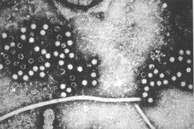
 TEM micrograph of hepatitis E virions.
| ||||||||
| Virus classification | ||||||||
|
|
Hepatitis E Microchapters |
|
Diagnosis |
|---|
|
Treatment |
|
Hepatitis E pathophysiology On the Web |
|
American Roentgen Ray Society Images of Hepatitis E pathophysiology |
|
Risk calculators and risk factors for Hepatitis E pathophysiology |
Editor-In-Chief: C. Michael Gibson, M.S., M.D. [1]; Associate Editor(s)-in-Chief: João André Alves Silva, M.D. [2] Jolanta Marszalek, M.D. [3]
Overview
Virology
The viral particles are 27 to 34 nanometers in diameter, are non-enveloped and contain a single-strand of positive-sense RNA that is approximately 7300 bases in length. The virus particle was first visualised in 1983[1] but was only molecularly cloned in 1990.[2]
It was previously classified family Caliciviridae. However, its genome more closely resembles the rubella virus. It is now classified in a new virus family, named as Hepeviridae.
References
- ↑ Balayan MS, Andjaparidze AG, Savinskaya SS; et al. (1983). "Evidence for a virus in non-A, non-B hepatitis transmitted via the fecal-oral route". Intervirology. 20 (1): 23–31. PMID 6409836.
- ↑ Reyes GR, Purdy MA, Kim JP; et al. (1990). "Isolation of a cDNA from the virus responsible for enterically transmitted non-A, non-B hepatitis". Science. 247 (4948): 1335–9. doi:10.1126/science.2107574. PMID 2107574.