Hepatitis D physical examination
|
Hepatitis D |
|
Diagnosis |
|
Treatment |
|
Hepatitis D physical examination On the Web |
|
American Roentgen Ray Society Images of Hepatitis D physical examination |
|
Risk calculators and risk factors for Hepatitis D physical examination |
Editor-In-Chief: C. Michael Gibson, M.S., M.D. [1]; Associate Editor(s)-In-Chief: Varun Kumar, M.B.B.S. [2]
Physical Examination
Physical examination to look for signs of advanced liver disease should be done. The findings may include:
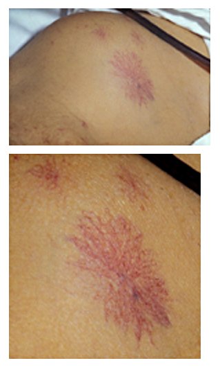
Spider angiomata in 47-year-old patient had longstanding jaundice and ascites consequent to biopsy-proven hepatic cirrhosis.[1]

- Hepatomegaly
- Splenomegaly
- Ascites
- Encephalopathy
- Esophageal varices
- Asterixis as demonstrated in the video below
{{#ev:youtube|pAOWjYo-sX4}}
References
- ↑ Fred, H.; van Dijk, H. Images of Memorable Cases: Case 114, Connexions Web site. http://cnx.org/content/m14900/1.3/, Feb 16, 2012