Hepatitis B physical examination
|
Hepatitis B |
|
Diagnosis |
|
Treatment |
|
Case Studies |
|
Hepatitis B physical examination On the Web |
|
American Roentgen Ray Society Images of Hepatitis B physical examination |
|
Risk calculators and risk factors for Hepatitis B physical examination |
Editor-In-Chief: C. Michael Gibson, M.S., M.D. [1]
Overview
For the majority of patients with acute and chronic hepatitis B, the physical examination is normal.[1]
Physical Examination
The aim of the initial physical examination is to observe for the presence of signs of chronic liver disease which include the following:[1]
Skin
HEENT
Abdomen
Extremities
Neurologic
- Asterixis
- Neurologic manifestations in cases of HBV progression to hepatic encephalopathy include a wide spectrum of cognitive impairment and motor system abnormalities,
Extrahepatic Manifestations
- Arthritis-dermatitis syndrome:[2]
- Urticaria
- Petechiae
- Palpable purpura
- Arthralgia
- Arthritis of small joints
- Neuropathy
References
- ↑ 1.0 1.1 Rotman Y, Brown TA, Hoofnagle JH (2009). "Evaluation of the patient with hepatitis B." Hepatology. 49 (5 Suppl): S22–7. doi:10.1002/hep.22976. PMC 2881483. PMID 19399815.
- ↑ Han SH (2004). "Extrahepatic manifestations of chronic hepatitis B." Clin Liver Dis. 8 (2): 403–18. PMID 15481347.
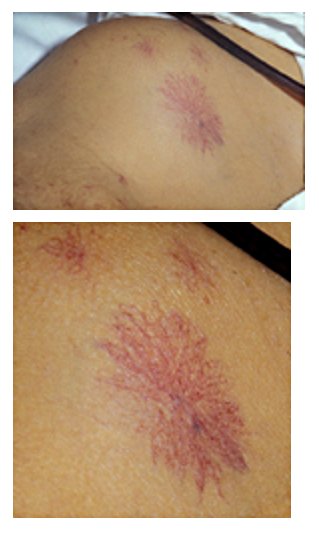
Spider angiomata in 47-year-old patient had longstanding jaundice and ascites consequent to biopsy-proven hepatic cirrhosis.[1]

- ↑ Fred, H.; van Dijk, H. Images of Memorable Cases: Case 114, Connexions Web site. http://cnx.org/content/m14900/1.3/, Feb 16, 2012