Granulocytic sarcoma (GS, also known as chloroma) was first discovered by Allen Burns, a British physician, in 1811. The term chloroma was first used by King to address the greenish appearance of the tumor due to [[myeloperoxidase]]. The association of the GS with [[Acute myeloid leukemia|acute myeloid leukemia (AML)]] was first recognized by Dock in 1902. GS can be classified into two categories based on its co-occurence with other malignancies. Infiltration of the tumor with [[myeloblasts]] is the main characteristic of the tumor on [[H&E stain]]. GS rises from primitive precursors of [[granulocytes]]. The disease is an extramedullary manifestation of myeloid diseases, however, it can occur as a primary disease. Aggregation of [[myeloblasts]], [[promyelocytes]] and [[Myelocyte|myelocytes]] outside of the bone marrow presents itself as these solid tumors. Tumors can occur at any site and can appear as green, gray, white or brown masses. GS must be differentiated from other diseases that can present as extramedullary solid tumors. All patients with GS must be evaluated for concurrent or future malignancies as GS can occur in the course of or prior to other malignancies. The prevalence of GS is approximately 2 per 1,000,000 individuals worldwide. The most important risk factor for development of GS is genetic mutations and susceptibility. Symptoms of GS may include the following: Symptoms due to mass effect such as [[deafness]], [[ptosis]], altered vision, intestinal obstruction, headache, neck pain, abdominal pain, and constitutional symptoms. [[Chemotherapy]] is the main stain of treatment in patients with GS. Even patients with isolated GS must receive systemic treatment to better the prognosis.
==Historical Perspective==
==Historical Perspective==
*Granulocytic sarcoma (GS, also known as chloroma) was first discovered by Allen Burns, a British physician, in 1811 <ref>{{Cite journal|last=Burns|first=Allen|date=|title=Observations of surgical anatomy, in Head andNeck.|url=|journal=London, England, Royce, 1811|volume=|pages=364-366|via=}}</ref>.
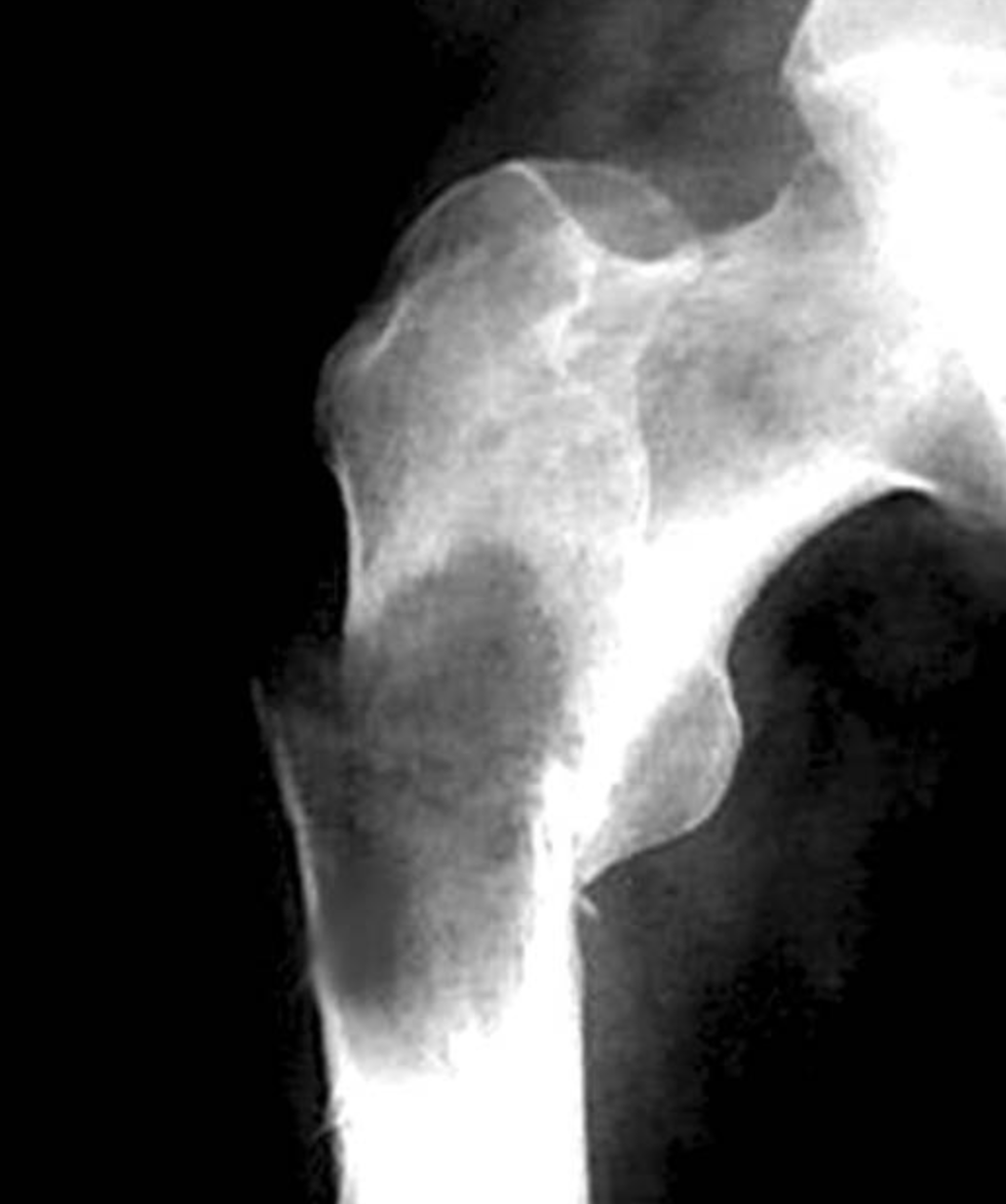
*[[File:Screenshot 2019-05-02 Myeloid sarcoma.png|thumb|Plain radiograph of femur in a patient with GS. Courtesy of image: pathology outlines (http://www.pathologyoutlines.com/topic/bonemyeloidsarcoma.html)]]Granulocytic sarcoma (GS, also known as chloroma) was first discovered by Allen Burns, a British physician, in 1811 <ref>{{Cite journal|last=Burns|first=Allen|date=|title=Observations of surgical anatomy, in Head andNeck.|url=|journal=London, England, Royce, 1811|volume=|pages=364-366|via=}}</ref>.
*The term chloroma was first used by King to address the greenish appearance of the tumor due to myeloperoxidase.
*The term chloroma was first used by King to address the greenish appearance of the tumor due to [[myeloperoxidase]].
*The association of the GS with acute myeloid leukemia was first recognized bt Dock in 1902 <ref>{{Cite journal|last=Dock G, Warthin AS|first=|date=|title=A new case of chloroma withleukemia.|url=|journal=Trans Assoc Am Phys, 1904|volume=19:64|pages=115|via=}}</ref>.
*The association of the GS with [[Acute myeloid leukemia|acute myeloid leukemia (AML)]] was first recognized by Dock in 1902 <ref>{{Cite journal|last=Dock G, Warthin AS|first=|date=|title=A new case of chloroma withleukemia.|url=|journal=Trans Assoc Am Phys, 1904|volume=19:64|pages=115|via=}}</ref>.
*The term granulocytic sarcoma was suggested by Rappaport in 1967 to grant generalisability to it <ref>{{Cite book|title=Tumors of the hematopoietic system, inAtlas of Tumor Pathology, Section III|last=Rappaport H|first=|publisher=Fascicle 8. ArmedForces Institute of Pathology|year=1967|isbn=|location=Washington|pages=241-247}}</ref>.
*The term "granulocytic sarcoma" was suggested by Rappaport in 1967 to grant generalisability to it <ref>{{Cite book|title=Tumors of the hematopoietic system, inAtlas of Tumor Pathology, Section III|last=Rappaport H|first=|publisher=Fascicle 8. ArmedForces Institute of Pathology|year=1967|isbn=|location=Washington|pages=241-247}}</ref>.
==Classification==
==Classification==
:* GS can be classified into two categories based on its co-occurence with other malignancies:
* GS can be classified into two categories based on its co-occurence with other malignancies:
* Infiltration of the tumor with myeloblasts is the main characteristic of the tumor on H&E stain.
* Infiltration of the tumor with [[myeloblasts]] is the main characteristic of the tumor on [[H&E stain]].
* GS rises from primitive precursors of granulocytes.
* GS rises from primitive precursors of [[granulocytes]].
* The disease is an extramedullary manifestation of myeloid diseases, however, it can occur as a primary disease.
* The disease is an extramedullary manifestation of myeloid diseases, however, it can occur as a primary disease.
* Aggregation of myeloblasts, promyelocytes and myelocytes outside of the bone marrow presents itself as these solid tumors.
* Aggregation of [[myeloblasts]], [[Promyelocyte|promyelocytes]] and [[Myelocyte|myelocytes]] outside of the bone marrow presents itself as these solid tumors.
* Tumors can occur at any site and can appear as green, gray, white or brown masses.
* Tumors can occur at any site and can appear as green, gray, white or brown masses.
==Differentiating GS from other Diseases==
GS must be differentiated from other diseases that can present as extramedullary solid tumors, such as:
* [[Lymphoma|Large cell lymphoma]]
* Non-Hodgkin lymphoma
* [[Thymoma]]
* [[Myeloma]]
* [[Sarcoma|Esosinophilic sarcoma]]
* [[Ewing sarcoma]]
* Extramedullary sites of [[hematopoiesis]]
* [[Burkitt lymphoma]]
* [[Hypereosinophilic syndrome]]
* [[Polycythemia vera]]
All patients with GS must be evaluated for concurrent or future malignancies as GS can occur in the course of or prior to other malignancies.
==Clinical Features==
==Differentiating granulocytic sarcoma from other Diseases==
*Granulocytic sarcoma must be differentiated from other diseases that can present as extramedullary solid tumors, such as:
:*Large cell lymphoma
:*Non-Hodgkin lymphoma
:*Thymoma
:*Myeloma
:*Esosinophilic sarcoma
:*Ewing sarcoma
:*Extramedullary sites of hematopoiesis
:All patients with granulocytic sarcoma must be evaluated for concurrent or future malignancies as granulocytic sarcoma can occur in the course of or prior to other malignancies.
==Epidemiology and Demographics==
==Epidemiology and Demographics==
* The prevalence ofgranulocytic sarcoma is approximately 2 per 1,000,000 individuals worldwide.
* The prevalence of GS is approximately 2 per 1,000,000 individuals worldwide.
* Most of the cases of granulocytic sarcoma are case reports and the disease is extremely rare.
* Most of the cases of GS are case reports and the disease is extremely rare.
===Age===
===Age===
* Patients of all age groups may develop granulocytic sarcoma.
* Patients of all age groups may develop GS.
* GS associated with [[AML]] occurs more commonly in children.
* Granulocytic sarcoma associated with acute myleloid leukemia occurs more commonly in children.
===Gender===
===Gender===
*Granulocytic sarcoma affects both men and women.
* GS affects both men and women.
* Due to the rarity of the disease it is not clear whether there is a gender predilection for it.
*Due to the rarity of the disease it is not clear whether there is a gender predilection for it.
===Race===
===Race===
*There is no racial predilection for granulocytic sarcoma.
* There is no racial predilection for GS.
==Risk Factors==
==Risk Factors==
*Risk factors for granulocytic sarcoma are usually chromosomal aberrations and include:
* Risk factors for GS are usually chromosomal aberrations and include<ref name=":0">{{Cite journal|last=Daniela Dörfel et al.|first=|date=2016|title=Cardiac Myeloid Sarcoma: Multimodality Radiologic Imaging Features and Pathologic Correlation|url=https://www.amjmed.com/article/S0002-9343(16)30356-4/fulltext|journal=The American Journal of Medicine|volume=129|pages=e117-e120|via=}}</ref>:
**Trisomy 8
** [[Trisomy 8]]
**Monosomy 7
** Monosomy 7
**MLL gene rearrangement
** MLL gene rearrangement
**NPM1 mutations
** [[NPM1]] mutations
**FLT3 mutations
** [[FLT3]] mutations
== Natural History, Complications and Prognosis==
== Natural History, Complications and Prognosis==
*How granulocytic sarcoma evolves over time depends on the co-occurence of the disease with other malignancies.
* GS evolve over time, that depends on the co-occurence of the disease with other malignancies.
*Granulocytic sarcoma may present before evidences of other malignancies manifest or after these malignancies are evident.
* GS may present before evidences of other malignancies manifest or after these malignancies are evident.
*Symptoms of the isolated granulocytic sarcoma depends on the location and the site of the tumor.
* Symptoms of the isolated GS depends on the location and the site of the tumor.
*Majority of cases are associated with acute myeloid leukemia or other myeloproliferative/myelodysplastic syndromes.
* Majority of cases are associated with [[AML]] or other [[myeloproliferative]]/[[Myelodysplastic syndrome|myelodysplastic syndromes]].
*Majority of granulocytic sarcoma tumors are found in the soft tissues such as the peritoneum, lymph nodes, CNS and skin. They are also found in bone and periosteum.
* Majority of GS tumors are found in the soft tissues such as the peritoneum, lymph nodes, CNS and skin. They are also found in bone and [[periosteum]].
*Early clinical features include weight loss, weakness. Other manifestations of the tumor depend on its size and location.
* Early clinical features include weight loss, fatigue. Other manifestations of the tumor depend on its size and location.
*Prognosis of granulocytic sarcoma depends on its association with other malignacies. In cases of isolated granulocytic sarcoma the prognosis is good. However, granulocytic sarcoma associated with myeloproliferative disorders has poor prognosis.
* Prognosis of GS depends on its association with other malignacies. In cases of isolated GS the prognosis is good. However, GS associated with [[myeloproliferative disorders]] has poor prognosis.
*Prognosis of isolated granulocytic sarcoma with chromosome 8 abnormalities is worse than other cases of isolated granulocytic sarcoma.
* Prognosis of isolated GS with chromosome 8 abnormalities is worse than other cases of isolated GS.
== Diagnosis ==
== Diagnosis ==
===Diagnostic Criteria===
===Diagnostic Criteria===
*There are no predefined criteria for diagnosis of granulocytic sarcoma.
* There are no predefined criteria for diagnosis of granulocytic sarcoma.
*Granulocytic sarcoma must be suspected in patients with AML or myelodysplatic syndromes. Diagnosis must be confirmed with histopathologic study of the specimen.
* Granulocytic sarcoma must be suspected in patients with [[AML]] or [[Myelodysplastic syndrome|myelodysplastic syndromes]]. Diagnosis must be confirmed with histopathologic study of the specimen.
=== Symptoms ===
=== Symptoms ===
*[Disease name] is usually asymptomatic.
Symptoms of GS may include the following:
*Symptoms of [disease name] may include the following:
* Symptoms due to mass effect such as [[deafness]], [[ptosis]], altered vision, intestinal obstruction, etc.
:*[symptom 1]
* [[Headache]], neck pain, abdominal pain,etc. based on the site of the tumor
:*[symptom 2]
* Constitutional symptoms such as fever, fatigue, etc.
:*[symptom 3]
:*[symptom 4]
:*[symptom 5]
:*[symptom 6]
=== Physical Examination ===
=== Physical Examination ===
*Patients with [disease name] usually appear [general appearance].
Patients with GS can present with varying presentations.
*Physical examination may be remarkable for:
:*[finding 1]
Physical examination may be remarkable for:
:*[finding 2]
* [[Lymphadenopathy]] (in cases associated with [[AML]] and other [[Myeloproliferative syndrome|myeloproliferative syndromes]])
:*[finding 3]
* Skin lesions (of varying colors such as green, grey, brown, etc.)
:*[finding 4]
* Organ enlargement such as [[hepatosplenomegaly]]
:*[finding 5]
* [[Petechiae]] in patients with [[thrombocytopenia]]
:*[finding 6]
* [[Hearing loss]]
* [[Heart murmurs]] in cases with heart chamber masses
* [[Crackles]] on lung auscultation
* [[Abdominal distension|Abdominal distention]]/tenderness in cases with intestinal obstruction
* Limb swelling due to different pathologies such as [[Deep vein thrombosis|deep vein thrombosis (DVT)]]
=== Laboratory Findings ===
=== Laboratory Findings ===
*There are no specific laboratory findings associated with [disease name].
* In cases associated with [[AML]]/[[CML]], [[anemia]], [[thrombocytopenia]] with normal, low or high white blood cells can be present.
* In cases associated with [[Polycythemia vera CT|polycythemia vera]], [[thrombocytosis]] and high levels of hemoglobin is present in complete blood count (CBC).
* High [[eosinophil]] levels can be present in CBC.
*A [positive/negative] [test name] is diagnostic of [disease name].
*An [elevated/reduced] concentration of [serum/blood/urinary/CSF/other] [lab test] is diagnostic of [disease name].
*Other laboratory findings consistent with the diagnosis of [disease name] include [abnormal test 1], [abnormal test 2], and [abnormal test 3].
===Imaging Findings===
===Imaging Findings===
*There are no [imaging study] findings associated with [disease name].
* In cases of CNS involvement, magnetic resonance imaging (MRI) or CT scan of the CNS can reveal extra-axial masses.
*[Imaging study 1] is the imaging modality of choice for [disease name].
* In cases with soft tissue involvement, sonogram of the tissue can reveal the mass.
*On [imaging study 1], [disease name] is characterized by [finding 1], [finding 2], and [finding 3].
GS appears as<ref>{{Cite journal|last=Guermazi et al.|first=|date=2002|title=Granulocytic sarcoma (chloroma): imaging findings in adults and children|url=https://www.ajronline.org/doi/full/10.2214/ajr.178.2.1780319|journal=American Journal of Roentgenology|volume=178|pages=319-25|via=}}</ref>:
*[Imaging study 2] may demonstrate [finding 1], [finding 2], and [finding 3].
* Hyperdense/isodence to brain/muscle in CT scan without enhancement
* Isointense/hyperintense on T2-weighted MRI
* Isointemse/hypointense on T1-weighted MRI
[[File:Chloroma histopathology micrograph (H&E stain).jpeg|thumb|Histopathologic micrograph of GS. Infiltration with myeloblasts which can be seen in the course of AML. Courtesy of image: Wikipedia (By Nephron - Own work, CC BY-SA 3.0, <nowiki>https://commons.wikimedia.org/w/index.php?curid=15893726</nowiki>)]]Abdominal plain radiogram can reveal obstruction, [[intussusception]], etc.
Echocardiogram may reveal mobile masses in any heart chamber in cases of heart involvement.
Chest radiographs can show lymph node enlargement and consolidation.
=== Other Diagnostic Studies ===
=== Other Diagnostic Studies ===
*[Disease name] may also be diagnosed using [diagnostic study name].
* Peripheral blood smear can reveal circulating [[Blast|blasts]].
*Findings on [diagnostic study name] include [finding 1], [finding 2], and [finding 3].
* [[Flow cytometry]] can help differentiate [[AML]] from [[Acute lymphoblastic leukemia|acute lymphoblastic leukemia (ALL)]].
* Histopathologic analysis of biopsy specimen retrieved by excision or [[fine needle aspiration]] shows abundant [[myeloblasts]].
* Histopathologic analysis of GS lesions reveals high infiltration with [[myeloblasts]].
== Treatment ==
== Treatment ==
=== Medical Therapy ===
=== Medical Therapy ===
*There is no treatment for [disease name]; the mainstay of therapy is supportive care.
* [[Chemotherapy]] is the main stain of treatment in patients with GS. Even patients with isolated GS must receive systemic treatment to better the prognosis.
* Patients receiving [[cytarabine]] have better prognosis<ref name=":0" />.
*The mainstay of therapy for [disease name] is [medical therapy 1] and [medical therapy 2].
* Patients with chemotherpeutic regimens accepted for [[AML]] had longer period of progression to [[AML]].
*[Medical therapy 1] acts by [mechanism of action 1].
Treatment includes different regimens<ref>{{Cite journal|last=Yilmaz, A. F., Saydam, G., Sahin, F., & Baran, Y.|first=|date=2013|title=Granulocytic sarcoma: a systematic review|url=https://www.ncbi.nlm.nih.gov/pmc/articles/PMC3875275/|journal=American Journal of Blood Research|volume=|pages=|via=}}</ref>:
*Response to [medical therapy 1] can be monitored with [test/physical finding/imaging] every [frequency/duration].
* Idarubicine and [[cytarabine]]
* [[Fludarabine]] and [[cytarabine]]
* Idarubicine and [[G-CSF]]
* [[Cyclophosphamide]], [[cytarabine]], [[topotecan]] and [[G-CSF]]
All of these therapeutic agents act through DNA damage and interferes with DNA synthesis.
===Bone marrow transplantation===
* [[Hematopoietic stem cell transplantation]] can be considered as a treatment option following induction [[chemotherapy]] in patients with [[AML]].<ref name=":1">{{Cite journal|last=Richard L. Bakst, Martin S. Tallman, Dan Douer and Joachim Yahalom|first=|date=2011|title=How I treat extramedullary acute myeloid leukemia|url=http://www.bloodjournal.org/content/118/14/3785?sso-checked=true|journal=Blood|volume=118|pages=3785-3793|via=}}</ref>
===Radiation===
* Radiation can be considered as an adjunctive therapy<ref name=":1" />.
* Combination of chemotherapy and radiotherapy can be considered in patients with CNS involvement or when rapid regression of symptoms is required.
=== Surgery ===
=== Surgery ===
*Surgery is the mainstay of therapy for [disease name].
* Surgery alone is not a good treatment strategy for GS.
*[Surgical procedure] in conjunction with [chemotherapy/radiation] is the most common approach to the treatment of [disease name].
* Surgery can be considered prior to chemotherapy in patients where debulking can better the prognosis and help with symptom relief.<ref>{{Cite journal|last=Antic D et al.|first=|date=2013|title=Is there a "gold" standard treatment for patients with isolated myeloid sarcoma?|url=https://www.sciencedirect.com/science/article/abs/pii/S0753332212001060?via%3Dihub|journal=Biomed Pharmacother|volume=67|pages=72-77|via=}}</ref>
*[Surgical procedure] can only be performed for patients with [disease stage] [disease name].
* Surgery can also have a diagnostic role in cases where excision of the mass provides specimen for histopatjologic diagnosis.
=== Prevention ===
=== Prevention ===
*There are no primary preventive measures available for [disease name].
* There are no primary preventive measures available for GS.
*Effective measures for the primary prevention of [disease name] include [measure1], [measure2], and [measure3].
*Once diagnosed and successfully treated, patients with [disease name] are followed-up every [duration]. Follow-up testing includes [test 1], [test 2], and [test 3].
Granulocytic sarcoma (GS, also known as chloroma) was first discovered by Allen Burns, a British physician, in 1811. The term chloroma was first used by King to address the greenish appearance of the tumor due to myeloperoxidase. The association of the GS with acute myeloid leukemia (AML) was first recognized by Dock in 1902. GS can be classified into two categories based on its co-occurence with other malignancies. Infiltration of the tumor with myeloblasts is the main characteristic of the tumor on H&E stain. GS rises from primitive precursors of granulocytes. The disease is an extramedullary manifestation of myeloid diseases, however, it can occur as a primary disease. Aggregation of myeloblasts, promyelocytes and myelocytes outside of the bone marrow presents itself as these solid tumors. Tumors can occur at any site and can appear as green, gray, white or brown masses. GS must be differentiated from other diseases that can present as extramedullary solid tumors. All patients with GS must be evaluated for concurrent or future malignancies as GS can occur in the course of or prior to other malignancies. The prevalence of GS is approximately 2 per 1,000,000 individuals worldwide. The most important risk factor for development of GS is genetic mutations and susceptibility. Symptoms of GS may include the following: Symptoms due to mass effect such as deafness, ptosis, altered vision, intestinal obstruction, headache, neck pain, abdominal pain, and constitutional symptoms. Chemotherapy is the main stain of treatment in patients with GS. Even patients with isolated GS must receive systemic treatment to better the prognosis.
Historical Perspective
Plain radiograph of femur in a patient with GS. Courtesy of image: pathology outlines (http://www.pathologyoutlines.com/topic/bonemyeloidsarcoma.html)Granulocytic sarcoma (GS, also known as chloroma) was first discovered by Allen Burns, a British physician, in 1811 [1].
The term chloroma was first used by King to address the greenish appearance of the tumor due to myeloperoxidase.
Majority of GS tumors are found in the soft tissues such as the peritoneum, lymph nodes, CNS and skin. They are also found in bone and periosteum.
Early clinical features include weight loss, fatigue. Other manifestations of the tumor depend on its size and location.
Prognosis of GS depends on its association with other malignacies. In cases of isolated GS the prognosis is good. However, GS associated with myeloproliferative disorders has poor prognosis.
Prognosis of isolated GS with chromosome 8 abnormalities is worse than other cases of isolated GS.
Diagnosis
Diagnostic Criteria
There are no predefined criteria for diagnosis of granulocytic sarcoma.
Granulocytic sarcoma must be suspected in patients with AML or myelodysplastic syndromes. Diagnosis must be confirmed with histopathologic study of the specimen.
Symptoms
Symptoms of GS may include the following:
Symptoms due to mass effect such as deafness, ptosis, altered vision, intestinal obstruction, etc.
Headache, neck pain, abdominal pain,etc. based on the site of the tumor
Constitutional symptoms such as fever, fatigue, etc.
Physical Examination
Patients with GS can present with varying presentations.
Hyperdense/isodence to brain/muscle in CT scan without enhancement
Isointense/hyperintense on T2-weighted MRI
Isointemse/hypointense on T1-weighted MRI
Histopathologic micrograph of GS. Infiltration with myeloblasts which can be seen in the course of AML. Courtesy of image: Wikipedia (By Nephron - Own work, CC BY-SA 3.0, https://commons.wikimedia.org/w/index.php?curid=15893726)
Abdominal plain radiogram can reveal obstruction, intussusception, etc.
Echocardiogram may reveal mobile masses in any heart chamber in cases of heart involvement.
Chest radiographs can show lymph node enlargement and consolidation.
Other Diagnostic Studies
Peripheral blood smear can reveal circulating blasts.
Histopathologic analysis of GS lesions reveals high infiltration with myeloblasts.
Treatment
Medical Therapy
Chemotherapy is the main stain of treatment in patients with GS. Even patients with isolated GS must receive systemic treatment to better the prognosis.
Patients receiving cytarabine have better prognosis[4].
Patients with chemotherpeutic regimens accepted for AML had longer period of progression to AML.
Radiation can be considered as an adjunctive therapy[7].
Combination of chemotherapy and radiotherapy can be considered in patients with CNS involvement or when rapid regression of symptoms is required.
Surgery
Surgery alone is not a good treatment strategy for GS.
Surgery can be considered prior to chemotherapy in patients where debulking can better the prognosis and help with symptom relief.[8]
Surgery can also have a diagnostic role in cases where excision of the mass provides specimen for histopatjologic diagnosis.
Prevention
There are no primary preventive measures available for GS.
References
↑Burns, Allen. "Observations of surgical anatomy, in Head andNeck". London, England, Royce, 1811: 364–366.
↑Dock G, Warthin AS. "A new case of chloroma withleukemia". Trans Assoc Am Phys, 1904. 19:64: 115.
↑Rappaport H (1967). Tumors of the hematopoietic system, inAtlas of Tumor Pathology, Section III. Washington: Fascicle 8. ArmedForces Institute of Pathology. pp. 241–247.

.jpeg)