Gaucher's disease pathophysiology
|
Gaucher's disease Microchapters |
|
Diagnosis |
|---|
|
Treatment |
|
Case Studies |
|
Gaucher's disease pathophysiology On the Web |
|
American Roentgen Ray Society Images of Gaucher's disease pathophysiology |
|
Risk calculators and risk factors for Gaucher's disease pathophysiology |
Please help WikiDoc by adding more content here. It's easy! Click here to learn about editing.
Editor-In-Chief: C. Michael Gibson, M.S., M.D. [1]; Associate Editor-In-Chief: Cafer Zorkun, M.D., Ph.D. [2]
Overview
Pathophysiology

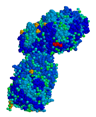
The disease is caused by a defect in the housekeeping gene lysosomal gluco-cerebrosidase (also known as β-glucosidase, EC 3.2.1.45, PDB: 1OGS) on the first chromosome (1q21).
The enzyme is a 55.6 KD, 497 amino acids long protein that catalyses the breakdown of glucocerebroside, a cell membrane constituent of red and white blood cells.
The macrophages that clear these cells are unable to eliminate the waste product, which accumulates in fibrils, and turn into Gaucher cells, which appear on light microscopy as appearing to contain crumpled-up paper.
Different mutations in the β-glucosidase determine the remaining activity of the enzyme, and, to a large extent, the phenotype.
In the brain (type II and III), glucocerebroside accumulates due to the turnover of complex lipids during brain development and the formation of the myelin sheath of nerves.
Research suggests that heterozygotes for particular acid β-glucosidase mutations are at an increased risk of Parkinson's disease.[1]
A study of 1525 Gaucher patients in the United States suggested that while cancer risk is not elevated, particular malignancies (non-Hodgkin lymphoma, melanoma and pancreatic cancer) occurred at a 2-3 times higher rate.[2]
References
- ↑ Aharon-Peretz J, Rosenbaum H, Gershoni-Baruch R (2004). "Mutations in the glucocerebrosidase gene and Parkinson's disease in Ashkenazi Jews". N. Engl. J. Med. 351 (19): 1972–7. doi:10.1056/NEJMoa033277. PMID 15525722.
- ↑ Landgren O, Turesson I, Gridley G, Caporaso NE (2007). "Risk of Malignant Disease Among 1525 Adult Male US Veterans With Gaucher Disease". 167 (11): 1189–1194. doi:10.1001/archinte.167.11.1189. PMID 17563029.