|
|
| (9 intermediate revisions by the same user not shown) |
| Line 15: |
Line 15: |
| MeshNumber = C06.405.748.280 | | | MeshNumber = C06.405.748.280 | |
| }} | | }} |
| {{SI}} | | {{Gastric antral vascular ectasia}} |
| | |
| {{CMG}} | | {{CMG}} |
|
| |
|
| {{SK}} Watermelon stomach; GAVE | | {{SK}} Watermelon stomach; GAVE |
|
| |
|
| ==Overview== | | ==[[Gastric antral vascular ectasia overview|Overview]]== |
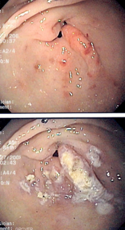
| Gastric antral vascular ectasia is an uncommon cause of chronic [[gastrointestinal bleeding]] or [[iron deficiency anemia]]. The condition is associated with dilated small blood vessels in the [[antrum]], or the last part of the [[stomach]]. It is also called watermelon stomach because streaky long red areas that are present in the stomach may resemble the markings on [[watermelon]]<ref>{{cite journal | author = Suit P, Petras R, Bauer T, Petrini J | title = Gastric antral vascular ectasia. A histologic and morphometric study of "the watermelon stomach". | journal = Am J Surg Pathol | volume = 11 | issue = 10 | pages = 750-7 | year = 1987 | id = PMID 3499091}}</ref>.
| |
| | |
| ==Historical Perspective==
| |
| The condition was first discovered in 1952, and reported in the literature in 1953.<ref name=Rider>{{cite journal |last1=Rider |first1=JA |last2=Klotz |first2=AP |last3=Kirsner |first3=JB |title=Gastritis with veno-capillary ectasia as a source of massive gastric hemorrhage |journal=Gastroenterology |volume=24 |issue=1 |pages=118–23 |year=1953 |pmid=13052170}}</ref> Watermelon disease was first diagnosed by Wheeler ''et al.'' in 1979, and definitively described in four living patients by Jabbari ''et al''. only in 1984. As of 2011, the etiology and pathogenesis are still not known.<ref name=Unusual>{{cite journal |last1=Tuveri |first1=Massimiliano |last2=Borsezio |first2=Valentina |last3=Gabbas |first3=Antonio |last4=Mura |first4=Guendalina |title=Gastric antral vascular ectasia—an unusual cause of gastric outlet obstruction: report of a case |journal=Surgery today |volume=37 |issue=6 |pages=503–5 |year=2007 |pmid=17522771 |doi=10.1007/s00595-006-3430-3}}</ref> However, there are several competing hypotheses as to various etiologies.
| |
| | |
| ==Pathophysiology==
| |
| GAVE is characterized by dilated capillaries in the lamina propria with fibrin thrombi.
| |
| | |
| ===Microscopic Pathology===
| |
| | |
| [[Image:Gastric_antral_vascular_ectasia.jpg|thumb|left|Micrograph showing gastric antral vascular ectasia. A large spherical, eosinophilic (i.e. pink) fibrin thrombus is seen off-center right. Stomach biopsy. H&E stain.]]
| |
| <br clear="left"/>
| |
| | |
| ===Asociated Conditions===
| |
| GAVE is associated with a number of conditions, including
| |
| * [[Portal hypertension]]
| |
| * [[Chronic renal failure]]
| |
| * [[Collagen vascular disease]]s, particularly [[scleroderma]]
| |
| * [[Pernicious anemia]]
| |
| * [[Liver cirrhosis]]
| |
| * [[Chronic renal failure]]
| |
| * [[Bone marrow transplantation]]
| |
| | |
| ==Causes==
| |
| 65% of patients with both cirrhosis and GAVE are male, but a total of 30% have both conditions. The causal connection between cirrhosis and GAVE has not been proven.
| |
| | |
| A [[connective tissue]] disease has been suspected in some cases.
| |
| | |
| Autoimmunity may have something to do with it,<ref name=RNA>{{cite journal |doi=10.1093/nar/24.7.1220 |last1=Valdez |first1=BC |last2=Henning |first2=D |last3=Busch |first3=RK |last4=Woods |first4=K |last5=Flores-Rozas |first5=H |last6=Hurwitz |first6=J |last7=Perlaky |first7=L |last8=Busch |first8=H |title=A nucleolar RNA helicase recognized by autoimmune antibodies from a patient with watermelon stomach disease |journal=Nucleic Acids Research |volume=24 |issue=7 |pages=1220–4 |year=1996 |pmid=8614622 |pmc=145780}}</ref> as 25% of all sclerosis patients who had a certain anti-RNA marker have GAVE. RNA autoimmunity has been suspected as a cause or marker since at least 1996.
| |
| | |
| One theory current since the 1990s focuses on a history of [[prolapse]] of the stomach into the [[small intestine]].
| |
|
| |
|
| [[Gastrin]] levels may indicate a hormonal connection. This may be due to vasoactive intestinal peptide and 5-hydroxy-tryptamine. | | ==[[Gastric antral vascular ectasia historical perspective|Historical Perspective]]== |
|
| |
|
| It is also possible that infection by ''[[H. pylori]]'' can cause it.
| | ==[[Gastric antral vascular ectasia pathophysiology|Pathophysiology]]== |
|
| |
|
| ==Differentiating Gastric antral vascular ectasia from other Diseases== | | ==[[Gastric antral vascular ectasia causes|Causes]]== |
| GAVE should be differentiated from other causes of intestinal bleeding such as
| |
| * [[Duodenal ulcer]]
| |
| * [[Portal hypertension]]<ref name=Portal>{{cite journal |last1=Spahr |first1=L |last2=Villeneuve |first2=J-P |last3=Dufresne |first3=M-P |last4=Tasse |first4=D |last5=Bui |first5=B |last6=Willems |first6=B |last7=Fenyves |first7=D |last8=Pomier-Layrargues |first8=G |title=Gastric antral vascular ectasia in cirrhotic patients: absence of relation with portal hypertension |journal=Gut |volume=44 |issue=5 |pages=739 |year=1999 |pmid=10205216 |pmc=1727493 |doi=10.1136/gut.44.5.739}}</ref>
| |
|
| |
|
| The differential diagnosis is important because treatments are different.
| | ==[[Gastric antral vascular ectasia differential diagnosis|Differentiating Gastric antral vascular ectasia from other Diseases]]== |
|
| |
|
| ==Epidemiology and Demographics== | | ==[[Gastric antral vascular ectasia epidemiology and demographics|Epidemiology and Demographics]]== |
| ===Age===
| |
| The average age of diagnosis for GAVE is 73 years of age for females, and 68 for males. Patients in their thirties have been found to have GAVE. It becomes more common in women in their eighties, rising to 4% of all such gastrointestinal conditions.
| |
|
| |
|
| ===Gender=== | | ==[[Gastric antral vascular ectasia natural history, complications and prognosis|Natural History, Complications and Prognosis]]== |
| Women are about twice as often diagnosed with gastric antral vascular ectasia than men. 71% of all cases of GAVE are diagnosed in females.
| |
|
| |
|
| ==Diagnosis== | | ==Diagnosis== |
| ===Symptoms===
| |
| * [[Fatigue]], [[tiredness]], [[palpitations]] - [[Anemia]]
| |
| * [[Blood loss]]
| |
| * [[GI bleeding]] - [[melena]] or [[hematochezia]]
| |
|
| |
|
| ===Endoscopy===
| | [[Gastric antral vascular ectasia history and symptoms|History and Symptoms ]] | [[ Gastric antral vascular ectasia physical examination|Physical Examination]] | [[Gastric antral vascular ectasia laboratory findings|Laboratory Findings]] | [[Gastric antral vascular ectasia ultrasound|Ultrasound]] | [[Gastric antral vascular ectasia endoscopy|Endoscopy]] | [[Gastric antral vascular ectasia other imaging findings|Other Imaging Findings]] | [[Gastric antral vascular ectasia other diagnostic studies|Other Diagnostic Studies]] |
| The endoscopic appearance of GAVE is similar to [[portal hypertensive gastropathy]]. Dilated capillaries are seen in the endoscopy which resemble the tell-tale watermelon stripes.
| |
|
| |
|
| ==Treatment== | | ==Treatment== |
| ===Medical Therapy===
| | [[Gastric antral vascular ectasia medical therapy|Medical Therapy]] | [[Gastric antral vascular ectasia surgery |Surgery]] | [[Gastric antral vascular ectasia primary prevention|Primary Prevention]] | [[Gastric antral vascular ectasia secondary prevention|Secondary Prevention]] | [[Gastric antral vascular ectasia cost-effectiveness of therapy|Cost-Effectiveness of Therapy]] | [[Gastric antral vascular ectasia future or investigational therapies|Future or Investigational Therapies]] |
| ====Traditional treatments====
| |
| GAVE is treated commonly by means of an endoscope, including [[argon plasma coagulation]] and electrocautery. Since endoscopy with argon photocoagulation is "usually effective", surgery is "usually not required."Coagulation therapy is well-tolerated but "tends to induce oozing and bleeding." "Endoscopy with thermal ablation" is favored [[medical treatment]] because of its low side effects and low mortality, but is "rarely curative."
| |
| ====Other treatments====
| |
| Other medical treatments have been tried and include [[estrogen]] and [[progesterone]] therapy, and anti-fibrinolytic drugs such as [[tranexamic acid]]. Corticosteroids are effective, but are "limited by their [[side effects]]."
| |
| | |
| ====Treatment of co-morbid conditions====
| |
| A [[transjugular intrahepatic portosystemic shunt]] (TIPS or TIPSS) procedure is used to treat portal hypertension when that is present as an associated condition. Unfortunately, the TIPSS, which has been used for similar conditions, may cause or exacerbate [[hepatic encephalopathy]].<ref name=Khan>{{cite journal |author=Khan S, Tudur Smith C, Williamson P, Sutton R |title=Cochrane Database of Systematic Reviews |journal=Cochrane Database Syst Rev |volume= |issue=4 |pages=CD000553 |year=2006 |pmid=17054131 |doi=10.1002/14651858.CD000553.pub2 |url=http://mrw.interscience.wiley.com/cochrane/clsysrev/articles/CD000553/frame.html |chapter=Portosystemic shunts versus endoscopic therapy for variceal rebleeding in patients with cirrhosis |editor1-last=Khan |editor1-first=Saboor A}}</ref><ref name=Saab>{{cite journal |author=Saab S, Nieto JM, Lewis SK, Runyon BA |title=TIPSS versus paracentesis for cirrhotic patients with refractory ascites |journal=Cochrane Database Syst Rev |volume= |issue=4 |pages=CD004889 |year=2006 |pmid=17054221 |doi=10.1002/14651858.CD004889.pub2 |url=http://mrw.interscience.wiley.com/cochrane/clsysrev/articles/CD004889/frame.html |chapter=TIPS versus paracentesis for cirrhotic patients with refractory ascites |editor1-last=Saab |editor1-first=Sammy}}</ref> TIPSS-related encephalopathy occurs in about 30% of cases, with the risk being higher in those with previous episodes of encephalopathy, higher age, female sex, and liver disease due to causes other than alcohol.<ref name=Sundaram>{{cite journal |author=Sundaram V, Shaikh OS |title=Hepatic encephalopathy: pathophysiology and emerging therapies |journal=Med. Clin. North Am. |volume=93 |issue=4 |pages=819–36, vii |year=2009 |month=July |pmid=19577116 |doi=10.1016/j.mcna.2009.03.009}}</ref> The patient, with his or her physician and family, must balance out a reduction in bleeding caused by TIPS with the significant risk of [[encephalopathy]]. Various shunts have been shown in a [[meta-study]] of 22 studies to be effective treatment to reduce bleeding, yet none have any demonstrated survival advantage.
| |
| | |
| If there is cirrhosis of the liver that has progressed to [[liver failure]], then [[lactulose]] may be prescribed for hepatic encephalopathy, especially for ''[[Hepatic_encephalopathy#Types|Type C encephalopathy]]'' with [[diabetes]]. Also, "antibiotics such as [[neomycin]], [[metronidazole]], and [[rifaximin]]" may be used effectively to treat the encephalopathy by removing nitrogen-producing bacteria from the gut.
| |
| | |
| [[Paracentesis]], a medical procedure involving needle drainage of fluid from a [[body cavity]],<ref>{{DorlandsDict|six/000078067|paracentesis}}</ref> may be used to remove fluid from the [[peritoneal cavity]] in the [[abdomen]] for such cases. This procedure uses a large needle, similar to the better-known [[amniocentesis]].
| |
| | |
| ===Surgery===
| |
| Surgery, consisting of excision of part of the lower stomach, also called ''antrectomy'', is another option. Antrectomy is "the resection, or surgical removal, of a part of the stomach known as the [[antrum]]."<ref name=Antrectomy>[http://www.surgeryencyclopedia.com/A-Ce/Antrectomy.html Surgery Encyclopedia website page on Antrectomy]. Accessed September 29, 2010.</ref> | |
| | |
| [[Laparoscopic surgery]] is possible in some cases, and as of 2003, was a "novel approach to treating watermelon stomach."<ref name=Lapro>{{cite journal |last1=Sherman |first1=V |last2=Klassen |first2=DR |last3=Feldman |first3=LS |last4=Jabbari |first4=M |last5=Marcus |first5=V |last6=Fried |first6=GM |title=Laparoscopic antrectomy: a novel approach to treating watermelon stomach |journal=Journal of the American College of Surgeons |volume=197 |issue=5 |pages=864 |year=2003 |pmid=14585429 |doi=10.1016/S1072-7515(03)00600-8}}</ref> | |
| | |
| A treatment used sometimes is endoscopic band ligation.<ref name=band>{{cite journal |last1=Wells |first1=C |last2=Harrison |first2=M |last3=Gurudu |first3=S |last4=Crowell |first4=M |last5=Byrne |first5=T |last6=Depetris |first6=G |last7=Sharma |first7=V |title=Treatment of gastric antral vascular ectasia (watermelon stomach) with endoscopic band ligation |journal=Gastrointestinal Endoscopy |volume=68 |issue=2 |pages=231 |year=2008 |pmid=18533150 |doi=10.1016/j.gie.2008.02.021}}</ref>
| |
| | |
| In 2010, a team of [[Japan]]ese surgeons performed a "novel endoscopic [[ablation]] of gastric antral vascular ectasia."<ref name=ablation>{{cite journal |last1=Komiyama |first1=Masae |title=A novel endoscopic ablation of gastric antral vascular ectasia |journal=World Journal of Gastrointestinal Endoscopy |volume=2 |pages=298 |year=2010 |doi=10.4253/wjge.v2.i8.298 |issue=8 |pmid=21160630 |last2=Fu |first2=K |last3=Morimoto |first3=T |last4=Konuma |first4=H |last5=Yamagata |first5=T |last6=Izumi |first6=Y |last7=Miyazaki |first7=A |last8=Watanabe |first8=S |pmc=2999147}}</ref> The experimental procedure resulted in "no complications."<ref name=ablation />
| |
| | |
| Relapse is possible, even after treatment by argon plasma coagulation and progesterone.<ref name=relapse>{{cite journal |last1=Shibukawa |first1=G |last2=Irisawa |first2=A |last3=Sakamoto |first3=N |last4=Takagi |first4=T |last5=Wakatsuki |first5=T |last6=Imamura |first6=H |last7=Takahashi |first7=Y |last8=Sato |first8=A |last9=Sato |first9=M |title=Gastric antral vascular ectasia (GAVE) associated with systemic sclerosis: relapse after endoscopic treatment by argon plasma coagulation |journal=Internal medicine (Tokyo, Japan) |volume=46 |issue=6 |pages=279–83 |year=2007 |pmid=17379994 |doi=10.2169/internalmedicine.46.6203}}</ref> In such cases of relapse, surgery may be the only option; in one case that involved "Endoscopic mucosal [[Segmental resection|resection]]".<ref name=resection>{{cite journal |last1=Katsinelos |first1=P |last2=Chatzimavroudis |first2=G |last3=Katsinelos |first3=T |last4=Panagiotopoulou |first4=K |last5=Kotakidou |first5=R |last6=Tsolkas |first6=G |last7=Triantafillidis |first7=I |last8=Papaziogas |first8=B |title=Endoscopic mucosal resection for recurrent gastric antral vascular ectasia |journal=VASA. Zeitschrift fur Gefasskrankheiten. Journal for vascular diseases |volume=37 |issue=3 |pages=289–92 |year=2008 |pmid=18690599 |doi=10.1024/0301-1526.37.3.289}}</ref>
| |
|
| |
|
| Antrectomy or other [[surgery]] is used as a last resort for GAVE. The risks of surgery should be considered. It is said that "surgery is the only cure" for GAVE.
| | ==Case Studies== |
|
| |
|
| ==References==
| | [[Gastric antral vascular ectasia case study one|Case #1]] |
| {{Reflist|2}}
| |
|
| |
|
| {{WikiDoc Help Menu}} | | {{WikiDoc Help Menu}} |
