Follicular thyroid cancer pathophysiology
|
Follicular thyroid cancer Microchapters |
|
Differentiating Follicular thyroid cancer from other Diseases |
|---|
|
Diagnosis |
|
Treatment |
|
Case Studies |
|
Follicular thyroid cancer pathophysiology On the Web |
|
American Roentgen Ray Society Images of Follicular thyroid cancer pathophysiology |
|
Risk calculators and risk factors for Follicular thyroid cancer pathophysiology |
Editor-In-Chief: C. Michael Gibson, M.S., M.D. [1]; Associate Editor(s)-in-Chief: Ammu Susheela, M.D. [2]
Overview
Follicular thyroid cancer arises from follicular cells of thyroid, which are secretory cells that are normally involved in production and secretion of thyroid hormones, thyroxine (T4) and triiodothyronine (T3). Genes involved in the pathogenesis of follicular thyroid cancer include RAS, PAX8/PPARγ, and PTEN.
Pathogenesis
- Follicular thyroid cancer is the second most common type of cancer. It constitutes about 15% of thyroid cancers.

- Follicular thyroid cancer occurs more commonly in women of over 50 years old.
- Thyroglobulin (Tg) can be used as a tumor marker for well-differentiated follicular thyroid cancer.
- Follicular carcinoma tends to metastasize to the lungs and bone via the bloodstream. Follicular thyroid cancer metastasize late to lymph nodes, with only 5-10% of patients having nodal metastases at the time of diagnosis. Hematogenous spread is however much more common with 20% of patients having distant hematogenous metastases at at the time of diagnosis.
- A Hürthle cell (also known as Askanazy cell) is an oncocytic cell in the thyroid that is often associated with Hashimoto's thyroiditis as well as follicular thyroid cancer. Hürthle cells are characterized as enlarged epithelial cells with abundant eosinophilic granular cytoplasm as a result of altered mitochondria.[1] They generally stain pink and are prominently found in histological sections of thyroid glands affected with Hashimoto's.
Genetics
- The Ras oncogene is positive in a significant proportion of individuals. The Ras oncogene acts through the RAF-MEK-MAPK kinase pathway.
- Other genes involved in the pathogenesis of follicular thyroid cancer are:
- RET/PTC (translocation) associated with MAPK and PI3K-AKT signaling pathways
- HRAS, KRAS, NRAS (mutation) associated with mitogen-activated protein kinase and PI3K-AKT signaling pathways
- PTEN (mutation) associated with PI3K-AKT signaling pathways
- PTEN (deletion) associated with PI3K-AKT signaling pathways
- IDH1 (mutation) assciated with IDH1-associated metabolic pathways signaling pathways
- Phosphatase and tensin homologue suppressor gene and the phosphatidylinositol 3-kinase pathway are also involved in the pathogenesis of follicular thyroid tumor.
- P53 (protein), c-myc, c-fos, and the thyrotropin (TSH) receptor are some other factors involved in the pathogenesis of follicular thyroid cancer.
- MicroRNAs namely miR-192, miR-197, miR-328, and miR-346 have increased expression in follicular cell carcinoma.

Associated Conditions
- Cowden disease
- Carney complex, type I
Gross Pathology
- Encapsulated tumors

Microscopic Pathology
- It is not possible to distinguish between follicular adenoma and carcinoma on cytological grounds. If fine needle aspiration cytology (FNAC) suggests follicular neoplasm, thyroid lobectomy should be performed to establish th histopathological diagnosis.
- On microscopic examination, trabecular, solid, follicular tumor cells that invade tumor capsule or surrounding vascular structures are found.
-
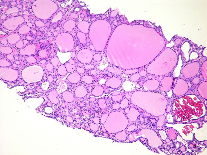
Metastatic follicular carcinoma<ref> Follicular thyroid cancer librepathology
-
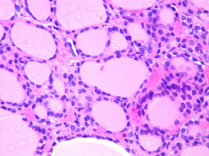
Metastatic follicular carcinoma<ref> Follicular thyroid cancer librepathology
.jpg)

Histopathological Video
Video
{{#ev:youtube|3_eCHeOkdgg}}
References
- ↑ Aytug, Serhat (June 13, 2006). "Hurthle Cell Carcinoma". eMedicine. Check date values in:
|date=(help)