File:Coronavirus16.jpeg
Size of this preview: 355 × 599 pixels. Other resolution: 700 × 1,182 pixels.
Original file (700 × 1,182 pixels, file size: 142 KB, MIME type: image/jpeg)
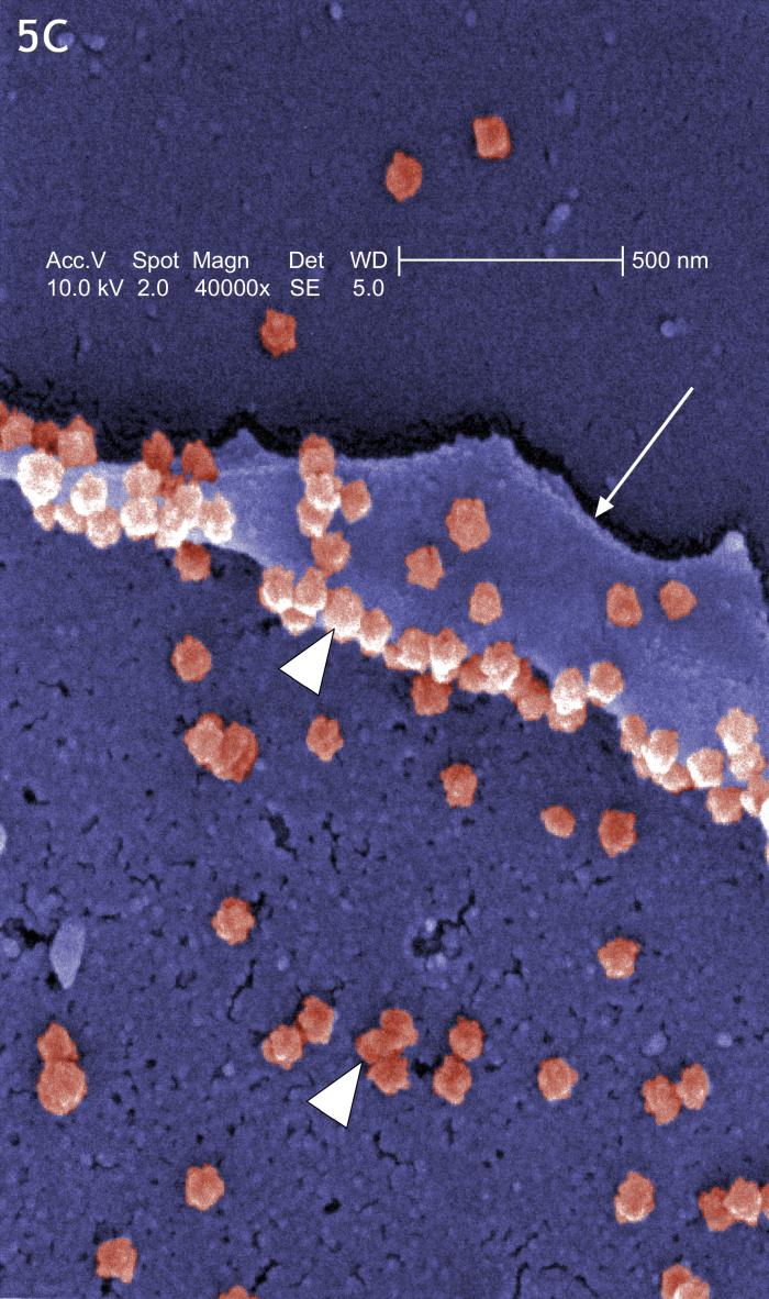
This digitally-colorized scanning electron micrograph reveals the prolific exportation of virus particles at the pseudopodial and cell surfaces.
File history
Click on a date/time to view the file as it appeared at that time.
| Date/Time | Thumbnail | Dimensions | User | Comment | |
|---|---|---|---|---|---|
| current | 20:47, 4 December 2014 | | 700 × 1,182 (142 KB) | Jesus Hernandez (talk | contribs) | This digitally-colorized scanning electron micrograph reveals the prolific exportation of virus particles at the pseudopodial and cell surfaces. |
You cannot overwrite this file.
File usage
The following 2 pages use this file: