Esophageal stricture physical examination
|
Esophageal stricture Microchapters |
|
Diagnosis |
|---|
|
Treatment |
|
Surgery |
|
Case Studies |
|
Esophageal stricture physical examination On the Web |
|
American Roentgen Ray Society Images of Esophageal stricture physical examination |
|
Risk calculators and risk factors for Esophageal stricture physical examination |
Editor-In-Chief: C. Michael Gibson, M.S., M.D. [1]; Associate Editor(s)-in-Chief:
Overview
Patients with [disease name] usually appear [general appearance]. Physical examination of patients with [disease name] is usually remarkable for [finding 1], [finding 2], and [finding 3].
OR
Common physical examination findings of [disease name] include [finding 1], [finding 2], and [finding 3].
OR
The presence of [finding(s)] on physical examination is diagnostic of [disease name].
OR
The presence of [finding(s)] on physical examination is highly suggestive of [disease name].
Physical Examination
Esophageal stricture has many causes. Physical examination due to different causes include:
- Physical examination of patients with [disease name] is usually remarkable for:[finding 1], [finding 2], and [finding 3].
- The presence of [finding(s)] on physical examination is diagnostic of [disease name].
- The presence of [finding(s)] on physical examination is highly suggestive of [disease name].
Appearance of the Patient
- Patients with esophageal stricture due to malignant causes usually appear cachectic and pale
Vital Signs
- Hypertension spicking specially in patients already with hypertension in gastroesophageal reflux disease[1]
- High-grade / low-grade fever
- Hypothermia / hyperthermia may be present
- Tachycardia with regular pulse or (ir)regularly irregular pulse
- Bradycardia with regular pulse or (ir)regularly irregular pulse
- Tachypnea / bradypnea
- Kussmal respirations may be present in _____ (advanced disease state)
- Weak/bounding pulse / pulsus alternans / paradoxical pulse / asymmetric pulse
- High/low blood pressure with normal pulse pressure / wide pulse pressure / narrow pulse pressure
Skin
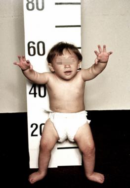
- Bullous skin eruptions in epidermolysis bullosa dystrophica as a cause of esophageal stricture [2]
- Pallor in malignant causes
-
![Epidermolysis bullosa pruriginosa. Adapted from Dermatology Atlas.[3]](/images/8/83/Epidermolysis_bullosa_pruriginosa01.jpg)
Epidermolysis bullosa pruriginosa. Adapted from Dermatology Atlas.[3]
-
Description (Adapted from Dermatology Atlas)
![Epidermolysis bullosa pruriginosa. Adapted from Dermatology Atlas.[3]](/index.php/File:Epidermolysis_bullosa_pruriginosa01.jpg)

HEENT
Esophageal stricture due to gastroesophageal reflux disease:
- Hoarse voice
- Oropharyngeal erythema
- Dental erosions
Neck
- Left supraclavicular lymphadenopathy (Virchow node) due to malignant causes of esophageal stricture
- Jugular venous distension
- Carotid bruits may be auscultated unilaterally/bilaterally using the bell/diaphragm of the otoscope
- Lymphadenopathy (describe location, size, tenderness, mobility, and symmetry)
- Thyromegaly / thyroid nodules
- Hepatojugular reflux
Lungs
- Wheezing
- Bronchitis[4]
- Asymmetric chest expansion / Decreased chest expansion
- Lungs are hypo/hyperresonant
- Fine/coarse crackles upon auscultation of the lung bases/apices unilaterally/bilaterally
- Rhonchi
- Vesicular breath sounds / Distant breath sounds
- Expiratory/inspiratory wheezing with normal / delayed expiratory phase
- Wheezing may be present
- Egophony present/absent
- Bronchophony present/absent
- Normal/reduced tactile fremitus
Heart
- Chest tenderness upon palpation
- PMI within 2 cm of the sternum (PMI) / Displaced point of maximal impulse (PMI) suggestive of ____
- Heave / thrill
- Friction rub
- S1
- S2
- S3
- S4
- Gallops
- A high/low grade early/late systolic murmur / diastolic murmur best heard at the base/apex/(specific valve region) may be heard using the bell/diaphgram of the otoscope
Abdomen
- Epigastric tenderness due to gastroesophageal reflux
- Abdominal distention
- Abdominal tenderness in the right/left upper/lower abdominal quadrant
- Rebound tenderness (positive Blumberg sign)
- A palpable abdominal mass in the right/left upper/lower abdominal quadrant
- Guarding may be present
- Hepatomegaly / splenomegaly / hepatosplenomegaly
- Additional findings, such as obturator test, psoas test, McBurney point test, Murphy test
Back
- Point tenderness over __ vertebrae (e.g. L3-L4)
- Sacral edema
- Costovertebral angle tenderness bilaterally/unilaterally
- Buffalo hump
Genitourinary
- A pelvic/adnexal mass may be palpated
- Inflamed mucosa
- Clear/(color), foul-smelling/odorless penile/vaginal discharge
Neuromuscular
- Patient is usually oriented to persons, place, and time
- Altered mental status
- Glasgow coma scale is ___ / 15
- Clonus may be present
- Hyperreflexia / hyporeflexia / areflexia
- Positive (abnormal) Babinski / plantar reflex unilaterally/bilaterally
- Muscle rigidity
- Proximal/distal muscle weakness unilaterally/bilaterally
- ____ (finding) suggestive of cranial nerve ___ (roman numerical) deficit (e.g. Dilated pupils suggestive of CN III deficit)
- Unilateral/bilateral upper/lower extremity weakness
- Unilateral/bilateral sensory loss in the upper/lower extremity
- Positive straight leg raise test
- Abnormal gait (describe gait: e.g. ataxic (cerebellar) gait / steppage gait / waddling gait / choeiform gait / Parkinsonian gait / sensory gait)
- Positive/negative Trendelenburg sign
- Unilateral/bilateral tremor (describe tremor, e.g. at rest, pill-rolling)
- Normal finger-to-nose test / Dysmetria
- Absent/present dysdiadochokinesia (palm tapping test)
Extremities
- Clubbing
- Cyanosis
- Pitting/non-pitting edema of the upper/lower extremities
- Muscle atrophy
- Fasciculations in the upper/lower extremity
References
- ↑ Li ZT, Ji F, Han XW, Wang L, Yue YQ, Wang ZG (2017). "The Role of Gastroesophageal Reflux in Provoking High Blood Pressure Episodes in Patients With Hypertension". J Clin Gastroenterol. doi:10.1097/MCG.0000000000000933. PMID 28961574.
- ↑ Luedtke P, Levine MS, Rubesin SE, Weinstein DS, Laufer I (2003). "Radiologic diagnosis of benign esophageal strictures: a pattern approach". Radiographics. 23 (4): 897–909. doi:10.1148/rg.234025717. PMID 12853664.
- ↑ "Dermatology Atlas".
- ↑ Hom C, Vaezi MF (2013). "Extraesophageal manifestations of gastroesophageal reflux disease". Gastroenterol Clin North Am. 42 (1): 71–91. doi:10.1016/j.gtc.2012.11.004. PMID 23452632.