Distal radius fracture x ray: Difference between revisions
Jump to navigation
Jump to search
No edit summary |
m (Bot: Removing from Primary care) |
||
| (5 intermediate revisions by 2 users not shown) | |||
| Line 1: | Line 1: | ||
__NOTOC__ | |||
{{Distal radius fracture}} | {{Distal radius fracture}} | ||
{{CMG}}; {{AE}} | {{CMG}}; {{AE}} {{Rohan}} | ||
==Overview== | ==Overview== | ||
[[Radiography|Radiographic]] imaging is important in diagnosis, classification, treatment and follow-up assessment of [[Distal radius fracture|distal radius fractures]]. The routine minimal evaluation for [[distal radius fracture]]<nowiki/>s must include two views - a postero-anterior (PA) view and lateral view. | |||
==X Ray== | ==X Ray== | ||
*Radiographic imaging is important in diagnosis, classification, treatment and follow-up assessment of distal radius fractures.<ref name="pmid8479720">{{cite journal| author=Metz VM, Gilula LA| title=Imaging techniques for distal radius fractures and related injuries. | journal=Orthop Clin North Am | year= 1993 | volume= 24 | issue= 2 | pages= 217-28 | pmid=8479720 | doi= | pmc= | url=https://www.ncbi.nlm.nih.gov/entrez/eutils/elink.fcgi?dbfrom=pubmed&tool=sumsearch.org/cite&retmode=ref&cmd=prlinks&id=8479720 }} </ref><ref name="pmid18762124">{{cite journal| author=Henry MH| title=Distal radius fractures: current concepts. | journal=J Hand Surg Am | year= 2008 | volume= 33 | issue= 7 | pages= 1215-27 | pmid=18762124 | doi=10.1016/j.jhsa.2008.07.013 | pmc= | url=https://www.ncbi.nlm.nih.gov/entrez/eutils/elink.fcgi?dbfrom=pubmed&tool=sumsearch.org/cite&retmode=ref&cmd=prlinks&id=18762124 }} </ref><ref name="pmid16039439">{{cite journal| author=Medoff RJ| title=Essential radiographic evaluation for distal radius fractures. | journal=Hand Clin | year= 2005 | volume= 21 | issue= 3 | pages= 279-88 | pmid=16039439 | doi=10.1016/j.hcl.2005.02.008 | pmc= | url=https://www.ncbi.nlm.nih.gov/entrez/eutils/elink.fcgi?dbfrom=pubmed&tool=sumsearch.org/cite&retmode=ref&cmd=prlinks&id=16039439 }} </ref><ref name="pmid16039440">{{cite journal| author=Slutsky DJ| title=Predicting the outcome of distal radius fractures. | journal=Hand Clin | year= 2005 | volume= 21 | issue= 3 | pages= 289-94 | pmid=16039440 | doi=10.1016/j.hcl.2005.03.001 | pmc= | url=https://www.ncbi.nlm.nih.gov/entrez/eutils/elink.fcgi?dbfrom=pubmed&tool=sumsearch.org/cite&retmode=ref&cmd=prlinks&id=16039440 }} </ref><ref name="pmid12679687">{{cite journal| author=Lill CA, Goldhahn J, Albrecht A, Eckstein F, Gatzka C, Schneider E| title=Impact of bone density on distal radius fracture patterns and comparison between five different fracture classifications. | journal=J Orthop Trauma | year= 2003 | volume= 17 | issue= 4 | pages= 271-8 | pmid=12679687 | doi= | pmc= | url=https://www.ncbi.nlm.nih.gov/entrez/eutils/elink.fcgi?dbfrom=pubmed&tool=sumsearch.org/cite&retmode=ref&cmd=prlinks&id=12679687 }} </ref><ref name="pmid15576227">{{cite journal| author=Nesbitt KS, Failla JM, Les C| title=Assessment of instability factors in adult distal radius fractures. | journal=J Hand Surg Am | year= 2004 | volume= 29 | issue= 6 | pages= 1128-38 | pmid=15576227 | doi=10.1016/j.jhsa.2004.06.008 | pmc= | url=https://www.ncbi.nlm.nih.gov/entrez/eutils/elink.fcgi?dbfrom=pubmed&tool=sumsearch.org/cite&retmode=ref&cmd=prlinks&id=15576227 }} </ref><ref name="pmid22036131">{{cite journal| author=Fujitani R, Omokawa S, Akahane M, Iida A, Ono H, Tanaka Y| title=Predictors of distal radioulnar joint instability in distal radius fractures. | journal=J Hand Surg Am | year= 2011 | volume= 36 | issue= 12 | pages= 1919-25 | pmid=22036131 | doi=10.1016/j.jhsa.2011.09.004 | pmc= | url=https://www.ncbi.nlm.nih.gov/entrez/eutils/elink.fcgi?dbfrom=pubmed&tool=sumsearch.org/cite&retmode=ref&cmd=prlinks&id=22036131 }} </ref> | *[[Radiography|Radiographic]] imaging is important in diagnosis, classification, treatment and follow-up assessment of [[Distal radius fracture|distal radius fractures]].<ref name="pmid8479720">{{cite journal| author=Metz VM, Gilula LA| title=Imaging techniques for distal radius fractures and related injuries. | journal=Orthop Clin North Am | year= 1993 | volume= 24 | issue= 2 | pages= 217-28 | pmid=8479720 | doi= | pmc= | url=https://www.ncbi.nlm.nih.gov/entrez/eutils/elink.fcgi?dbfrom=pubmed&tool=sumsearch.org/cite&retmode=ref&cmd=prlinks&id=8479720 }} </ref><ref name="pmid18762124">{{cite journal| author=Henry MH| title=Distal radius fractures: current concepts. | journal=J Hand Surg Am | year= 2008 | volume= 33 | issue= 7 | pages= 1215-27 | pmid=18762124 | doi=10.1016/j.jhsa.2008.07.013 | pmc= | url=https://www.ncbi.nlm.nih.gov/entrez/eutils/elink.fcgi?dbfrom=pubmed&tool=sumsearch.org/cite&retmode=ref&cmd=prlinks&id=18762124 }} </ref><ref name="pmid16039439">{{cite journal| author=Medoff RJ| title=Essential radiographic evaluation for distal radius fractures. | journal=Hand Clin | year= 2005 | volume= 21 | issue= 3 | pages= 279-88 | pmid=16039439 | doi=10.1016/j.hcl.2005.02.008 | pmc= | url=https://www.ncbi.nlm.nih.gov/entrez/eutils/elink.fcgi?dbfrom=pubmed&tool=sumsearch.org/cite&retmode=ref&cmd=prlinks&id=16039439 }} </ref><ref name="pmid16039440">{{cite journal| author=Slutsky DJ| title=Predicting the outcome of distal radius fractures. | journal=Hand Clin | year= 2005 | volume= 21 | issue= 3 | pages= 289-94 | pmid=16039440 | doi=10.1016/j.hcl.2005.03.001 | pmc= | url=https://www.ncbi.nlm.nih.gov/entrez/eutils/elink.fcgi?dbfrom=pubmed&tool=sumsearch.org/cite&retmode=ref&cmd=prlinks&id=16039440 }} </ref><ref name="pmid12679687">{{cite journal| author=Lill CA, Goldhahn J, Albrecht A, Eckstein F, Gatzka C, Schneider E| title=Impact of bone density on distal radius fracture patterns and comparison between five different fracture classifications. | journal=J Orthop Trauma | year= 2003 | volume= 17 | issue= 4 | pages= 271-8 | pmid=12679687 | doi= | pmc= | url=https://www.ncbi.nlm.nih.gov/entrez/eutils/elink.fcgi?dbfrom=pubmed&tool=sumsearch.org/cite&retmode=ref&cmd=prlinks&id=12679687 }} </ref><ref name="pmid15576227">{{cite journal| author=Nesbitt KS, Failla JM, Les C| title=Assessment of instability factors in adult distal radius fractures. | journal=J Hand Surg Am | year= 2004 | volume= 29 | issue= 6 | pages= 1128-38 | pmid=15576227 | doi=10.1016/j.jhsa.2004.06.008 | pmc= | url=https://www.ncbi.nlm.nih.gov/entrez/eutils/elink.fcgi?dbfrom=pubmed&tool=sumsearch.org/cite&retmode=ref&cmd=prlinks&id=15576227 }} </ref><ref name="pmid22036131">{{cite journal| author=Fujitani R, Omokawa S, Akahane M, Iida A, Ono H, Tanaka Y| title=Predictors of distal radioulnar joint instability in distal radius fractures. | journal=J Hand Surg Am | year= 2011 | volume= 36 | issue= 12 | pages= 1919-25 | pmid=22036131 | doi=10.1016/j.jhsa.2011.09.004 | pmc= | url=https://www.ncbi.nlm.nih.gov/entrez/eutils/elink.fcgi?dbfrom=pubmed&tool=sumsearch.org/cite&retmode=ref&cmd=prlinks&id=22036131 }} </ref> | ||
*The routine minimal evaluation for distal radius fractures must include two views-a postero-anterior (PA) view and lateral view. | *The routine minimal evaluation for [[Distal radius fracture|distal radius fractures]] must include two views - a postero-anterior (PA) view and lateral view. | ||
*'''Positioning for the x-rays''': | *'''Positioning for the x-rays''': | ||
**The posteroanterior view should be acquired with the patient’s elbow and shoulder at 90° and the forearm in neutral rotation. | **The posteroanterior view should be acquired with the patient’s [[elbow]] and [[shoulder]] at 90° and the [[forearm]] in neutral rotation. | ||
**When the lateral view is acquired correctly, i.e., in the absence of relative pronation or supination, the pisiform bone should be superimposed on the distal pole of the scaphoid. | **When the lateral view is acquired correctly, i.e., in the absence of relative [[pronation]] or [[supination]], the [[Pisiform bone|pisiform]] bone should be superimposed on the distal pole of the [[Scaphoid bone|scaphoid]]. | ||
===Posteroanterior View Inspection=== | ===Posteroanterior View Inspection=== | ||
===Radial Length=== | ===Radial Length=== | ||
*It is used when assessing shortening of the radius after fracture, can be obtained using the posteroanterior view. | *It is used when assessing shortening of the [[radius]] after [[Bone fracture|fracture]], can be obtained using the posteroanterior view. | ||
*'''Method''': | *'''Method''': | ||
**Two lines are drawn perpendicular to the long axis of the radius, one at the tip of the radial styloid and the second at the ulnar border of the distal radial articular surface. | **Two lines are drawn perpendicular to the long axis of the [[radius]], one at the tip of the [[Radial styloid process|radial styloid]] and the second at the ulnar border of the [[distal radial articular surface]]. | ||
*Normally, this length is about 12 mm. | **Normally, this length is about 12 mm. | ||
*Excessive radial shortening after fracture of the distal radius may be associated with tears of the triangular fibrocartilage complex (TFCC). | **Excessive radial [[shortening]] after fracture of the distal [[radius]] may be associated with tears of the [[triangular fibrocartilage]] complex (TFCC). | ||
===Radial Inclination=== | ===Radial Inclination=== | ||
| Line 28: | Line 28: | ||
|} | |} | ||
*'''Method''': | *'''Method''': | ||
**Radial inclination is the angle between a line perpendicular to the central axis of the radius and a line connecting the radial and ulnar limits of the articular surface of the distal radius. | **Radial inclination is the angle between a line perpendicular to the central axis of the [[radius]] and a line connecting the radial and ulnar limits of the articular surface of the distal [[radius]]. | ||
*The articular surface of the distal radius exhibits approximately 23° (range:13–30°) of normal radial inclination. | **The articular surface of the distal [[radius]] exhibits approximately 23° (range:13–30°) of normal radial inclination. | ||
===Ulnar Variance=== | ===Ulnar Variance=== | ||
*Ulnar variance, defined as neutral, positive, or negative, is evaluated on the frontal view. | *Ulnar variance, defined as neutral, positive, or negative, is evaluated on the frontal view. | ||
*'''Method''': | *'''Method''': | ||
**Ulnar variance, according to the method of perpendiculars, is the vertical distance between two tangential lines both perpendicular to the long axis of the radius. | **Ulnar variance, according to the method of perpendiculars, is the vertical distance between two tangential lines both perpendicular to the long axis of the [[radius]]. | ||
**One line is drawn at the level of the radial sigmoid notch and the second at the level of the lateral cortical margin of the distal ulna. | **One line is drawn at the level of the radial sigmoid notch and the second at the level of the lateral cortical margin of the distal [[ulna]]. | ||
*With excessive radial shortening, ulnar positive variance will be present. | **With excessive radial [[shortening]], ulnar positive variance will be present. | ||
===Radial translation ratio=== | ===Radial translation ratio=== | ||
*'''Method''': | *'''Method''': | ||
**The distal radioulnar joint gap is the distance between two longitudinal lines along the cortical rim of the sigmoid notch of the radius and the adjacent ulnar head. | **The distal radioulnar joint gap is the distance between two longitudinal lines along the cortical rim of the sigmoid notch of the [[radius]] and the adjacent [[ulnar]] head. | ||
**The fraction of the distal radioulnar joint gap relative to the radioulnar width of the proximal fracture fragment reflects the radial translation ratio. | **The fraction of the distal radioulnar joint gap relative to the radioulnar width of the proximal fracture fragment reflects the radial translation ratio. | ||
*This ratio was is a significant risk factor of distal radioulnar joint instability following unstable distal radius fracture and indicates | **This ratio was is a significant risk factor of distal radioulnar joint instability following unstable [[distal radius fracture]] and indicates tears of the [[triangular fibrocartilage]] complex (TFCC). | ||
===Lateral View Inspection=== | ===Lateral View Inspection=== | ||
| Line 51: | Line 51: | ||
[[File:Dorsal tilt of distal radius fracture.jpg|200px|thumb|none|Dorsal tilt of distal radius fracture. [https://upload.wikimedia.org/wikipedia/commons/e/ee/Dorsal_tilt_of_distal_radius_fracture.jpg Source: Case courtesy of Mikael Häggström]]] | [[File:Dorsal tilt of distal radius fracture.jpg|200px|thumb|none|Dorsal tilt of distal radius fracture. [https://upload.wikimedia.org/wikipedia/commons/e/ee/Dorsal_tilt_of_distal_radius_fracture.jpg Source: Case courtesy of Mikael Häggström]]] | ||
|} | |} | ||
*The normal distal radius shows relative volar tilt. | *The normal distal [[radius]] shows relative volar tilt. | ||
*'''Method''': | *'''Method''': | ||
**Volar tilt is the angle between a line perpendicular to the central axis of the radius and a line connecting the dorsal and volar margins of the articular surface of the distal radius on the lateral view. | **Volar tilt is the angle between a line perpendicular to the central axis of the [[radius]] and a line connecting the dorsal and volar margins of the articular surface of the distal [[radius]] on the lateral view. | ||
*Loss of the normal volar tilt can accompany fractures of the distal radius. | **Loss of the normal volar tilt can accompany [[Bone fracture|fractures]] of the distal [[radius]]. | ||
*Extreme dorsal angulation may be associated with injury to the TFCC. | **Extreme dorsal angulation may be associated with injury to the [[TFCC]]. | ||
===Teardrop angle=== | ===Teardrop angle=== | ||
*The volar rim of the lunate facet of the distal radius forms a teardrop shape along the distal, volar surface of the radius on the lateral view. | *The volar rim of the [[lunate]] facet of the distal radius forms a teardrop shape along the distal, volar surface of the radius on the lateral view. | ||
*'''Method''': | *'''Method''': | ||
**A teardrop angle can be acquired by drawing a line down the long axis of the radius that intersects a line drawn through the center of the lunate facet–teardrop. | **A teardrop angle can be acquired by drawing a line down the long axis of the radius that intersects a line drawn through the center of the lunate facet–teardrop. | ||
*A normal teardrop angle is approximately 70°. | **A normal teardrop angle is approximately 70°. | ||
*This angle is used to determine whether there is persistent articular incongruity after reduction of a fractured volar rim fragment. | **This angle is used to determine whether there is persistent articular incongruity after reduction of a [[Fracture|fractured]] volar rim fragment. | ||
==References== | ==References== | ||
| Line 71: | Line 68: | ||
{{WH}} | {{WH}} | ||
{{WS}} | {{WS}} | ||
[[Category:Needs overview]] | [[Category:Needs overview]] | ||
[[Category:Orthopedics]] | [[Category:Orthopedics]] | ||
[[Category:Orthopedic surgery]] | [[Category:Orthopedic surgery]] | ||
[[Category:Fractures]] | [[Category:Fractures]] | ||
[[Category:Bone fractures]] | [[Category:Bone fractures]] | ||
Latest revision as of 21:25, 29 July 2020
|
Distal radius fracture Microchapters |
|
Diagnosis |
|---|
|
Treatment |
|
Case Studies |
|
Distal radius fracture x ray On the Web |
|
American Roentgen Ray Society Images of Distal radius fracture x ray |
|
Risk calculators and risk factors for Distal radius fracture x ray |
Editor-In-Chief: C. Michael Gibson, M.S., M.D. [1]; Associate Editor(s)-in-Chief: Rohan A. Bhimani, M.B.B.S., D.N.B., M.Ch.[2]
Overview
Radiographic imaging is important in diagnosis, classification, treatment and follow-up assessment of distal radius fractures. The routine minimal evaluation for distal radius fractures must include two views - a postero-anterior (PA) view and lateral view.
X Ray
- Radiographic imaging is important in diagnosis, classification, treatment and follow-up assessment of distal radius fractures.[1][2][3][4][5][6][7]
- The routine minimal evaluation for distal radius fractures must include two views - a postero-anterior (PA) view and lateral view.
- Positioning for the x-rays:
- The posteroanterior view should be acquired with the patient’s elbow and shoulder at 90° and the forearm in neutral rotation.
- When the lateral view is acquired correctly, i.e., in the absence of relative pronation or supination, the pisiform bone should be superimposed on the distal pole of the scaphoid.
Posteroanterior View Inspection
Radial Length
- It is used when assessing shortening of the radius after fracture, can be obtained using the posteroanterior view.
- Method:
- Two lines are drawn perpendicular to the long axis of the radius, one at the tip of the radial styloid and the second at the ulnar border of the distal radial articular surface.
- Normally, this length is about 12 mm.
- Excessive radial shortening after fracture of the distal radius may be associated with tears of the triangular fibrocartilage complex (TFCC).
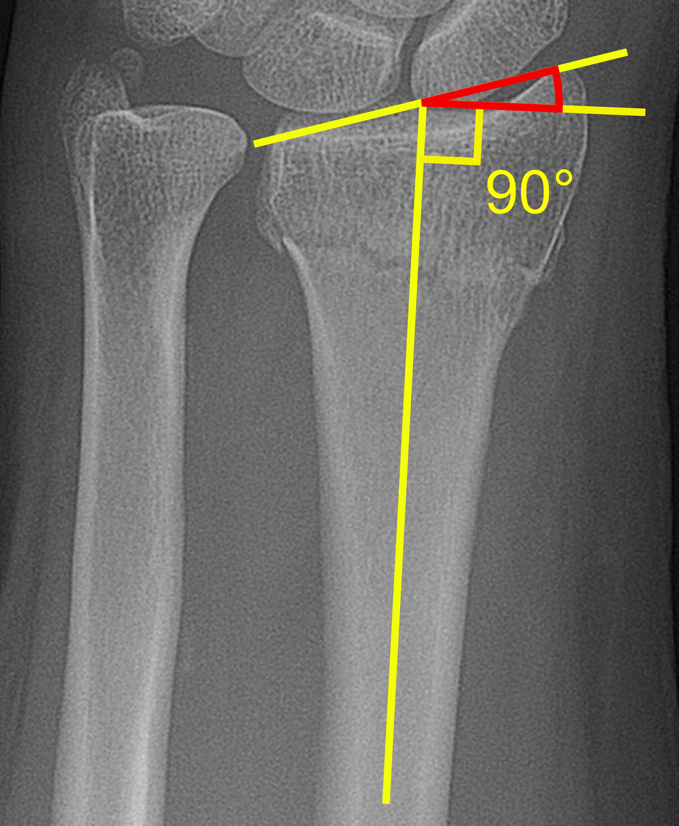
Radial Inclination
 |
- Method:
- Radial inclination is the angle between a line perpendicular to the central axis of the radius and a line connecting the radial and ulnar limits of the articular surface of the distal radius.
- The articular surface of the distal radius exhibits approximately 23° (range:13–30°) of normal radial inclination.
Ulnar Variance
- Ulnar variance, defined as neutral, positive, or negative, is evaluated on the frontal view.
- Method:
- Ulnar variance, according to the method of perpendiculars, is the vertical distance between two tangential lines both perpendicular to the long axis of the radius.
- One line is drawn at the level of the radial sigmoid notch and the second at the level of the lateral cortical margin of the distal ulna.
- With excessive radial shortening, ulnar positive variance will be present.
Radial translation ratio
- Method:
- The distal radioulnar joint gap is the distance between two longitudinal lines along the cortical rim of the sigmoid notch of the radius and the adjacent ulnar head.
- The fraction of the distal radioulnar joint gap relative to the radioulnar width of the proximal fracture fragment reflects the radial translation ratio.
- This ratio was is a significant risk factor of distal radioulnar joint instability following unstable distal radius fracture and indicates tears of the triangular fibrocartilage complex (TFCC).
Lateral View Inspection
Volar Tilt
 |
- The normal distal radius shows relative volar tilt.
- Method:
- Volar tilt is the angle between a line perpendicular to the central axis of the radius and a line connecting the dorsal and volar margins of the articular surface of the distal radius on the lateral view.
- Loss of the normal volar tilt can accompany fractures of the distal radius.
- Extreme dorsal angulation may be associated with injury to the TFCC.
Teardrop angle
- The volar rim of the lunate facet of the distal radius forms a teardrop shape along the distal, volar surface of the radius on the lateral view.
- Method:
- A teardrop angle can be acquired by drawing a line down the long axis of the radius that intersects a line drawn through the center of the lunate facet–teardrop.
- A normal teardrop angle is approximately 70°.
- This angle is used to determine whether there is persistent articular incongruity after reduction of a fractured volar rim fragment.
References
- ↑ Metz VM, Gilula LA (1993). "Imaging techniques for distal radius fractures and related injuries". Orthop Clin North Am. 24 (2): 217–28. PMID 8479720.
- ↑ Henry MH (2008). "Distal radius fractures: current concepts". J Hand Surg Am. 33 (7): 1215–27. doi:10.1016/j.jhsa.2008.07.013. PMID 18762124.
- ↑ Medoff RJ (2005). "Essential radiographic evaluation for distal radius fractures". Hand Clin. 21 (3): 279–88. doi:10.1016/j.hcl.2005.02.008. PMID 16039439.
- ↑ Slutsky DJ (2005). "Predicting the outcome of distal radius fractures". Hand Clin. 21 (3): 289–94. doi:10.1016/j.hcl.2005.03.001. PMID 16039440.
- ↑ Lill CA, Goldhahn J, Albrecht A, Eckstein F, Gatzka C, Schneider E (2003). "Impact of bone density on distal radius fracture patterns and comparison between five different fracture classifications". J Orthop Trauma. 17 (4): 271–8. PMID 12679687.
- ↑ Nesbitt KS, Failla JM, Les C (2004). "Assessment of instability factors in adult distal radius fractures". J Hand Surg Am. 29 (6): 1128–38. doi:10.1016/j.jhsa.2004.06.008. PMID 15576227.
- ↑ Fujitani R, Omokawa S, Akahane M, Iida A, Ono H, Tanaka Y (2011). "Predictors of distal radioulnar joint instability in distal radius fractures". J Hand Surg Am. 36 (12): 1919–25. doi:10.1016/j.jhsa.2011.09.004. PMID 22036131.