Digastric muscle: Difference between revisions
No edit summary |
m (Bot: Automated text replacement (-{{SIB}} + & -{{EH}} + & -{{EJ}} + & -{{Editor Help}} + & -{{Editor Join}} +)) |
||
| Line 21: | Line 21: | ||
{{CMG}} | {{CMG}} | ||
The '''digastric muscle''' (named ''digastric'' as it has two bellies) is a small [[muscle]] located under the [[jaw]]. | The '''digastric muscle''' (named ''digastric'' as it has two bellies) is a small [[muscle]] located under the [[jaw]]. | ||
| Line 91: | Line 91: | ||
[[Category:Muscles of the head and neck]] | [[Category:Muscles of the head and neck]] | ||
[[de:Musculus digastricus]] | [[de:Musculus digastricus]] | ||
Latest revision as of 00:45, 9 August 2012
|
WikiDoc Resources for Digastric muscle |
|
Articles |
|---|
|
Most recent articles on Digastric muscle Most cited articles on Digastric muscle |
|
Media |
|
Powerpoint slides on Digastric muscle |
|
Evidence Based Medicine |
|
Clinical Trials |
|
Ongoing Trials on Digastric muscle at Clinical Trials.gov Trial results on Digastric muscle Clinical Trials on Digastric muscle at Google
|
|
Guidelines / Policies / Govt |
|
US National Guidelines Clearinghouse on Digastric muscle NICE Guidance on Digastric muscle
|
|
Books |
|
News |
|
Commentary |
|
Definitions |
|
Patient Resources / Community |
|
Patient resources on Digastric muscle Discussion groups on Digastric muscle Patient Handouts on Digastric muscle Directions to Hospitals Treating Digastric muscle Risk calculators and risk factors for Digastric muscle
|
|
Healthcare Provider Resources |
|
Causes & Risk Factors for Digastric muscle |
|
Continuing Medical Education (CME) |
|
International |
|
|
|
Business |
|
Experimental / Informatics |
Editor-In-Chief: C. Michael Gibson, M.S., M.D. [1]
The digastric muscle (named digastric as it has two bellies) is a small muscle located under the jaw.
It lies below the body of the mandible, and extends, in a curved form, from the mastoid process to the symphysis menti. It belongs to the suprahyoid muscles group.
A broad aponeurotic layer is given off from the tendon of the Digastricus on either side, to be attached to the body and greater cornu of the hyoid bone; this is termed the suprahyoid aponeurosis.
Structure
The Digastricus (Digastric muscle) consists of two fleshy bellies united by an intermediate rounded tendon.
The two bellies of the digastric muscle have different embryological origins, and are supplied by different cranial nerves.
Posterior belly
The posterior belly, longer than the anterior, arises on the inferior surface of the skull, from the medial surface of the mastoid process of the temporal bone and a deep groove between the mastoid process and the styloid process called the digastric groove.
The posterior belly is supplied by a branch of the facial nerve.
Anterior belly
The anterior belly arises from a depression on the inner side of the lower border of the mandible, close to the symphysis, and passes downward and backward.
The anterior body supplied by the trigeminal via the mylohyoid nerve.
Intermediate tendon
The two bellies end in an intermediate tendon which perforates the Stylohyoideus muscle, and is held in connection with the side of the body and the greater cornu of the hyoid bone by a fibrous loop, which is sometimes lined by a mucous sheath.
Action
When the digastric muscle contracts, it acts to elevate the hyoid bone.
If the hyoid is being held in place (by the infrahyoid muscles), it will tend to depress the mandible (open the mouth).
Variations
Variations are numerous.
The posterior belly may arise partly or entirely from the styloid process, or be connected by a slip to the middle or inferior constrictor; the anterior belly may be double or extra slips from this belly may pass to the jaw or Mylohyoideus or decussate with a similar slip on opposite side; anterior belly may be absent and posterior belly inserted into the middle of the jaw or hyoid bone.
The tendon may pass in front, more rarely behind the Stylohoideus. The Mentohyoideus muscle passes from the body of hyoid bone to chin.
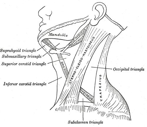
Triangles
The Digastricus divides the anterior triangle of the neck into three smaller triangles.
- (1) the submaxillary triangle, bounded above by the lower border of the body of the mandible, and a line drawn from its angle to the Sternocleidomastoideus, below by the posterior belly of the Digastricus and the Stylohyoideus, in front by the anterior belly of the Diagastricus;
- (2) the carotid triangle, bounded above by the posterior belly of the Digastricus and Stylohyoideus, behind by the Sternocleidomastoideus, below by the Omohyoideus;
- (3) the suprahyoid or submental triangle, bounded laterally by the anterior belly of the Digastricus, medially by the middle line of the neck from the hyoid bone to the symphysis menti, and inferiorly by the body of the hyoid bone.
Additional images
-
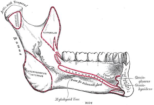
Mandible. Inner surface. Side view.
-
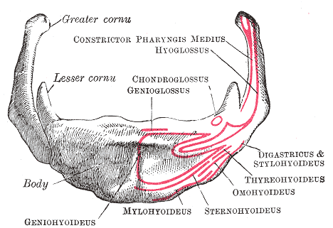
Hyoid bone. Anterior surface. Enlarged.
-
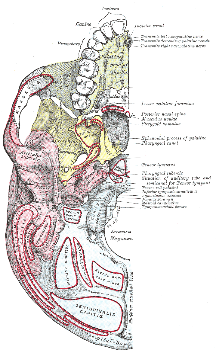
Base of skull. Inferior surface.
-
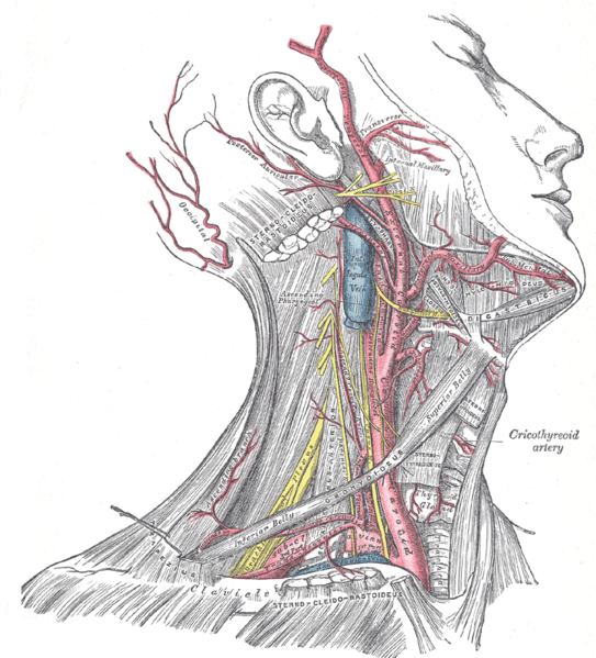
Superficial dissection of the right side of the neck, showing the carotid and subclavian arteries.
-
-
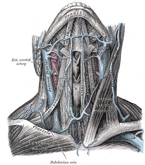
The veins of the neck, viewed from in front.
-
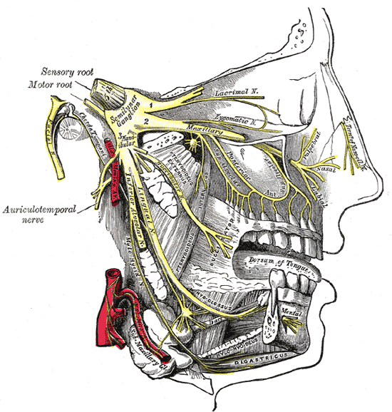
Distribution of the maxillary and mandibular nerves, and the submaxillary ganglion.
-
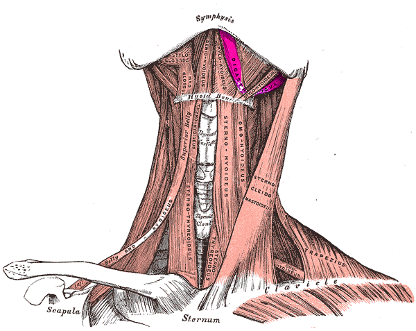
Anterior view of digastric muscle


External links
- Template:GPnotebook
- Template:MuscleLoyola
- Frontal section
- Template:SUNYAnatomyFigs
- Template:SUNYAnatomyLabs
- Template:RocheLexicon
Template:Gray's Template:Muscles of neck