Cryptococcosis CT
Jump to navigation
Jump to search
|
Cryptococcosis Microchapters |
|
Diagnosis |
|---|
|
Treatment |
|
Case Studies |
|
Cryptococcosis CT On the Web |
|
American Roentgen Ray Society Images of Cryptococcosis CT |
Editor-In-Chief: C. Michael Gibson, M.S., M.D. [1]; Associate Editor(s)-in-Chief: Aravind Kuchkuntla, M.B.B.S[2]
Overview
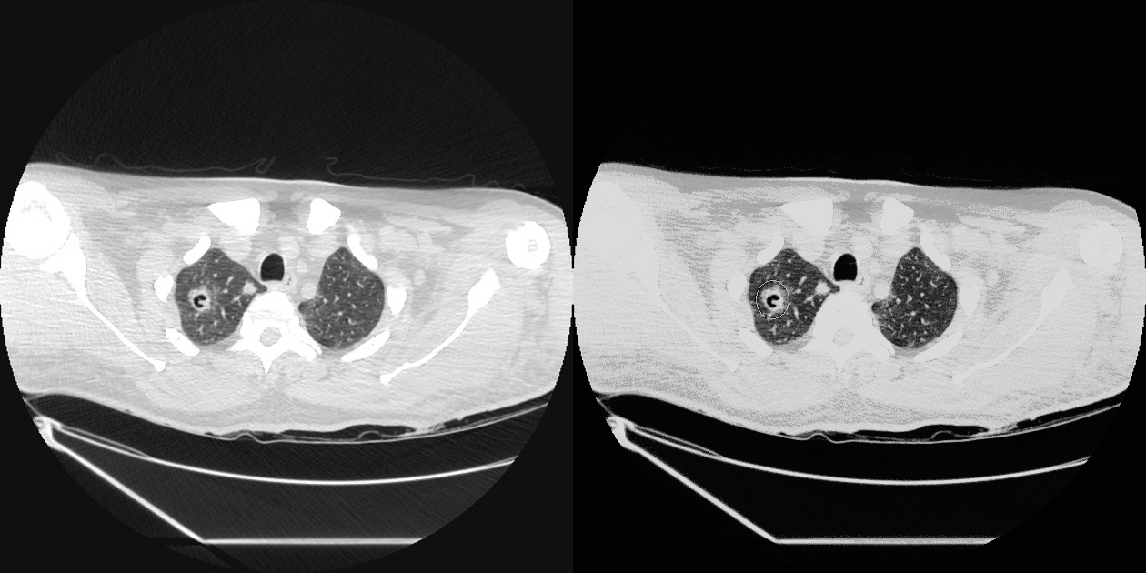
The most common CT findings in patients with pulmonary cryptococcosis are pulmonary nodules and pulmonary opacities that range from a perihilar interstitial pattern to an area of dense alveolar consolidation.
CT scan
The most common CT scan findings in immunocompromised patients with pulmonary cryptococcosis are pulmonary nodules.[1][2]
- The nodules are most often multiple, smaller than 10 mm in diameter, and have well defined smooth margins.
- The nodules usually involve less than 10% of the parenchyma and tend to be distributed peripherally in the middle and upper zones.

References
- ↑ Hu Z, Xu C, Wei H, Zhong Y, Bo C, Chi Y; et al. (2013). "Solitary cavitary pulmonary nodule may be a common CT finding in AIDS-associated pulmonary cryptococcosis". Scand J Infect Dis. 45 (5): 378–89. doi:10.3109/00365548.2012.749422. PMID 23244589.
- ↑ Sider L, Westcott MA (1994). "Pulmonary manifestations of cryptococcosis in patients with AIDS: CT features". J Thorac Imaging. 9 (2): 78–84. PMID 8207784.