Conjunctivitis physical examination: Difference between revisions
No edit summary |
No edit summary |
||
| Line 8: | Line 8: | ||
==Physical Examination== | ==Physical Examination== | ||
===Viral Conjunctivitis=== | ===Viral Conjunctivitis=== | ||
Patients with | Patients with viral conjunctivitis usually appear [[febrile]], and they have preauricular [[adenopathy]]. [[Ophthalmologic]] examination of patients with viral conjunctivitis is usually remarkable for:<ref name="pmid26077630">{{cite journal| author=Jhanji V, Chan TC, Li EY, Agarwal K, Vajpayee RB| title=Adenoviral keratoconjunctivitis. | journal=Surv Ophthalmol | year= 2015 | volume= 60 | issue= 5 | pages= 435-43 | pmid=26077630 | doi=10.1016/j.survophthal.2015.04.001 | pmc= | url=http://www.ncbi.nlm.nih.gov/entrez/eutils/elink.fcgi?dbfrom=pubmed&tool=sumsearch.org/cite&retmode=ref&cmd=prlinks&id=26077630 }} </ref><ref name="pmid24150468">{{cite journal| author=Azari AA, Barney NP| title=Conjunctivitis: a systematic review of diagnosis and treatment. | journal=JAMA | year= 2013 | volume= 310 | issue= 16 | pages= 1721-9 | pmid=24150468 | doi=10.1001/jama.2013.280318 | pmc=4049531 | url=http://www.ncbi.nlm.nih.gov/entrez/eutils/elink.fcgi?dbfrom=pubmed&tool=sumsearch.org/cite&retmode=ref&cmd=prlinks&id=24150468 }} </ref> | ||
*[[Epiphora]] | *[[Epiphora]] | ||
*[[Hyperemia]] | *[[Hyperemia]] | ||
| Line 19: | Line 19: | ||
*[[Corneal]] [[epithelial]] defect (severe cases) | *[[Corneal]] [[epithelial]] defect (severe cases) | ||
===Acute Hemorrhagic Conjunctivitis=== | ===Acute Hemorrhagic Conjunctivitis=== | ||
[[Ophthalmologic]] examination of patients with | [[Ophthalmologic]] examination of patients with acute hemorrhagic conjunctivitis is usually remarkable for:<ref name="pmid1088513">{{cite journal| author=Yin-Murphy M| title=Simple tests for the diagnosis of picornavirus epidemic conjunctivitis (acute haemorrhagic conjunctivitis). | journal=Bull World Health Organ | year= 1976 | volume= 54 | issue= 6 | pages= 675-9 | pmid=1088513 | doi= | pmc=2366581 | url=http://www.ncbi.nlm.nih.gov/entrez/eutils/elink.fcgi?dbfrom=pubmed&tool=sumsearch.org/cite&retmode=ref&cmd=prlinks&id=1088513 }} </ref> | ||
*Eyelid [[edema]] | *Eyelid [[edema]] | ||
*Eye pain in [[palpation]] | *Eye pain in [[palpation]] | ||
*[[Bulbar]] [[conjunctiva]] [[hemorrhage]] | *[[Bulbar]] [[conjunctiva]] [[hemorrhage]] | ||
===Bacterial Conjunctivitis=== | ===Bacterial Conjunctivitis=== | ||
Ophthalmologic examination of patients with bacterial conjunctivitis is usually remarkable for:<ref name="pmid10922425">{{cite journal| author=Leibowitz HM| title=The red eye. | journal=N Engl J Med | year= 2000 | volume= 343 | issue= 5 | pages= 345-51 | pmid=10922425 | doi=10.1056/NEJM200008033430507 | pmc= | url=http://www.ncbi.nlm.nih.gov/entrez/eutils/elink.fcgi?dbfrom=pubmed&tool=sumsearch.org/cite&retmode=ref&cmd=prlinks&id=10922425 }} </ref><ref name="pmid21160459">{{cite journal| author=Workowski KA, Berman S, Centers for Disease Control and Prevention (CDC)| title=Sexually transmitted diseases treatment guidelines, 2010. | journal=MMWR Recomm Rep | year= 2010 | volume= 59 | issue= RR-12 | pages= 1-110 | pmid=21160459 | doi= | pmc= | url=http://www.ncbi.nlm.nih.gov/entrez/eutils/elink.fcgi?dbfrom=pubmed&tool=sumsearch.org/cite&retmode=ref&cmd=prlinks&id=21160459 }} </ref> | |||
*[[Bulbar]] [[conjunctival]] [[injection]] | *[[Bulbar]] [[conjunctival]] [[injection]] | ||
*Palpebral [[conjunctival]] [[papillary]] reaction | *Palpebral [[conjunctival]] [[papillary]] reaction | ||
| Line 32: | Line 32: | ||
*[[Corneal]] involvement (''[[Neisseria gonorrhea]]'') | *[[Corneal]] involvement (''[[Neisseria gonorrhea]]'') | ||
===Neonatal Conjunctivitis=== | ===Neonatal Conjunctivitis=== | ||
Ophthalmologic examination of patients with neonatal conjunctivitis or [[ophthalmia neonatorum]] is usually remarkable for:<ref name="pmid25606121">{{cite journal| author=Mallika P, Asok T, Faisal H, Aziz S, Tan A, Intan G| title=Neonatal conjunctivitis - a review. | journal=Malays Fam Physician | year= 2008 | volume= 3 | issue= 2 | pages= 77-81 | pmid=25606121 | doi= | pmc=4170304 | url=http://www.ncbi.nlm.nih.gov/entrez/eutils/elink.fcgi?dbfrom=pubmed&tool=sumsearch.org/cite&retmode=ref&cmd=prlinks&id=25606121 }} </ref> | |||
*''[[Neisseria gonorrhea]]'' | *''[[Neisseria gonorrhea]]'' | ||
**Chemosis | **Chemosis | ||
| Line 47: | Line 47: | ||
**[[Epiphora]] | **[[Epiphora]] | ||
===Allergic Conjunctivitis=== | ===Allergic Conjunctivitis=== | ||
Ophthalmologic examination of patients with allergic conjunctivitis is usually remarkable for:<ref name="pmid23497516">{{cite journal| author=La Rosa M, Lionetti E, Reibaldi M, Russo A, Longo A, Leonardi S et al.| title=Allergic conjunctivitis: a comprehensive review of the literature. | journal=Ital J Pediatr | year= 2013 | volume= 39 | issue= | pages= 18 | pmid=23497516 | doi=10.1186/1824-7288-39-18 | pmc=3640929 | url=http://www.ncbi.nlm.nih.gov/entrez/eutils/elink.fcgi?dbfrom=pubmed&tool=sumsearch.org/cite&retmode=ref&cmd=prlinks&id=23497516 }} </ref> | Ophthalmologic examination of patients with [[allergic conjunctivitis]] is usually remarkable for:<ref name="pmid23497516">{{cite journal| author=La Rosa M, Lionetti E, Reibaldi M, Russo A, Longo A, Leonardi S et al.| title=Allergic conjunctivitis: a comprehensive review of the literature. | journal=Ital J Pediatr | year= 2013 | volume= 39 | issue= | pages= 18 | pmid=23497516 | doi=10.1186/1824-7288-39-18 | pmc=3640929 | url=http://www.ncbi.nlm.nih.gov/entrez/eutils/elink.fcgi?dbfrom=pubmed&tool=sumsearch.org/cite&retmode=ref&cmd=prlinks&id=23497516 }} </ref> | ||
*[[Bilateral]] conjunctival [[injection]] | *[[Bilateral]] conjunctival [[injection]] | ||
*[[Chemosis]] | *[[Chemosis]] | ||
| Line 54: | Line 54: | ||
===Keratoconjunctivitis Sicaa=== | ===Keratoconjunctivitis Sicaa=== | ||
Examination should include evaluation of the face, eyelids, blinking patterns, eyelid margins, [[eyelashes]], [[conjunctiva]], [[cornea]], and tear film. | Examination should include evaluation of the face, eyelids, blinking patterns, eyelid margins, [[eyelashes]], [[conjunctiva]], [[cornea]], and tear film. | ||
Examination of patients with keratoconjunctivitis sicaa is usually remarkable for:<ref name="pmid9820935">{{cite journal| author=Stern ME, Beuerman RW, Fox RI, Gao J, Mircheff AK, Pflugfelder SC| title=The pathology of dry eye: the interaction between the ocular surface and lacrimal glands. | journal=Cornea | year= 1998 | volume= 17 | issue= 6 | pages= 584-9 | pmid=9820935 | doi= | pmc= | url=http://www.ncbi.nlm.nih.gov/entrez/eutils/elink.fcgi?dbfrom=pubmed&tool=sumsearch.org/cite&retmode=ref&cmd=prlinks&id=9820935 }} </ref><ref name="pmid19506195">{{cite journal| author=Schaumberg DA, Dana R, Buring JE, Sullivan DA| title=Prevalence of dry eye disease among US men: estimates from the Physicians' Health Studies. | journal=Arch Ophthalmol | year= 2009 | volume= 127 | issue= 6 | pages= 763-8 | pmid=19506195 | doi=10.1001/archophthalmol.2009.103 | pmc=2836718 | url=http://www.ncbi.nlm.nih.gov/entrez/eutils/elink.fcgi?dbfrom=pubmed&tool=sumsearch.org/cite&retmode=ref&cmd=prlinks&id=19506195 }} </ref> | Examination of patients with [[keratoconjunctivitis sicaa]] is usually remarkable for:<ref name="pmid9820935">{{cite journal| author=Stern ME, Beuerman RW, Fox RI, Gao J, Mircheff AK, Pflugfelder SC| title=The pathology of dry eye: the interaction between the ocular surface and lacrimal glands. | journal=Cornea | year= 1998 | volume= 17 | issue= 6 | pages= 584-9 | pmid=9820935 | doi= | pmc= | url=http://www.ncbi.nlm.nih.gov/entrez/eutils/elink.fcgi?dbfrom=pubmed&tool=sumsearch.org/cite&retmode=ref&cmd=prlinks&id=9820935 }} </ref><ref name="pmid19506195">{{cite journal| author=Schaumberg DA, Dana R, Buring JE, Sullivan DA| title=Prevalence of dry eye disease among US men: estimates from the Physicians' Health Studies. | journal=Arch Ophthalmol | year= 2009 | volume= 127 | issue= 6 | pages= 763-8 | pmid=19506195 | doi=10.1001/archophthalmol.2009.103 | pmc=2836718 | url=http://www.ncbi.nlm.nih.gov/entrez/eutils/elink.fcgi?dbfrom=pubmed&tool=sumsearch.org/cite&retmode=ref&cmd=prlinks&id=19506195 }} </ref> | ||
*More or less pronounced [[conjunctival]] [[redness]] | *More or less pronounced [[conjunctival]] [[redness]] | ||
*Damage to the ocular surface with punctate [[epithelial]] [erosions (superficial punctate [[keratitis]]) | *Damage to the ocular surface with punctate [[epithelial]] [erosions (superficial punctate [[keratitis]]) | ||
Revision as of 18:46, 18 July 2016
|
Conjunctivitis Microchapters |
|
Diagnosis |
|---|
|
Treatment |
|
Case Studies |
|
Conjunctivitis physical examination On the Web |
|
American Roentgen Ray Society Images of Conjunctivitis physical examination |
|
Risk calculators and risk factors for Conjunctivitis physical examination |
Editor-In-Chief: C. Michael Gibson, M.S., M.D. [1] Associate Editor(s)-in-Chief: Sara Mehrsefat, M.D. [2]
Overview
Physical examination of patients with conjunctivitis is usually remarkable for conjunctival injections, epiphora, hyperemia, chemosis and muco-purulent or watery discharge. However, ophthalmologic examination may be varies based on conjunctivitis subtypes.
Physical Examination
Viral Conjunctivitis
Patients with viral conjunctivitis usually appear febrile, and they have preauricular adenopathy. Ophthalmologic examination of patients with viral conjunctivitis is usually remarkable for:[1][2]
- Epiphora
- Hyperemia
- Chemosis
- Lymphoid follicle on the undersurface of the eyelid
- Follicular conjunctival reaction
- Pseudomembranous (occasionally)
- Cicatricial conjunctival reaction
- Eyelids ecchymosis
- Corneal epithelial defect (severe cases)
Acute Hemorrhagic Conjunctivitis
Ophthalmologic examination of patients with acute hemorrhagic conjunctivitis is usually remarkable for:[3]
- Eyelid edema
- Eye pain in palpation
- Bulbar conjunctiva hemorrhage
Bacterial Conjunctivitis
Ophthalmologic examination of patients with bacterial conjunctivitis is usually remarkable for:[4][5]
- Bulbar conjunctival injection
- Palpebral conjunctival papillary reaction
- Muco-purulent or watery discharge
- Chemosis
- Lid erythema
- Corneal involvement (Neisseria gonorrhea)
Neonatal Conjunctivitis
Ophthalmologic examination of patients with neonatal conjunctivitis or ophthalmia neonatorum is usually remarkable for:[6]
- Neisseria gonorrhea
- Chemosis
- Severe lid edema
- Mucopurulent discharge
- Corneal involvement (diffuse epithelial edema, ulceration, corneal perforation, and endophthalmitis
- Chlamydia trachomatis
- Chemical
Allergic Conjunctivitis
Ophthalmologic examination of patients with allergic conjunctivitis is usually remarkable for:[7]
- Bilateral conjunctival injection
- Chemosis
- Watery discharge or mild mucous discharge
- Large cobblestone papillae under upper eyelid
Keratoconjunctivitis Sicaa
Examination should include evaluation of the face, eyelids, blinking patterns, eyelid margins, eyelashes, conjunctiva, cornea, and tear film. Examination of patients with keratoconjunctivitis sicaa is usually remarkable for:[8][9]
- More or less pronounced conjunctival redness
- Damage to the ocular surface with punctate epithelial [erosions (superficial punctate keratitis)
- Thickened eyelid margins and telangiectasia (signs of meibomian gland dysfunction)
- Meibomian gland orifices are obstructed with a cloudy, granular or solid secretion (expressed by exerting considerable pressure on the lower lid)
- Blepharitis (associated with meibomian gland dysfunction)
- Meibomitis (inflammation of the meibomian glands)
Superior Limbic Keratoconjunctivitis
Ophthalmologic examination of patients with superior limbic keratoconjunctivitis (SLK) is usually remarkable for:[10]
- Hyperemia
- Micro-papillary reaction in the upper tarsal conjunctiva,
- Thickening of the superior bulbar conjunctiva
- Ciliary injection in the upper bulbar conjunctiva
- Corneal erosions in the upper quadrants
- Diffuse superficial corneal erosions
- Eyelid edema
Images
The following are gross images associated with conjunctivitis.[11]
-
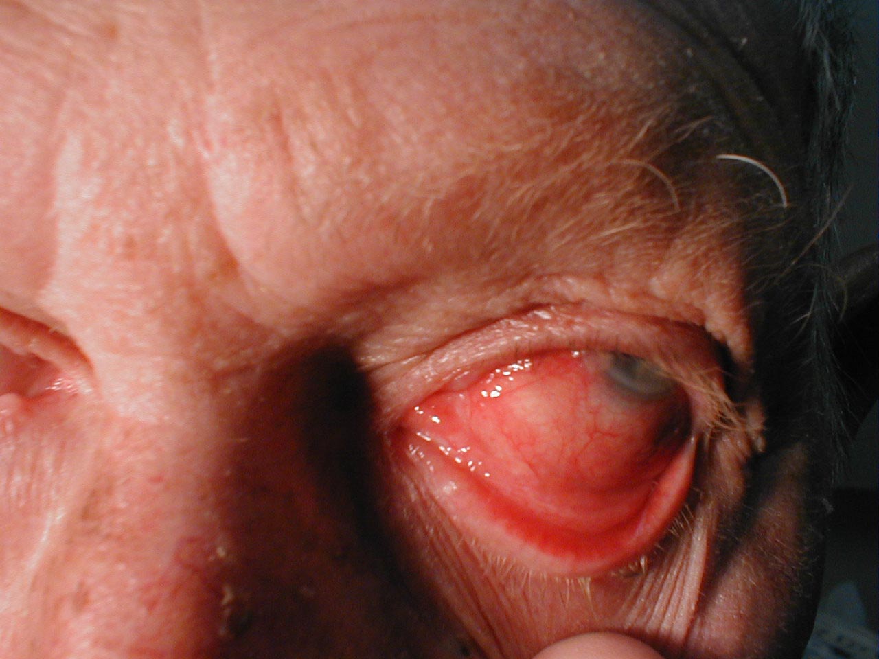
Conjunctivitis: Note inflamed conjunctiva of sclera and reflection onto underside of eyelid.
-
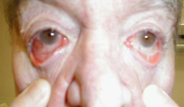
Conjunctivitis: Marked bilateral inflammation involving conjunctiva that covers sclera and under surface of eyelid. Thick exudate can also be seen.
-
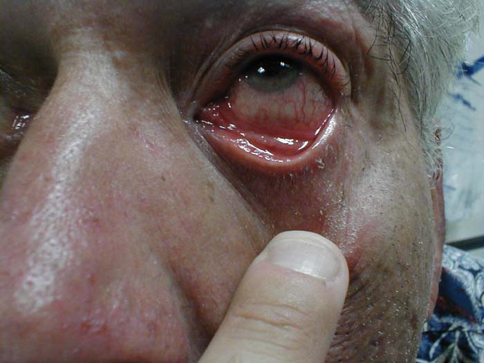
Conjunctivitis: Inflammation of conjunctiva covering sclera and under surface of eyelid.


References
- ↑ Jhanji V, Chan TC, Li EY, Agarwal K, Vajpayee RB (2015). "Adenoviral keratoconjunctivitis". Surv Ophthalmol. 60 (5): 435–43. doi:10.1016/j.survophthal.2015.04.001. PMID 26077630.
- ↑ Azari AA, Barney NP (2013). "Conjunctivitis: a systematic review of diagnosis and treatment". JAMA. 310 (16): 1721–9. doi:10.1001/jama.2013.280318. PMC 4049531. PMID 24150468.
- ↑ Yin-Murphy M (1976). "Simple tests for the diagnosis of picornavirus epidemic conjunctivitis (acute haemorrhagic conjunctivitis)". Bull World Health Organ. 54 (6): 675–9. PMC 2366581. PMID 1088513.
- ↑ Leibowitz HM (2000). "The red eye". N Engl J Med. 343 (5): 345–51. doi:10.1056/NEJM200008033430507. PMID 10922425.
- ↑ Workowski KA, Berman S, Centers for Disease Control and Prevention (CDC) (2010). "Sexually transmitted diseases treatment guidelines, 2010". MMWR Recomm Rep. 59 (RR-12): 1–110. PMID 21160459.
- ↑ Mallika P, Asok T, Faisal H, Aziz S, Tan A, Intan G (2008). "Neonatal conjunctivitis - a review". Malays Fam Physician. 3 (2): 77–81. PMC 4170304. PMID 25606121.
- ↑ La Rosa M, Lionetti E, Reibaldi M, Russo A, Longo A, Leonardi S; et al. (2013). "Allergic conjunctivitis: a comprehensive review of the literature". Ital J Pediatr. 39: 18. doi:10.1186/1824-7288-39-18. PMC 3640929. PMID 23497516.
- ↑ Stern ME, Beuerman RW, Fox RI, Gao J, Mircheff AK, Pflugfelder SC (1998). "The pathology of dry eye: the interaction between the ocular surface and lacrimal glands". Cornea. 17 (6): 584–9. PMID 9820935.
- ↑ Schaumberg DA, Dana R, Buring JE, Sullivan DA (2009). "Prevalence of dry eye disease among US men: estimates from the Physicians' Health Studies". Arch Ophthalmol. 127 (6): 763–8. doi:10.1001/archophthalmol.2009.103. PMC 2836718. PMID 19506195.
- ↑ Nelson JD (1989). "Superior limbic keratoconjunctivitis (SLK)". Eye (Lond). 3 ( Pt 2): 180–9. doi:10.1038/eye.1989.26. PMID 2695351.
- ↑ courtesy of Charlie Goldberg, M.D., UCSD School of Medicine and VA Medical Center, San Diego, CA)