Cervix
|
WikiDoc Resources for Cervix |
|
Articles |
|---|
|
Media |
|
Evidence Based Medicine |
|
Clinical Trials |
|
Ongoing Trials on Cervix at Clinical Trials.gov Clinical Trials on Cervix at Google
|
|
Guidelines / Policies / Govt |
|
US National Guidelines Clearinghouse on Cervix
|
|
Books |
|
News |
|
Commentary |
|
Definitions |
|
Patient Resources / Community |
|
Directions to Hospitals Treating Cervix Risk calculators and risk factors for Cervix
|
|
Healthcare Provider Resources |
|
Continuing Medical Education (CME) |
|
International |
|
|
|
Business |
|
Experimental / Informatics |
Editor-In-Chief: C. Michael Gibson, M.S., M.D. [1]
Overview
The cervix (from Latin "neck") is the lower, narrow portion of the uterus where it joins with the top end of the vagina. It is cylindrical or conical in shape and protrudes through the upper anterior vaginal wall. Approximately half its length is visible with appropriate medical equipment; the remainder lies above the vagina beyond view. It is occasionally called "cervix uteri", or "neck of the uterus".
Anatomy
Ectocervix
The portion projecting into the vagina is referred to as the portio vaginalis or ectocervix. On average, the ectocervix is 3 cm long and 2.5 cm wide. It has a convex, elliptical surface and is divided into anterior and posterior lips.
External os
The ectocervix's opening is called the external os. The size and shape of the external os and the ectocervix varies widely with age, hormonal state, and whether the woman has had a vaginal birth. In women who have not had a vaginal birth the external os appears as a small, circular opening. In women who have had a vaginal birth, the ectocervix appears bulkier and the external os appears wider, more slit-like and gaping.
Endocervical canal
The passageway between the external os and the uterine cavity is referred to as the endocervical canal. It varies widely in length and width, along with the cervix overall. Flattened anterior to posterior, the endocervical canal measures 7 to 8 mm at its widest in reproductive-aged women.
Internal os
The endocervical canal terminates at the internal os which is the opening of the cervix inside the uterine cavity.
Cervical crypts
There are pockets in the lining of the cervix known as cervical crypts. They function to produce cervical fluid.[1]
Histology
The epithelium of the cervix is nonkeratinized stratified squamous epithelium at the ectocervix, and simple columnar epithelium at the cervix proper.[2][3] At certain times of life, the columnar epithelium is replaced by metaplastic squamous epithelium, and is then known as the transformation zone.
Nabothian cysts are often found in the cervix.[4]
Cervical mucus
After a menstrual period ends, the external os is blocked by mucus that is thick and acidic. This "infertile" mucus blocks spermatozoa from entering the uterus.[5] For several days around the time of ovulation, "fertile" types of mucus are produced: they have a higher water content, are less acidic, and have a ferning pattern that helps guide spermatozoa through the cervix.[6] This ferning is a branching pattern seen in the mucus when observed with low magnification.
Some methods of fertility awareness involve estimating a woman's periods of fertility and infertility by observing changes in her body. Among these changes are several involving the quality of her cervical mucus: the sensation it causes at the vulva, its elasticity (spinnbarkeit), its transparency, and the presence of ferning.[6]
Most methods of hormonal contraception work primarily by preventing ovulation, but their effectiveness is increased because they prevent the fertile types of cervical mucus from being produced. Conversely, methods of thinning the mucus may help to achieve pregnancy. One suggested method is to take guaifenesin in the few days before ovulation.[7]
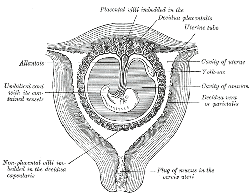
During pregnancy the cervix is blocked by a special antibacterial mucosal plug which prevents infection, somewhat similar to its state during the infertile portion of the menstrual cycle. The mucus plug comes out as the cervix dilates in labor or shortly before.
Cervical position
After menstruation and directly under the influence of estrogen, the cervix undergoes a series of changes in position and texture.
- During most of the menstrual cycle, the cervix remains firm, like the tip of the nose, and is positioned low and closed.
- However, as a woman approaches ovulation, the cervix becomes softer, and rises and opens in response to the high levels of estrogen present at ovulation.[1] These changes, accompanied by the production of fertile types of cervical mucus, support the survival and movement of sperm.
Functionality
During menstruation the cervix stretches open slightly to allow the endometrium to be shed. This stretching is believed to be part of the cramping pain that many women experience. Evidence for this is given by the fact that some women's cramps subside or disappear after their first vaginal birth because the cervical opening has widened.
During childbirth, contractions of the uterus will dilate the cervix up to 10 cm in diameter to allow the fetus to pass through.
During orgasm, the cervix convulses and the external os dilates. Dr. R. Robin Baker and Dr. Mark A. Bellis, both at the University of Manchester, first proposed that this behavior worked in such a way as to draw any semen in the vagina into the uterus, increasing the likelihood of conception. Later researchers, most notably Elisabeth A. Lloyd, have questioned the logic of this theory and the quality of the experimental data used to back it.
Cervical cancer
In humans the cervix may be affected by cervical cancer, a particular form of cancer which is detectable by cytological study of epithelial cells removed from the cervix in a process known as the pap smear. Evidence now shows that those with exposure to HPV (human papilloma virus) are at increased risk for cervical cancer. These viruses are related to the viruses that causes warts. The incidence of new cases of cervical cancer in the United States was observed to be 7 per 100,000 women in 2004.[8]
Cervix: Squamous cell carcinoma
{{#ev:youtube|zB47nE-i8dQ}}
{{#ev:youtube|J3kULzKGzws}}
Lymphatic drainage
The lymphatic drainage of the cervix is along the uterine arteries and cardinal ligaments to the parametrial, external iliac vein, internal iliac vein, and obturator and presacral lymph nodes. From these pelvic lymph nodes, drainage then proceeds to the paraaortic lymph nodes. In some women, the lymphatics drain directly to the paraaortic nodes.
Additional images
-
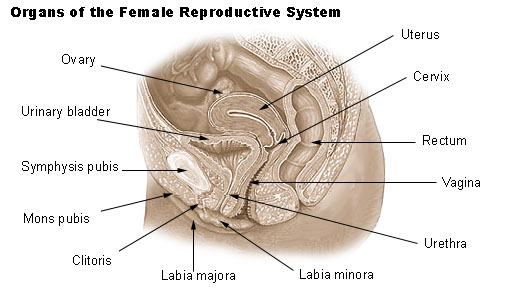
Organs of the female reproductive system.
-
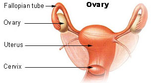
Ovary


-
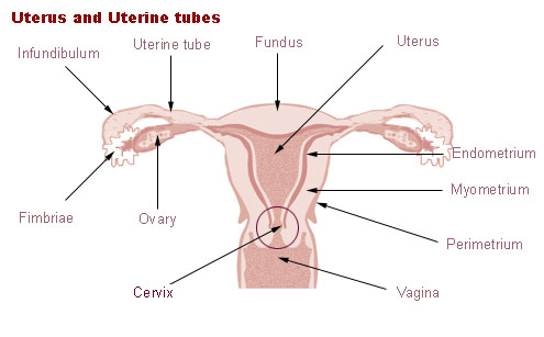
Uterus and uterine tubes.
-
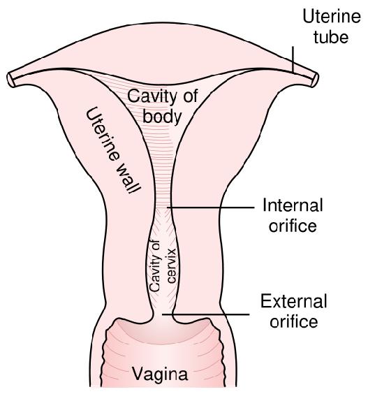
Posterior half of uterus and upper part of vagina.
-
Mucus plug


Histopathological Findings in Cervix Diseases
Cervix: Bacterial vaginosis
{{#ev:youtube|19QWI9PUXnA}}
Cervix: Candida
{{#ev:youtube|01j6qd9Puvg}}
Cervix: Trichomonas
{{#ev:youtube|ho-STuDLLKE}}
References
- ↑ 1.0 1.1 Weschler, Toni, MPH, Taking Charge of Your Fertility, Second Edition, 2002, pp. 59,64
- ↑ Histology image: 19404loa – Histology Learning System at Boston University
- ↑ Histology at University of Southern California rep/c_48
- ↑ Weschler, pp. 227-228
- ↑ Ann Westinore; Evelyn, Dr Billings (1998). The Billings Method: Controlling Fertility Without Drugs or Devices. Toronto: Life Cycle Books. p. 37. ISBN 0-919225-17-9.
- ↑ 6.0 6.1 Weschler, pp. 58-59
- ↑ Weschler, p. 173
- ↑ SEER cancer statistics
Template:Female reproductive system
ar:عنق الرحم bg:Маточна шийка cs:Děložní hrdlo cy:Ceg y groth da:Livmoderhals de:Cervix uteri he:צוואר הרחם lt:Gimdos kaklelis nl:Baarmoederhals no:Livmorhals simple:Cervix sv:Livmoderhals