Candida vulvovaginitis physical examination
|
Candidiasis Main page |
Editor-In-Chief: C. Michael Gibson, M.S., M.D. [1]; Associate Editor(s)-in-Chief: Kiran Singh, M.D. [2], Dima Nimri, M.D. [3]
Overview
Physical Examination
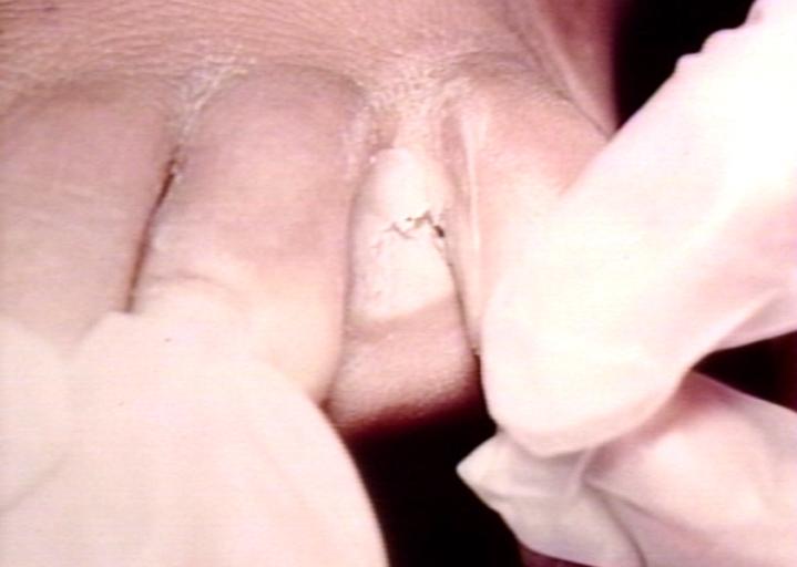
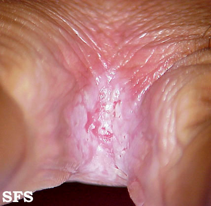
Candida vulvovaginitis requires a careful examination of the external genitalia, the vaginal sidewalls, as well as the cervix. Signs include:[1][2]
- Vulvar edema or erythema
- Fissures and excoriations of the external genitalia, and/or
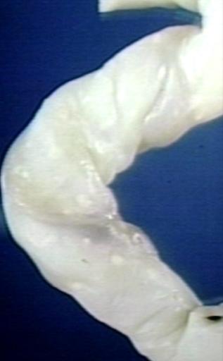
- Thick, white vaginal discharge.
Gallery
-
-
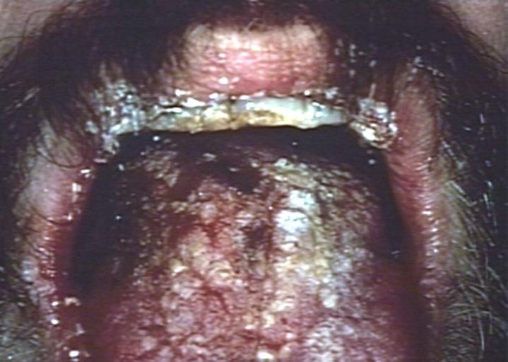
Oral manifestations of HIV infection and AIDS. Chronic oral candidiasis in patient with AIDS. Image courtesy of Professor Peter Anderson DVM PhD and published with permission. © PEIR, University of Alabama at Birmingham, Department of Pathology


-
Soft palate showing extensive oral candidiasis in patient with AIDS. Image courtesy of Professor Peter Anderson DVM PhD and published with permission. © PEIR, University of Alabama at Birmingham, Department of Pathology
-
Oral candidiasis Image courtesy of Professor Peter Anderson DVM PhD and published with permission. © PEIR, University of Alabama at Birmingham, Department of Pathology


-
Eczema secondary to candidiasis. Image courtesy of Professor Peter Anderson DVM PhD and published with permission. © PEIR, University of Alabama at Birmingham, Department of Pathology
-
Candidiasis; skinfold. Image courtesy of Professor Peter Anderson DVM PhD and published with permission. © PEIR, University of Alabama at Birmingham, Department of Pathology


-
Erythematous candidiasis. Image courtesy of Professor Peter Anderson DVM PhD and published with permission. © PEIR, University of Alabama at Birmingham, Department of Pathology
-
Genital candidiasis. Image courtesy of Professor Peter Anderson DVM PhD and published with permission. © PEIR, University of Alabama at Birmingham, Department of Pathology


-
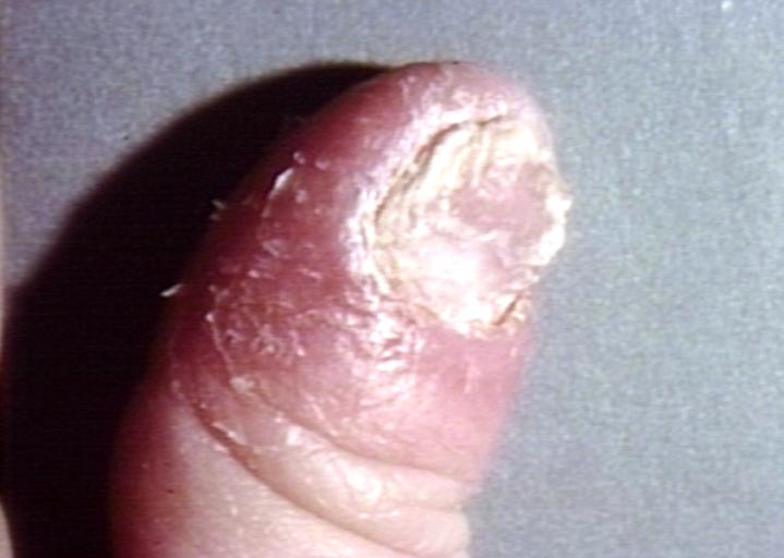
Paronychia: Another manifestation of candidiasis. Image courtesy of Professor Peter Anderson DVM PhD and published with permission. © PEIR, University of Alabama at Birmingham, Department of Pathology
-
Interdigital candidiasis. Image courtesy of Professor Peter Anderson DVM PhD and published with permission. © PEIR, University of Alabama at Birmingham, Department of Pathology


-
Candidiasis of umblical cord. White spots of colonies are present. Image courtesy of Professor Peter Anderson DVM PhD and published with permission. © PEIR, University of Alabama at Birmingham, Department of Pathology
-
Lung: Candidiasis. Postmortem findings. Image courtesy of Professor Peter Anderson DVM PhD and published with permission. © PEIR, University of Alabama at Birmingham, Department of Pathology


Image: Candidiasis 17.jpeg| Candidiasis of the fingernail caused by a fungus of the genus Candida. From Public Health Image Library (PHIL). [3]
Skin folds
-
![Candidiasis. Adapted from Dermatology Atlas.[4]](/images/1/10/Candidiasis06.jpg)
Candidiasis. Adapted from Dermatology Atlas.[4]
-
![Candidiasis. Adapted from Dermatology Atlas.[4]](/images/3/33/Candidiasis07.jpg)
Candidiasis. Adapted from Dermatology Atlas.[4]
![Candidiasis. Adapted from Dermatology Atlas.[4]](/index.php/File:Candidiasis06.jpg)
![Candidiasis. Adapted from Dermatology Atlas.[4]](/index.php/File:Candidiasis07.jpg)
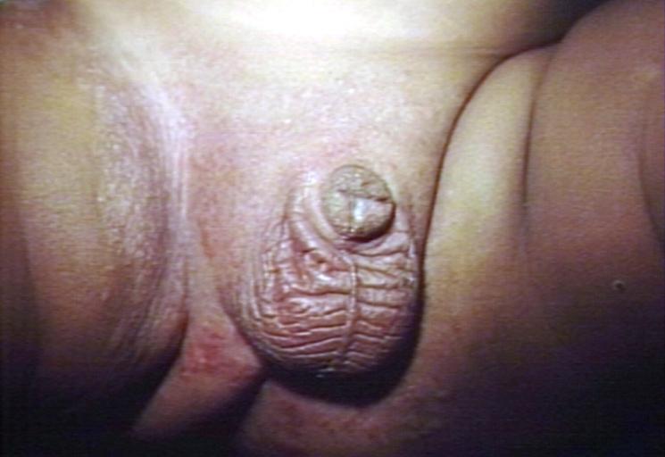
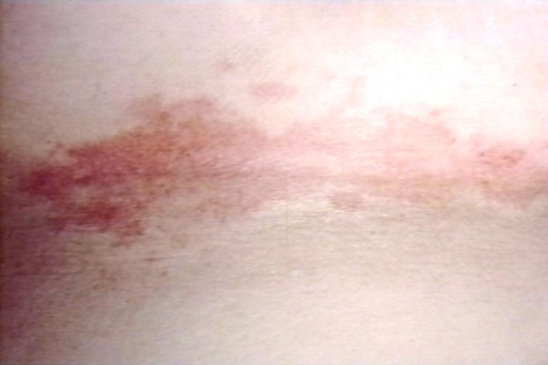
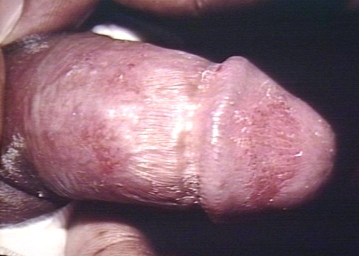
Genitourinary
-
![Infant presented with a rash formerly known as “Moniliasis” now called Candidiasis caused by the fungus Candida sp. From Public Health Image Library (PHIL). [3]](/images/6/6d/Moniliasis04.jpeg)
Infant presented with a rash formerly known as “Moniliasis” now called Candidiasis caused by the fungus Candida sp. From Public Health Image Library (PHIL). [3]
-
![Wet mounted vaginal smear specimen, revealed the presence of Candida albicans, which had been extracted from a patient with vaginal candidiasis. From Public Health Image Library (PHIL). [3]](/images/f/f5/Moniliasis02.jpeg)
Wet mounted vaginal smear specimen, revealed the presence of Candida albicans, which had been extracted from a patient with vaginal candidiasis. From Public Health Image Library (PHIL). [3]
-
![Candidiasis. Adapted from Dermatology Atlas.[4]](/images/c/c4/Candidiasis01.jpg)
Candidiasis. Adapted from Dermatology Atlas.[4]
-
![Candidiasis. Adapted from Dermatology Atlas.[4]](/images/5/5f/Candidiasis02.jpg)
Candidiasis. Adapted from Dermatology Atlas.[4]
-
Candidiasis. Adapted from Dermatology Atlas.<ref name="Dermatology Atlas">
-
![Candidiasis. Adapted from Dermatology Atlas.[4]](/images/7/78/Candidiasis13.jpg)
Candidiasis. Adapted from Dermatology Atlas.[4]
-
![Candidiasis. Adapted from Dermatology Atlas.[4]](/images/8/8c/Candidiasis04.jpg)
Candidiasis. Adapted from Dermatology Atlas.[4]
![Infant presented with a rash formerly known as “Moniliasis” now called Candidiasis caused by the fungus Candida sp. From Public Health Image Library (PHIL). [3]](/index.php/File:Moniliasis04.jpeg)
![Wet mounted vaginal smear specimen, revealed the presence of Candida albicans, which had been extracted from a patient with vaginal candidiasis. From Public Health Image Library (PHIL). [3]](/index.php/File:Moniliasis02.jpeg)
![Candidiasis. Adapted from Dermatology Atlas.[4]](/index.php/File:Candidiasis01.jpg)
![Candidiasis. Adapted from Dermatology Atlas.[4]](/index.php/File:Candidiasis02.jpg)

![Candidiasis. Adapted from Dermatology Atlas.[4]](/index.php/File:Candidiasis13.jpg)
![Candidiasis. Adapted from Dermatology Atlas.[4]](/index.php/File:Candidiasis04.jpg)
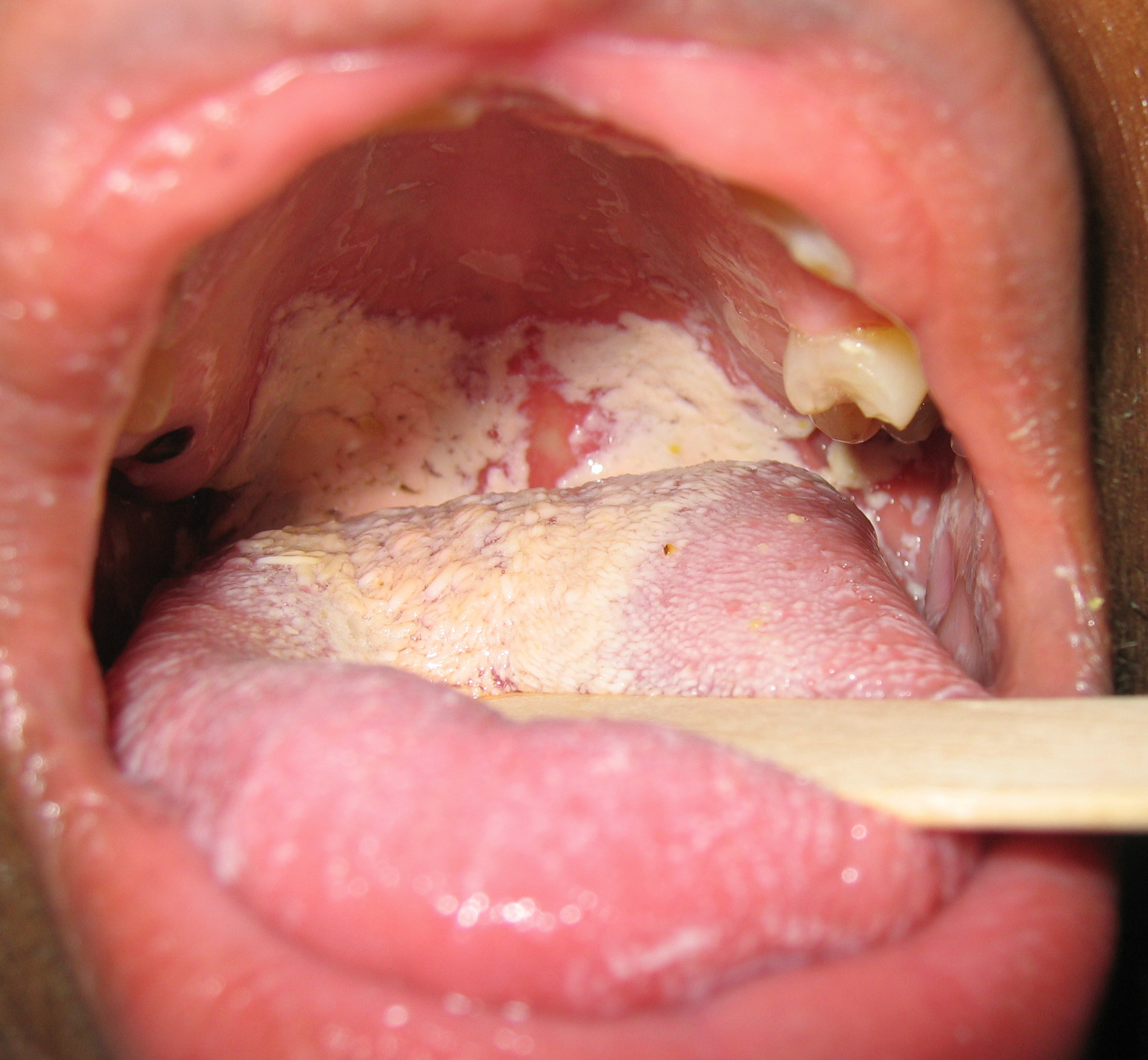
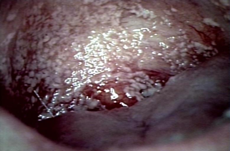
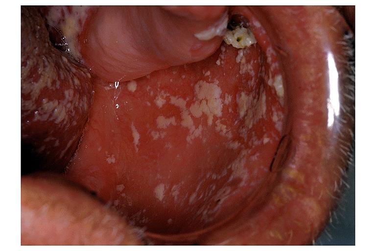
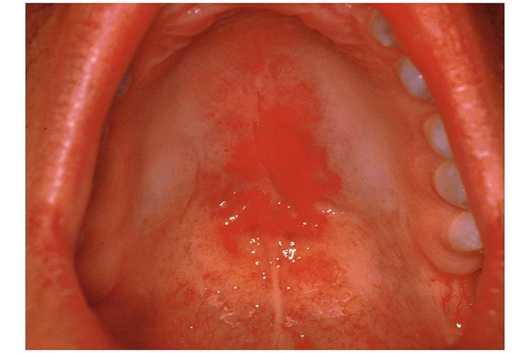
Oral cavity
-
![Candidiasis. Adapted from Dermatology Atlas.[4]](/images/c/ca/Candidiasis10.jpg)
Candidiasis. Adapted from Dermatology Atlas.[4]
-
![Candidiasis. Adapted from Dermatology Atlas.[4]](/images/4/42/Candidiasis11.jpg)
Candidiasis. Adapted from Dermatology Atlas.[4]
-
![Candidiasis. Adapted from Dermatology Atlas.[4]](/images/6/67/Candidiasis12.jpg)
Candidiasis. Adapted from Dermatology Atlas.[4]
![Candidiasis. Adapted from Dermatology Atlas.[4]](/index.php/File:Candidiasis10.jpg)
![Candidiasis. Adapted from Dermatology Atlas.[4]](/index.php/File:Candidiasis11.jpg)
![Candidiasis. Adapted from Dermatology Atlas.[4]](/index.php/File:Candidiasis12.jpg)
References
- ↑ Eckert LO, Hawes SE, Stevens CE, Koutsky LA, Eschenbach DA, Holmes KK (1998). "Vulvovaginal candidiasis: clinical manifestations, risk factors, management algorithm". Obstet Gynecol. 92 (5): 757–65. PMID 9794664.
- ↑ Eckert LO (2006). "Clinical practice. Acute vulvovaginitis". N. Engl. J. Med. 355 (12): 1244–52. doi:10.1056/NEJMcp053720. PMID 16990387.
- ↑ 3.0 3.1 3.2 "Public Health Image Library (PHIL)".
- ↑ 4.0 4.1 4.2 4.3 4.4 4.5 4.6 4.7 4.8 "Dermatology Atlas".