Brucellosis pathophysiology: Difference between revisions
No edit summary |
No edit summary |
||
| Line 1: | Line 1: | ||
__NOTOC__ | |||
== Overview == | == Overview == | ||
''[[Brucella]]'' is usually transmitted via the digestive route to the human host. Following transmission, [[white blood cells]] phagocyte the pathogen and transports it via the hematologic or lymphatic route to different organs, specially to those of the [[reticuloendothelial system]].<sup>[1][2]</sup> | ''[[Brucella]]'' is usually transmitted via the digestive route to the human host. Following transmission, [[white blood cells]] phagocyte the pathogen and transports it via the hematologic or lymphatic route to different organs, specially to those of the [[reticuloendothelial system]].<sup>[1][2]</sup> | ||
| Line 72: | Line 73: | ||
* On microscopic histopathological analysis of the liver, common findings are: | * On microscopic histopathological analysis of the liver, common findings are: | ||
** [[Granulomas]] with centrilobular necrosis or focal necrosis and parenchyma destruction.<sup>[10]</sup> | ** [[Granulomas]] with centrilobular necrosis or focal necrosis and parenchyma destruction.<sup>[10]</sup> | ||
** ** | ** ** | ||
<gallery> | |||
Image:Brucella-histo.jpg|thumb|200px|Brucella spp. are poorly staining, small gram-negative coccobacilli (0.5-0.7 x 0.6-1.5 µm), and are seen mostly as single cells and appearing like “fine sand”. | |||
Image:Bruce-granulomanecrosis.jpg|thumb|200px|Histopathology of guinea pig liver in experimental Brucella suis infection. Granuloma with necrosis. | |||
</gallery> | |||
==Reference== | |||
{{reflist|2}} | {{reflist|2}} | ||
[[Category:Needs overview]] | |||
[[Category:Bacterial diseases]] | |||
[[Category:Occupational diseases]] | |||
[[Category:Zoonoses]] | |||
[[Category:Infectious disease]] | |||
[[Category:Biological weapons]] | |||
[[Category:Disease]] | |||
Revision as of 02:36, 5 January 2017
Overview
Brucella is usually transmitted via the digestive route to the human host. Following transmission, white blood cells phagocyte the pathogen and transports it via the hematologic or lymphatic route to different organs, specially to those of the reticuloendothelial system.[1][2]
Pathophysiology
The pathophysiology of brucellosis may be described in the following steps:
Transmission:
| Humans are generally infected with brucellosis in one of three ways | |
|---|---|
Eating undercooked meat or consuming unpasteurized/raw dairy products |
The most common way to be infected is by eating or drinking unpasteurized/raw dairy products. When sheep, goats, cows, or camels are infected, their milk becomes contaminated with the bacteria.
If the milk from infected animals is not pasteurized, the infection will be transmitted to people who consume the milk and/or cheese products. |
Breathing in the bacteria that cause brucellosis (inhalation) |
Breathing in the bacteria that causes brucellosis may also lead to infection. This risk is generally greater for people in laboratories that work with the bacteria. In addition, slaughterhouse and meat-packing employees have also been known to be exposed to the bacteria and ultimately become infected. |
Bacteria entering the body through skin wounds or mucous membranes |
Bacteria can also enter wounds in the skin/mucous membranes through contact with infected animals.
This poses a problem for workers who have close contact with animals or animal excretions (newborn animals, fetuses, and excretions that may result from birth). Such workers may include:
|
| |
Incubation
Incubation period of brucellosis varies from one to four weeks. But occasionally, it may be as long as several months. [3][4]
Dissemination
Following transmission, brucellae is ingested by macrophages and polymorphonuclear cells. On ingestion, they replicate intracellularly inside the lysed cells, infecting the other cells or disseminating systemically. [5]
Seeding
- On transmission, Neutrophilic granulocytes and monocytes actively phagocytosed the bacteria
- On entry into the body, brucellae multiply in the neutrophilic granulocytes ad monocytes, initially in lymph nodes, which is followed by systemic hematogenous spread resulting in multiple localizing infection
Immune response
Brucellosis elicits both humoral and cell-mediated immune responses:[6]
Humoral immune response
Antibodies are produced by body immune system aganist the extracellular brucellae not only promote the clearance of extracellular brucellae , but also facilitate the phagocytosis of the brucellae
Cell mediates immune response
- Tumor necrosis factor α (TNF-α) produce on activation of cell mediated immunity, stimulates T lymphocytes and macrophages, which help in eliminating intracellular brucellae. Virulent brucellar tend to supress the activity of Tumor necrosis factor α (TNF-α)
- Cytokines such as interleukin (IL) 12 promote production ofvation and interferon γ (IFN-γ) responses. IFN-γ, which drives TH1-type responses and stimulates macrophage activation. Inflammatory cytokines, including IL-4, IL-6, and IL-10, downregulate the protective response.
Pathogenesis
The pathogenesis of brucellosis involve the following:[7][8]
By avoiding innate immunity, brucella survive with in monocytic cells.
- Endotoxic lipopolysaccharide LPS, plays a key role in survival of bacteria inside monocytic cell.
- LPS helps in survival of the bacteria inside the monocytic cell, by suppressing phagosome–lysosome fusion, and internalizing bacteria into endoplasmic reticulum
- Type IV secretion system (VirB) and type III secretion system, that regulates intracellular survival and trafficking has been identified, although type 3 not yet confirmed.
- Acid-stable proteins produced by brucella, facilitates the survival in phogosomes
- Cu-Zn superoxide dismutase, produced by brucellae, gives them resistance from reactive oxygen intermediates
Genetics
There is no known genetic association to brucellosis.
Microscopic Pathology
- Brucella spp. are poorly staining, small gram-negative coccobacilli (0.5-0.7 x 0.6-1.5 µm).
- Brucella spp. are seen mostly as single cells and appearing like “fine sand”.[9]
- On microscopic histopathological analysis of the liver, common findings are:
- Granulomas with centrilobular necrosis or focal necrosis and parenchyma destruction.[10]
- **
-
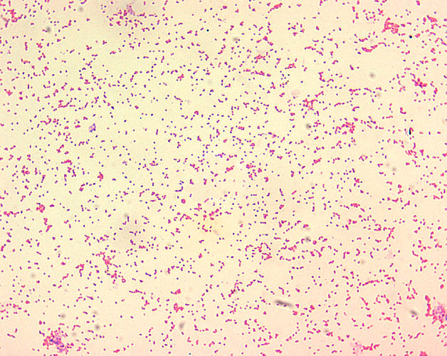
Brucella spp. are poorly staining, small gram-negative coccobacilli (0.5-0.7 x 0.6-1.5 µm), and are seen mostly as single cells and appearing like “fine sand”.
-
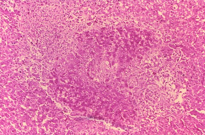
Histopathology of guinea pig liver in experimental Brucella suis infection. Granuloma with necrosis.

