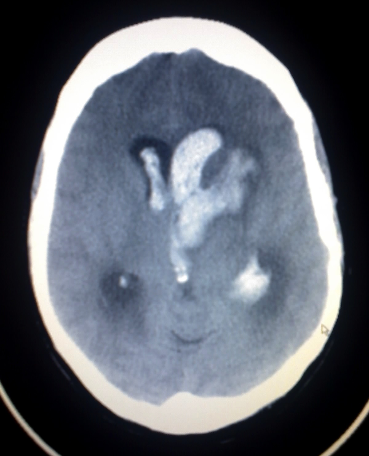
Bonnet-Dechaume-Blanc syndrome
Editor-In-Chief: C. Michael Gibson, M.S., M.D. [1] Associate Editor(s)-in-Chief: Jyostna Chouturi, M.B.B.S [2]
Synonyms and keywords: Wyburn mason's syndrome; Retinoencephalofacial angiomatosis

Overview
.
Signs and symptoms
Causes
Mechanism
Epidemiology
Diagnosis
Treatment
If lesions are present, they are watched closely for changes in size. Prognosis is best when lesions are less than 3 cm in length. Most complications occur when the lesions are greater than 6 cm in size.[1] Surgical intervention for intracranial lesions has been done successfully. Nonsurgical treatments include embolization, radiation therapy, and continued observation.[2] Arterial vascular malformations may be treated with the cyberknife treatment. Possible treatment for cerebral arterial vascular malformations include stereotactic radiosurgery, endovascular embolization, and microsurgical resection.[1]
When pursuing treatment, it is important to consider the size of the malformations, their locations, and the neurological involvement.[3] Because it is a congenital disorder, there are not preventative steps to take aside from regular follow ups with a doctor to keep an eye on the symptoms so that future complications are avoided.
References
- ↑ 1.0 1.1 SINGH, A; RUNDLE, P; RENNIE, I (March 2005). "Retinal Vascular Tumors". Ophthalmology Clinics of North America. 18 (1): 167–176. doi:10.1016/j.ohc.2004.07.005.
- ↑ Dayani, P. N.; Sadun, A. A. (18 January 2007). "A case report of Wyburn-Mason syndrome and review of the literature". Neuroradiology. 49 (5): 445–456. doi:10.1007/s00234-006-0205-x.
- ↑ Lester, Jacobo; Ruano-Calderon, Luis Angel; Gonzalez-Olhovich, Irene (July 2005). "Wyburn-Mason Syndrome". Journal of Neuroimaging. 15 (3): 284–285. doi:10.1111/j.1552-6569.2005.tb00324.x.