Blastomycosis laboratory findings: Difference between revisions
No edit summary |
No edit summary |
||
| Line 29: | Line 29: | ||
</gallery> | </gallery> | ||
==Gallery== | |||
<gallery> | |||
Image: Blastomycosis02.jpeg| This composite photomicrograph reveals some of the ultrastructural histopathology in a tissue specimen from a patient with a keloidean blastomycosis infection, which was caused by the fungus, Blastomyces dermatitidis. The specimen originated from tissue scraping samplings. It’s important to note the abundance of large budding cells that had been configured in chains. <SMALL><SMALL>''[http://phil.cdc.gov/phil/home.asp From Public Health Image Library (PHIL).] ''<ref name=PHIL> {{Cite web | title = Public Health Image Library (PHIL) | url = http://phil.cdc.gov/phil/home.asp}}</ref></SMALL></SMALL> | |||
Image: Blastomycosis03.jpeg| This photomicrograph reveals some of the ultrastructural histopathology in a tissue specimen from a patient with a keloidean blastomycosis infection, which was caused by the fungus, Blastomyces dermatitidis. The specimen originated from a sample of tissue scrapings. In this particular section note the abundance of large budding cells.<SMALL><SMALL>''[http://phil.cdc.gov/phil/home.asp From Public Health Image Library (PHIL).] ''<ref name=PHIL> {{Cite web | title = Public Health Image Library (PHIL) | url = http://phil.cdc.gov/phil/home.asp}}</ref></SMALL></SMALL> | |||
Image: Blastomycosis04.jpeg| This photomicrograph reveals some of the ultrastructural histopathology in a tissue specimen from a patient with a keloidean blastomycosis infection, which was caused by the fungus, Blastomyces dermatitidis. In this particular section, you’ll note fungal invasion of a crural lymph node. <SMALL><SMALL>''[http://phil.cdc.gov/phil/home.asp From Public Health Image Library (PHIL).] ''<ref name=PHIL> {{Cite web | title = Public Health Image Library (PHIL) | url = http://phil.cdc.gov/phil/home.asp}}</ref></SMALL></SMALL> | |||
Image: Blastomycosis05.jpeg| This hematoxylin-eosin (H&E)-stained photomicrograph reveals some of the ultrastructural histopathology in an dermal skin tissue specimen in a patient with an intradermal keloidean blastomycosis infection, which was caused by the fungus, Blastomyces dermatitidis. In this particular section, you’ll note presence of a parasitized multinucleated giant cell. <SMALL><SMALL>''[http://phil.cdc.gov/phil/home.asp From Public Health Image Library (PHIL).] ''<ref name=PHIL> {{Cite web | title = Public Health Image Library (PHIL) | url = http://phil.cdc.gov/phil/home.asp}}</ref></SMALL></SMALL> | |||
Image: Blastomycosis08.jpeg| This image depicts the morphologic changes that took place upon a patient’s arm, which included keloidal scarring brought on due to a case of cutaneous blastomycosis, that was caused by the fungus, Blastomyces dermatitidis. <SMALL><SMALL>''[http://phil.cdc.gov/phil/home.asp From Public Health Image Library (PHIL).] ''<ref name=PHIL> {{Cite web | title = Public Health Image Library (PHIL) | url = http://phil.cdc.gov/phil/home.asp}}</ref></SMALL></SMALL> | |||
Image: Blastomycosis09.jpeg| This image depicts the morphologic changes that took place upon a patient’s arm, which included keloidal scarring brought on due to a case of cutaneous blastomycosis, that was caused by the fungus, Blastomyces dermatitidis. <SMALL><SMALL>''[http://phil.cdc.gov/phil/home.asp From Public Health Image Library (PHIL).] ''<ref name=PHIL> {{Cite web | title = Public Health Image Library (PHIL) | url = http://phil.cdc.gov/phil/home.asp}}</ref></SMALL></SMALL> | |||
Image: Blastomycosis10.jpeg| This photomicrograph reveals some of the ultrastructural histopathology in an dermal skin tissue specimen in a patient with an intradermal keloidal blastomycosis infection, which had been long standing, and resulted in a chronic granulomatous inflammatory response. <SMALL><SMALL>''[http://phil.cdc.gov/phil/home.asp From Public Health Image Library (PHIL).] ''<ref name=PHIL> {{Cite web | title = Public Health Image Library (PHIL) | url = http://phil.cdc.gov/phil/home.asp}}</ref></SMALL></SMALL> | |||
Image: Blastomycosis11.jpeg| This anterior view of a patient’s right knee revealed the keloidal scarring brought on due to a case of cutaneous blastomycosis, which was caused by the fungus, Blastomyces dermatitidis. <SMALL><SMALL>''[http://phil.cdc.gov/phil/home.asp From Public Health Image Library (PHIL).] ''<ref name=PHIL> {{Cite web | title = Public Health Image Library (PHIL) | url = http://phil.cdc.gov/phil/home.asp}}</ref></SMALL></SMALL> | |||
Image: Blastomycosis13.jpeg| This anterior view of a patient’s right knee revealed the keloidal scarring brought on due to a case of cutaneous blastomycosis, which was caused by the fungus, Blastomyces dermatitidis. <SMALL><SMALL>''[http://phil.cdc.gov/phil/home.asp From Public Health Image Library (PHIL).] ''<ref name=PHIL> {{Cite web | title = Public Health Image Library (PHIL) | url = http://phil.cdc.gov/phil/home.asp}}</ref></SMALL></SMALL> | |||
Image: Blastomycosis15.jpeg|Seen from the medial perspective, this patient’s right ankle displayed keloidal scarring brought on due to a case of cutaneous blastomycosis, which was caused by the fungus, Blastomyces dermatitidis. <SMALL><SMALL>''[http://phil.cdc.gov/phil/home.asp From Public Health Image Library (PHIL).] ''<ref name=PHIL> {{Cite web | title = Public Health Image Library (PHIL) | url = http://phil.cdc.gov/phil/home.asp}}</ref></SMALL></SMALL> | |||
Image: Blastomycosis16.jpeg| This illustration depicts the ultrastructural details found in the dimorphic fungal organism, Blastomyces dermatitidis including the organism’s aerial hypha, developing sporangia, which would eventually contain mature sporangiospores, and the sporangiophore, which gives rise to the developing sporangia. <SMALL><SMALL>''[http://phil.cdc.gov/phil/home.asp From Public Health Image Library (PHIL).] ''<ref name=PHIL> {{Cite web | title = Public Health Image Library (PHIL) | url = http://phil.cdc.gov/phil/home.asp}}</ref></SMALL></SMALL> | |||
Image: Blastomycosis17.jpeg|Magnified 562X, this "digested", and fluorescent antibody-stained photomicrograph reveals the presence of Blastomyces dermatitidis antigens in this human lung tissue specimen. <SMALL><SMALL>''[http://phil.cdc.gov/phil/home.asp From Public Health Image Library (PHIL).] ''<ref name=PHIL> {{Cite web | title = Public Health Image Library (PHIL) | url = http://phil.cdc.gov/phil/home.asp}}</ref></SMALL></SMALL> | |||
Image: Blastomycosis18.jpeg| Magnified 562X, this "digested", and fluorescent antibody-stained photomicrograph reveals the presence of Blastomyces dermatitidis antigens in this human lung tissue specimen. <SMALL><SMALL>''[http://phil.cdc.gov/phil/home.asp From Public Health Image Library (PHIL).] ''<ref name=PHIL> {{Cite web | title = Public Health Image Library (PHIL) | url = http://phil.cdc.gov/phil/home.asp}}</ref></SMALL></SMALL> | |||
Image: Blastomycosis20.jpeg| Magnified 125X, this "undigested", and fluorescent antibody-stained photomicrograph reveals the presence of Blastomyces dermatitidis antigens in this human lung tissue specimen. <SMALL><SMALL>''[http://phil.cdc.gov/phil/home.asp From Public Health Image Library (PHIL).] ''<ref name=PHIL> {{Cite web | title = Public Health Image Library (PHIL) | url = http://phil.cdc.gov/phil/home.asp}}</ref></SMALL></SMALL> | |||
Image: Blastomycosis21.jpeg|Magnified 500X, this Gamori-stained photomicrograph of a canine liver tissue specimen, revealed the presence of budding Blastomyces dermatitidis fungal cells of various sizes. Note the accompanying filaments, or mycelium. <SMALL><SMALL>''[http://phil.cdc.gov/phil/home.asp From Public Health Image Library (PHIL).] ''<ref name=PHIL> {{Cite web | title = Public Health Image Library (PHIL) | url = http://phil.cdc.gov/phil/home.asp}}</ref></SMALL></SMALL> | |||
Image: Blastomycosis22.jpeg| Magnified 1150X, this Gram-stained photomicrograph reveals the presence of a number of hyphae of the fungal organism, Exophiala castellanii. These fungi were harvested from a pus-laden lesion located on a patient’s buttock. <SMALL><SMALL>''[http://phil.cdc.gov/phil/home.asp From Public Health Image Library (PHIL).] ''<ref name=PHIL> {{Cite web | title = Public Health Image Library (PHIL) | url = http://phil.cdc.gov/phil/home.asp}}</ref></SMALL></SMALL> | |||
Image: Blastomycosis23.jpeg|Magnified 1188X, this H&E-stained photomicrograph revealed histopathologic changes, which were indicative of the chronic fungal disease process known as chromoblastomycosis, or chromomycosis. The tissue sample was harvested from an Indian patient. Predominantly, this infection affects the skin, or cutaneous tissues, and usually begins at the site of a puncture wound, which introduces the pathologic fungal organism into the recipient’s tissues. <SMALL><SMALL>''[http://phil.cdc.gov/phil/home.asp From Public Health Image Library (PHIL).] ''<ref name=PHIL> {{Cite web | title = Public Health Image Library (PHIL) | url = http://phil.cdc.gov/phil/home.asp}}</ref></SMALL></SMALL> | |||
Image: Blastomycosis25.jpeg| Note the histopathologic changes seen in blastomycosis due to Blastomyces dermatitidis using methenamine silver stain. <SMALL><SMALL>''[http://phil.cdc.gov/phil/home.asp From Public Health Image Library (PHIL).] ''<ref name=PHIL> {{Cite web | title = Public Health Image Library (PHIL) | url = http://phil.cdc.gov/phil/home.asp}}</ref></SMALL></SMALL> | |||
Image: Blastomycosis26.jpeg| Note the histopathologic changes seen in blastomycosis due to Blastomyces dermatitidis using methenamine silver stain.<SMALL><SMALL>''[http://phil.cdc.gov/phil/home.asp From Public Health Image Library (PHIL).] ''<ref name=PHIL> {{Cite web | title = Public Health Image Library (PHIL) | url = http://phil.cdc.gov/phil/home.asp}}</ref></SMALL></SMALL> | |||
Image: Blastomycosis27.jpeg| Direct FA stain revealing the histopathology of lung tissue blastomycosis due to the organism Blastomyces dermatitidis <SMALL><SMALL>''[http://phil.cdc.gov/phil/home.asp From Public Health Image Library (PHIL).] ''<ref name=PHIL> {{Cite web | title = Public Health Image Library (PHIL) | url = http://phil.cdc.gov/phil/home.asp}}</ref></SMALL></SMALL> | |||
Image: Blastomycosis28.jpeg| Direct FA stain revealing the histopathology of lung tissue blastomycosis due to the organism Blastomyces dermatitidis. <SMALL><SMALL>''[http://phil.cdc.gov/phil/home.asp From Public Health Image Library (PHIL).] ''<ref name=PHIL> {{Cite web | title = Public Health Image Library (PHIL) | url = http://phil.cdc.gov/phil/home.asp}}</ref></SMALL></SMALL> | |||
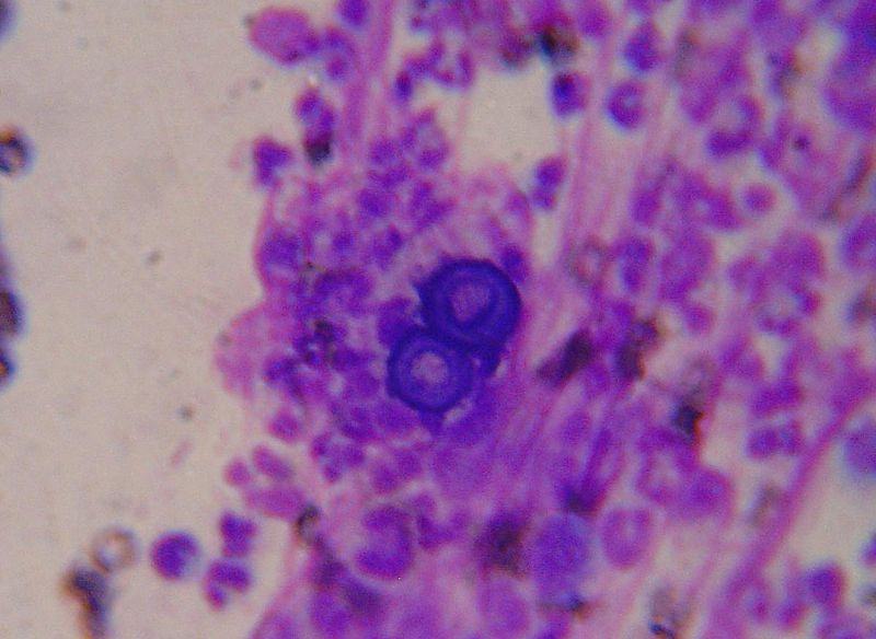
Image: Blastomycosis29.jpeg| This is a photomicrograph of Blastomyces dermatitidis using a cotton blue staining technique. <SMALL><SMALL>''[http://phil.cdc.gov/phil/home.asp From Public Health Image Library (PHIL).] ''<ref name=PHIL> {{Cite web | title = Public Health Image Library (PHIL) | url = http://phil.cdc.gov/phil/home.asp}}</ref></SMALL></SMALL> | |||
Image: Blastomycosis30.jpeg|This micrograph shows histopathologic changes that reveal the presence of the fungal agent Blastomyces dermatitidis. <SMALL><SMALL>''[http://phil.cdc.gov/phil/home.asp From Public Health Image Library (PHIL).] ''<ref name=PHIL> {{Cite web | title = Public Health Image Library (PHIL) | url = http://phil.cdc.gov/phil/home.asp}}</ref></SMALL></SMALL> | |||
Image: Blastomycosis30.jpeg|This micrograph shows histopathologic changes that reveal the presence of the fungal agent Blastomyces dermatitidis. <SMALL><SMALL>''[http://phil.cdc.gov/phil/home.asp From Public Health Image Library (PHIL).] ''<ref name=PHIL> {{Cite web | title = Public Health Image Library (PHIL) | url = http://phil.cdc.gov/phil/home.asp}}</ref></SMALL></SMALL> | |||
Image: Blastomycosis30.jpeg|This micrograph shows histopathologic changes that reveal the presence of the fungal agent Blastomyces dermatitidis. <SMALL><SMALL>''[http://phil.cdc.gov/phil/home.asp From Public Health Image Library (PHIL).] ''<ref name=PHIL> {{Cite web | title = Public Health Image Library (PHIL) | url = http://phil.cdc.gov/phil/home.asp}}</ref></SMALL></SMALL> | |||
Image: Blastomycosis30.jpeg|This micrograph shows histopathologic changes that reveal the presence of the fungal agent Blastomyces dermatitidis. <SMALL><SMALL>''[http://phil.cdc.gov/phil/home.asp From Public Health Image Library (PHIL).] ''<ref name=PHIL> {{Cite web | title = Public Health Image Library (PHIL) | url = http://phil.cdc.gov/phil/home.asp}}</ref></SMALL></SMALL> | |||
Image: Blastomycosis30.jpeg|This micrograph shows histopathologic changes that reveal the presence of the fungal agent Blastomyces dermatitidis. <SMALL><SMALL>''[http://phil.cdc.gov/phil/home.asp From Public Health Image Library (PHIL).] ''<ref name=PHIL> {{Cite web | title = Public Health Image Library (PHIL) | url = http://phil.cdc.gov/phil/home.asp}}</ref></SMALL></SMALL> | |||
Image: Blastomycosis30.jpeg|This micrograph shows histopathologic changes that reveal the presence of the fungal agent Blastomyces dermatitidis. <SMALL><SMALL>''[http://phil.cdc.gov/phil/home.asp From Public Health Image Library (PHIL).] ''<ref name=PHIL> {{Cite web | title = Public Health Image Library (PHIL) | url = http://phil.cdc.gov/phil/home.asp}}</ref></SMALL></SMALL> | |||
Image: Blastomycosis30.jpeg|This micrograph shows histopathologic changes that reveal the presence of the fungal agent Blastomyces dermatitidis. <SMALL><SMALL>''[http://phil.cdc.gov/phil/home.asp From Public Health Image Library (PHIL).] ''<ref name=PHIL> {{Cite web | title = Public Health Image Library (PHIL) | url = http://phil.cdc.gov/phil/home.asp}}</ref></SMALL></SMALL> | |||
</gallery> | |||
==References== | ==References== | ||
{{Reflist|2}} | {{Reflist|2}} | ||
Revision as of 18:49, 11 June 2015
|
Blastomycosis Microchapters |
|
Diagnosis |
|---|
|
Treatment |
|
Case Studies |
|
Blastomycosis laboratory findings On the Web |
|
American Roentgen Ray Society Images of Blastomycosis laboratory findings |
|
Risk calculators and risk factors for Blastomycosis laboratory findings |
Editor-In-Chief: C. Michael Gibson, M.S., M.D. [1]; Associate Editor(s)-in-Chief: ; Vidit Bhargava, M.B.B.S [2]
Overview
Once suspected, the diagnosis of blastomycosis can usually be confirmed by demonstration of the characteristic broad based budding organisms in sputum or tissues by KOH prep, cytology, or histology.
Laboratory Findings
Commonly performed tests include:
- KOH preparation - shows a broad based budding yeast as shown in the microscopic picture above. sputum, blood, pleural and other body fluids may be used, however this process has a low clinical yield.
- Tissue biopsy of skin or other organs may be required in order to diagnose extra-pulmonary disease. A granulomatous inflammation might be suggestive of fungal presence but is not diagnostic.
- Commercially available urine antigen testing appears to be quite sensitive in suggesting the diagnosis in cases where the organism is not readily detected. It appears to be more helpful than serum antigen testing.
- While culture of the organism remains the definitive diagnostic standard, its slow growing nature can lead to delays in treatment of up to several weeks. Culture on dextrose sabourd agar at 37ºC can be used for diagnosis. Highest diagnostic yield is of bronchoscopy derived fluid, followed by sputum. Real time PCR is being experimentally tested for direct diagnosis from culture or tissue. [1]
- Serological testing is limited in utility by the fact that there is a considerable overlap with other fungal antigens.
However, sometimes blood and sputum cultures may not detect blastomycosis; lung biopsy is another option, and results will be shown promptly.
Microscopy
-
Broad based budding yeast
-

Smear from a foot lesion showing a Blastomyces dermatitidis yeast cell.


Gallery
-
![This composite photomicrograph reveals some of the ultrastructural histopathology in a tissue specimen from a patient with a keloidean blastomycosis infection, which was caused by the fungus, Blastomyces dermatitidis. The specimen originated from tissue scraping samplings. It’s important to note the abundance of large budding cells that had been configured in chains. From Public Health Image Library (PHIL). [2]](/images/b/b6/Blastomycosis02.jpeg)
This composite photomicrograph reveals some of the ultrastructural histopathology in a tissue specimen from a patient with a keloidean blastomycosis infection, which was caused by the fungus, Blastomyces dermatitidis. The specimen originated from tissue scraping samplings. It’s important to note the abundance of large budding cells that had been configured in chains. From Public Health Image Library (PHIL). [2]
-
![This photomicrograph reveals some of the ultrastructural histopathology in a tissue specimen from a patient with a keloidean blastomycosis infection, which was caused by the fungus, Blastomyces dermatitidis. The specimen originated from a sample of tissue scrapings. In this particular section note the abundance of large budding cells.From Public Health Image Library (PHIL). [2]](/images/e/ea/Blastomycosis03.jpeg)
This photomicrograph reveals some of the ultrastructural histopathology in a tissue specimen from a patient with a keloidean blastomycosis infection, which was caused by the fungus, Blastomyces dermatitidis. The specimen originated from a sample of tissue scrapings. In this particular section note the abundance of large budding cells.From Public Health Image Library (PHIL). [2]
-
![This photomicrograph reveals some of the ultrastructural histopathology in a tissue specimen from a patient with a keloidean blastomycosis infection, which was caused by the fungus, Blastomyces dermatitidis. In this particular section, you’ll note fungal invasion of a crural lymph node. From Public Health Image Library (PHIL). [2]](/images/8/87/Blastomycosis04.jpeg)
This photomicrograph reveals some of the ultrastructural histopathology in a tissue specimen from a patient with a keloidean blastomycosis infection, which was caused by the fungus, Blastomyces dermatitidis. In this particular section, you’ll note fungal invasion of a crural lymph node. From Public Health Image Library (PHIL). [2]
-
![This hematoxylin-eosin (H&E)-stained photomicrograph reveals some of the ultrastructural histopathology in an dermal skin tissue specimen in a patient with an intradermal keloidean blastomycosis infection, which was caused by the fungus, Blastomyces dermatitidis. In this particular section, you’ll note presence of a parasitized multinucleated giant cell. From Public Health Image Library (PHIL). [2]](/images/b/ba/Blastomycosis05.jpeg)
This hematoxylin-eosin (H&E)-stained photomicrograph reveals some of the ultrastructural histopathology in an dermal skin tissue specimen in a patient with an intradermal keloidean blastomycosis infection, which was caused by the fungus, Blastomyces dermatitidis. In this particular section, you’ll note presence of a parasitized multinucleated giant cell. From Public Health Image Library (PHIL). [2]
-
![This image depicts the morphologic changes that took place upon a patient’s arm, which included keloidal scarring brought on due to a case of cutaneous blastomycosis, that was caused by the fungus, Blastomyces dermatitidis. From Public Health Image Library (PHIL). [2]](/images/b/ba/Blastomycosis08.jpeg)
This image depicts the morphologic changes that took place upon a patient’s arm, which included keloidal scarring brought on due to a case of cutaneous blastomycosis, that was caused by the fungus, Blastomyces dermatitidis. From Public Health Image Library (PHIL). [2]
-
![This image depicts the morphologic changes that took place upon a patient’s arm, which included keloidal scarring brought on due to a case of cutaneous blastomycosis, that was caused by the fungus, Blastomyces dermatitidis. From Public Health Image Library (PHIL). [2]](/images/1/17/Blastomycosis09.jpeg)
This image depicts the morphologic changes that took place upon a patient’s arm, which included keloidal scarring brought on due to a case of cutaneous blastomycosis, that was caused by the fungus, Blastomyces dermatitidis. From Public Health Image Library (PHIL). [2]
-
![This photomicrograph reveals some of the ultrastructural histopathology in an dermal skin tissue specimen in a patient with an intradermal keloidal blastomycosis infection, which had been long standing, and resulted in a chronic granulomatous inflammatory response. From Public Health Image Library (PHIL). [2]](/images/0/03/Blastomycosis10.jpeg)
This photomicrograph reveals some of the ultrastructural histopathology in an dermal skin tissue specimen in a patient with an intradermal keloidal blastomycosis infection, which had been long standing, and resulted in a chronic granulomatous inflammatory response. From Public Health Image Library (PHIL). [2]
-
![This anterior view of a patient’s right knee revealed the keloidal scarring brought on due to a case of cutaneous blastomycosis, which was caused by the fungus, Blastomyces dermatitidis. From Public Health Image Library (PHIL). [2]](/images/e/e4/Blastomycosis11.jpeg)
This anterior view of a patient’s right knee revealed the keloidal scarring brought on due to a case of cutaneous blastomycosis, which was caused by the fungus, Blastomyces dermatitidis. From Public Health Image Library (PHIL). [2]
-
![This anterior view of a patient’s right knee revealed the keloidal scarring brought on due to a case of cutaneous blastomycosis, which was caused by the fungus, Blastomyces dermatitidis. From Public Health Image Library (PHIL). [2]](/images/7/7e/Blastomycosis13.jpeg)
This anterior view of a patient’s right knee revealed the keloidal scarring brought on due to a case of cutaneous blastomycosis, which was caused by the fungus, Blastomyces dermatitidis. From Public Health Image Library (PHIL). [2]
-
![Seen from the medial perspective, this patient’s right ankle displayed keloidal scarring brought on due to a case of cutaneous blastomycosis, which was caused by the fungus, Blastomyces dermatitidis. From Public Health Image Library (PHIL). [2]](/images/e/e8/Blastomycosis15.jpeg)
Seen from the medial perspective, this patient’s right ankle displayed keloidal scarring brought on due to a case of cutaneous blastomycosis, which was caused by the fungus, Blastomyces dermatitidis. From Public Health Image Library (PHIL). [2]
-
![This illustration depicts the ultrastructural details found in the dimorphic fungal organism, Blastomyces dermatitidis including the organism’s aerial hypha, developing sporangia, which would eventually contain mature sporangiospores, and the sporangiophore, which gives rise to the developing sporangia. From Public Health Image Library (PHIL). [2]](/images/7/77/Blastomycosis16.jpeg)
This illustration depicts the ultrastructural details found in the dimorphic fungal organism, Blastomyces dermatitidis including the organism’s aerial hypha, developing sporangia, which would eventually contain mature sporangiospores, and the sporangiophore, which gives rise to the developing sporangia. From Public Health Image Library (PHIL). [2]
-
![Magnified 562X, this "digested", and fluorescent antibody-stained photomicrograph reveals the presence of Blastomyces dermatitidis antigens in this human lung tissue specimen. From Public Health Image Library (PHIL). [2]](/images/b/ba/Blastomycosis17.jpeg)
Magnified 562X, this "digested", and fluorescent antibody-stained photomicrograph reveals the presence of Blastomyces dermatitidis antigens in this human lung tissue specimen. From Public Health Image Library (PHIL). [2]
-
![Magnified 562X, this "digested", and fluorescent antibody-stained photomicrograph reveals the presence of Blastomyces dermatitidis antigens in this human lung tissue specimen. From Public Health Image Library (PHIL). [2]](/images/e/e9/Blastomycosis18.jpeg)
Magnified 562X, this "digested", and fluorescent antibody-stained photomicrograph reveals the presence of Blastomyces dermatitidis antigens in this human lung tissue specimen. From Public Health Image Library (PHIL). [2]
-
![Magnified 125X, this "undigested", and fluorescent antibody-stained photomicrograph reveals the presence of Blastomyces dermatitidis antigens in this human lung tissue specimen. From Public Health Image Library (PHIL). [2]](/images/2/24/Blastomycosis20.jpeg)
Magnified 125X, this "undigested", and fluorescent antibody-stained photomicrograph reveals the presence of Blastomyces dermatitidis antigens in this human lung tissue specimen. From Public Health Image Library (PHIL). [2]
-
![Magnified 500X, this Gamori-stained photomicrograph of a canine liver tissue specimen, revealed the presence of budding Blastomyces dermatitidis fungal cells of various sizes. Note the accompanying filaments, or mycelium. From Public Health Image Library (PHIL). [2]](/images/1/10/Blastomycosis21.jpeg)
Magnified 500X, this Gamori-stained photomicrograph of a canine liver tissue specimen, revealed the presence of budding Blastomyces dermatitidis fungal cells of various sizes. Note the accompanying filaments, or mycelium. From Public Health Image Library (PHIL). [2]
-
![Magnified 1150X, this Gram-stained photomicrograph reveals the presence of a number of hyphae of the fungal organism, Exophiala castellanii. These fungi were harvested from a pus-laden lesion located on a patient’s buttock. From Public Health Image Library (PHIL). [2]](/images/0/03/Blastomycosis22.jpeg)
Magnified 1150X, this Gram-stained photomicrograph reveals the presence of a number of hyphae of the fungal organism, Exophiala castellanii. These fungi were harvested from a pus-laden lesion located on a patient’s buttock. From Public Health Image Library (PHIL). [2]
-
![Magnified 1188X, this H&E-stained photomicrograph revealed histopathologic changes, which were indicative of the chronic fungal disease process known as chromoblastomycosis, or chromomycosis. The tissue sample was harvested from an Indian patient. Predominantly, this infection affects the skin, or cutaneous tissues, and usually begins at the site of a puncture wound, which introduces the pathologic fungal organism into the recipient’s tissues. From Public Health Image Library (PHIL). [2]](/images/2/2a/Blastomycosis23.jpeg)
Magnified 1188X, this H&E-stained photomicrograph revealed histopathologic changes, which were indicative of the chronic fungal disease process known as chromoblastomycosis, or chromomycosis. The tissue sample was harvested from an Indian patient. Predominantly, this infection affects the skin, or cutaneous tissues, and usually begins at the site of a puncture wound, which introduces the pathologic fungal organism into the recipient’s tissues. From Public Health Image Library (PHIL). [2]
-
![Note the histopathologic changes seen in blastomycosis due to Blastomyces dermatitidis using methenamine silver stain. From Public Health Image Library (PHIL). [2]](/images/9/97/Blastomycosis25.jpeg)
Note the histopathologic changes seen in blastomycosis due to Blastomyces dermatitidis using methenamine silver stain. From Public Health Image Library (PHIL). [2]
-
![Note the histopathologic changes seen in blastomycosis due to Blastomyces dermatitidis using methenamine silver stain.From Public Health Image Library (PHIL). [2]](/images/8/8d/Blastomycosis26.jpeg)
Note the histopathologic changes seen in blastomycosis due to Blastomyces dermatitidis using methenamine silver stain.From Public Health Image Library (PHIL). [2]
-
![Direct FA stain revealing the histopathology of lung tissue blastomycosis due to the organism Blastomyces dermatitidis From Public Health Image Library (PHIL). [2]](/images/d/d0/Blastomycosis27.jpeg)
Direct FA stain revealing the histopathology of lung tissue blastomycosis due to the organism Blastomyces dermatitidis From Public Health Image Library (PHIL). [2]
-
![Direct FA stain revealing the histopathology of lung tissue blastomycosis due to the organism Blastomyces dermatitidis. From Public Health Image Library (PHIL). [2]](/images/9/97/Blastomycosis28.jpeg)
Direct FA stain revealing the histopathology of lung tissue blastomycosis due to the organism Blastomyces dermatitidis. From Public Health Image Library (PHIL). [2]
-
![This is a photomicrograph of Blastomyces dermatitidis using a cotton blue staining technique. From Public Health Image Library (PHIL). [2]](/images/2/25/Blastomycosis29.jpeg)
This is a photomicrograph of Blastomyces dermatitidis using a cotton blue staining technique. From Public Health Image Library (PHIL). [2]
-
![This micrograph shows histopathologic changes that reveal the presence of the fungal agent Blastomyces dermatitidis. From Public Health Image Library (PHIL). [2]](/images/5/55/Blastomycosis30.jpeg)
This micrograph shows histopathologic changes that reveal the presence of the fungal agent Blastomyces dermatitidis. From Public Health Image Library (PHIL). [2]
-
This micrograph shows histopathologic changes that reveal the presence of the fungal agent Blastomyces dermatitidis. From Public Health Image Library (PHIL). [2]
-
This micrograph shows histopathologic changes that reveal the presence of the fungal agent Blastomyces dermatitidis. From Public Health Image Library (PHIL). [2]
-
This micrograph shows histopathologic changes that reveal the presence of the fungal agent Blastomyces dermatitidis. From Public Health Image Library (PHIL). [2]
-
This micrograph shows histopathologic changes that reveal the presence of the fungal agent Blastomyces dermatitidis. From Public Health Image Library (PHIL). [2]
-
This micrograph shows histopathologic changes that reveal the presence of the fungal agent Blastomyces dermatitidis. From Public Health Image Library (PHIL). [2]
-
This micrograph shows histopathologic changes that reveal the presence of the fungal agent Blastomyces dermatitidis. From Public Health Image Library (PHIL). [2]
![This composite photomicrograph reveals some of the ultrastructural histopathology in a tissue specimen from a patient with a keloidean blastomycosis infection, which was caused by the fungus, Blastomyces dermatitidis. The specimen originated from tissue scraping samplings. It’s important to note the abundance of large budding cells that had been configured in chains. From Public Health Image Library (PHIL). [2]](/index.php/File:Blastomycosis02.jpeg)
![This photomicrograph reveals some of the ultrastructural histopathology in a tissue specimen from a patient with a keloidean blastomycosis infection, which was caused by the fungus, Blastomyces dermatitidis. The specimen originated from a sample of tissue scrapings. In this particular section note the abundance of large budding cells.From Public Health Image Library (PHIL). [2]](/index.php/File:Blastomycosis03.jpeg)
![This photomicrograph reveals some of the ultrastructural histopathology in a tissue specimen from a patient with a keloidean blastomycosis infection, which was caused by the fungus, Blastomyces dermatitidis. In this particular section, you’ll note fungal invasion of a crural lymph node. From Public Health Image Library (PHIL). [2]](/index.php/File:Blastomycosis04.jpeg)
![This hematoxylin-eosin (H&E)-stained photomicrograph reveals some of the ultrastructural histopathology in an dermal skin tissue specimen in a patient with an intradermal keloidean blastomycosis infection, which was caused by the fungus, Blastomyces dermatitidis. In this particular section, you’ll note presence of a parasitized multinucleated giant cell. From Public Health Image Library (PHIL). [2]](/index.php/File:Blastomycosis05.jpeg)
![This image depicts the morphologic changes that took place upon a patient’s arm, which included keloidal scarring brought on due to a case of cutaneous blastomycosis, that was caused by the fungus, Blastomyces dermatitidis. From Public Health Image Library (PHIL). [2]](/index.php/File:Blastomycosis08.jpeg)
![This image depicts the morphologic changes that took place upon a patient’s arm, which included keloidal scarring brought on due to a case of cutaneous blastomycosis, that was caused by the fungus, Blastomyces dermatitidis. From Public Health Image Library (PHIL). [2]](/index.php/File:Blastomycosis09.jpeg)
![This photomicrograph reveals some of the ultrastructural histopathology in an dermal skin tissue specimen in a patient with an intradermal keloidal blastomycosis infection, which had been long standing, and resulted in a chronic granulomatous inflammatory response. From Public Health Image Library (PHIL). [2]](/index.php/File:Blastomycosis10.jpeg)
![This anterior view of a patient’s right knee revealed the keloidal scarring brought on due to a case of cutaneous blastomycosis, which was caused by the fungus, Blastomyces dermatitidis. From Public Health Image Library (PHIL). [2]](/index.php/File:Blastomycosis11.jpeg)
![This anterior view of a patient’s right knee revealed the keloidal scarring brought on due to a case of cutaneous blastomycosis, which was caused by the fungus, Blastomyces dermatitidis. From Public Health Image Library (PHIL). [2]](/index.php/File:Blastomycosis13.jpeg)
![Seen from the medial perspective, this patient’s right ankle displayed keloidal scarring brought on due to a case of cutaneous blastomycosis, which was caused by the fungus, Blastomyces dermatitidis. From Public Health Image Library (PHIL). [2]](/index.php/File:Blastomycosis15.jpeg)
![This illustration depicts the ultrastructural details found in the dimorphic fungal organism, Blastomyces dermatitidis including the organism’s aerial hypha, developing sporangia, which would eventually contain mature sporangiospores, and the sporangiophore, which gives rise to the developing sporangia. From Public Health Image Library (PHIL). [2]](/index.php/File:Blastomycosis16.jpeg)
![Magnified 562X, this "digested", and fluorescent antibody-stained photomicrograph reveals the presence of Blastomyces dermatitidis antigens in this human lung tissue specimen. From Public Health Image Library (PHIL). [2]](/index.php/File:Blastomycosis17.jpeg)
![Magnified 562X, this "digested", and fluorescent antibody-stained photomicrograph reveals the presence of Blastomyces dermatitidis antigens in this human lung tissue specimen. From Public Health Image Library (PHIL). [2]](/index.php/File:Blastomycosis18.jpeg)
![Magnified 125X, this "undigested", and fluorescent antibody-stained photomicrograph reveals the presence of Blastomyces dermatitidis antigens in this human lung tissue specimen. From Public Health Image Library (PHIL). [2]](/index.php/File:Blastomycosis20.jpeg)
![Magnified 500X, this Gamori-stained photomicrograph of a canine liver tissue specimen, revealed the presence of budding Blastomyces dermatitidis fungal cells of various sizes. Note the accompanying filaments, or mycelium. From Public Health Image Library (PHIL). [2]](/index.php/File:Blastomycosis21.jpeg)
![Magnified 1150X, this Gram-stained photomicrograph reveals the presence of a number of hyphae of the fungal organism, Exophiala castellanii. These fungi were harvested from a pus-laden lesion located on a patient’s buttock. From Public Health Image Library (PHIL). [2]](/index.php/File:Blastomycosis22.jpeg)
![Magnified 1188X, this H&E-stained photomicrograph revealed histopathologic changes, which were indicative of the chronic fungal disease process known as chromoblastomycosis, or chromomycosis. The tissue sample was harvested from an Indian patient. Predominantly, this infection affects the skin, or cutaneous tissues, and usually begins at the site of a puncture wound, which introduces the pathologic fungal organism into the recipient’s tissues. From Public Health Image Library (PHIL). [2]](/index.php/File:Blastomycosis23.jpeg)
![Note the histopathologic changes seen in blastomycosis due to Blastomyces dermatitidis using methenamine silver stain. From Public Health Image Library (PHIL). [2]](/index.php/File:Blastomycosis25.jpeg)
![Note the histopathologic changes seen in blastomycosis due to Blastomyces dermatitidis using methenamine silver stain.From Public Health Image Library (PHIL). [2]](/index.php/File:Blastomycosis26.jpeg)
![Direct FA stain revealing the histopathology of lung tissue blastomycosis due to the organism Blastomyces dermatitidis From Public Health Image Library (PHIL). [2]](/index.php/File:Blastomycosis27.jpeg)
![Direct FA stain revealing the histopathology of lung tissue blastomycosis due to the organism Blastomyces dermatitidis. From Public Health Image Library (PHIL). [2]](/index.php/File:Blastomycosis28.jpeg)
![This is a photomicrograph of Blastomyces dermatitidis using a cotton blue staining technique. From Public Health Image Library (PHIL). [2]](/index.php/File:Blastomycosis29.jpeg)
![This micrograph shows histopathologic changes that reveal the presence of the fungal agent Blastomyces dermatitidis. From Public Health Image Library (PHIL). [2]](/index.php/File:Blastomycosis30.jpeg)
References
- ↑ Sidamonidze, K.; Peck, MK.; Perez, M.; Baumgardner, D.; Smith, G.; Chaturvedi, V.; Chaturvedi, S. (2012). "Real-time PCR assay for identification of Blastomyces dermatitidis in culture and in tissue". J Clin Microbiol. 50 (5): 1783–6. doi:10.1128/JCM.00310-12. PMID 22403418. Unknown parameter
|month=ignored (help) - ↑ 2.00 2.01 2.02 2.03 2.04 2.05 2.06 2.07 2.08 2.09 2.10 2.11 2.12 2.13 2.14 2.15 2.16 2.17 2.18 2.19 2.20 2.21 2.22 2.23 2.24 2.25 2.26 2.27 2.28 "Public Health Image Library (PHIL)".