Basivertebral veins
| Cardiology Network |
 Discuss Basivertebral veins further in the WikiDoc Cardiology Network |
| Adult Congenital |
|---|
| Biomarkers |
| Cardiac Rehabilitation |
| Congestive Heart Failure |
| CT Angiography |
| Echocardiography |
| Electrophysiology |
| Cardiology General |
| Genetics |
| Health Economics |
| Hypertension |
| Interventional Cardiology |
| MRI |
| Nuclear Cardiology |
| Peripheral Arterial Disease |
| Prevention |
| Public Policy |
| Pulmonary Embolism |
| Stable Angina |
| Valvular Heart Disease |
| Vascular Medicine |
Editor-In-Chief: C. Michael Gibson, M.S., M.D. [1]
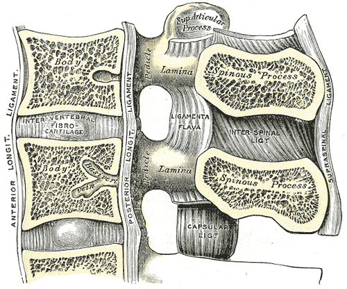
The basivertebral veins emerge from the foramina on the posterior surfaces of the vertebral bodies.
They are contained in large, tortuous channels in the substance of the bones, similar in every respect to those found in the diploë of the cranial bones.
They communicate through small openings on the front and sides of the bodies of the vertebræ with the anterior external vertebral plexuses, and converge behind to the principal canal, which is sometimes double toward its posterior part, and open by valved orifices into the transverse branches which unite the anterior internal vertebral plexuses.
They become greatly enlarged in advanced age.
Additional images
-
Gray301
