Asherman's syndrome other imaging findings: Difference between revisions
m (Robot: Automated text replacement (-mgibson@perfuse.org +charlesmichaelgibson@gmail.com & -kfeeney@perfuse.org +kfeeney@elon.edu)) |
No edit summary |
||
| Line 2: | Line 2: | ||
[[Image:HSG Ashermans syndrome.jpg|thumb|HSG view.]] | [[Image:HSG Ashermans syndrome.jpg|thumb|HSG view.]] | ||
{{Asherman's syndrome}} | {{Asherman's syndrome}} | ||
{{CMG}} '''Associate Editor-In-Chief: {{Skhan}}''' | |||
==Overview== | ==Overview== | ||
Saline sonography or hysterosalpingography may be used initially in the evaluation of Asherman's syndrome. Though hysteroscopy remains the gold standard. | |||
== Diagnosis == | == Diagnosis == | ||
Hysteroscopy is the gold standard for diagnosis <ref name="Valle">Valle RF, Sciarra JJ. Intrauterine adhesions: hysteroscopic diagnosis, classification, treatment, and reproductive outcome. Am J Obstet Gynecol 1988; 158:1459-1470.</ref>. Imaging by [[sonohysterography]] or [[hysterosalpingography]] will reveal the extent of the scar formation. Hormone studies show normal levels consistent with reproductive function. | Hysteroscopy is the gold standard for diagnosis <ref name="Valle">Valle RF, Sciarra JJ. Intrauterine adhesions: hysteroscopic diagnosis, classification, treatment, and reproductive outcome. Am J Obstet Gynecol 1988; 158:1459-1470.</ref>. Imaging by [[sonohysterography]] or [[hysterosalpingography]] will reveal the extent of the scar formation. Hormone studies show normal levels consistent with reproductive function. | ||
Revision as of 17:15, 1 August 2022

|
Asherman's syndrome Microchapters |
|
Diagnosis |
|---|
|
Treatment |
|
Case Studies |
|
Asherman's syndrome other imaging findings On the Web |
|
American Roentgen Ray Society Images of Asherman's syndrome other imaging findings |
|
Risk calculators and risk factors for Asherman's syndrome other imaging findings |
Editor-In-Chief: C. Michael Gibson, M.S., M.D. [1] Associate Editor-In-Chief: Saud Khan M.D.
Overview
Saline sonography or hysterosalpingography may be used initially in the evaluation of Asherman's syndrome. Though hysteroscopy remains the gold standard.
Diagnosis
Hysteroscopy is the gold standard for diagnosis [1]. Imaging by sonohysterography or hysterosalpingography will reveal the extent of the scar formation. Hormone studies show normal levels consistent with reproductive function.
Hysterosalpingography
(Images courtesy of RadsWiki)
-
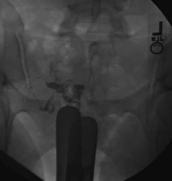
Hysterosalpingography: Asherman's syndrome
-
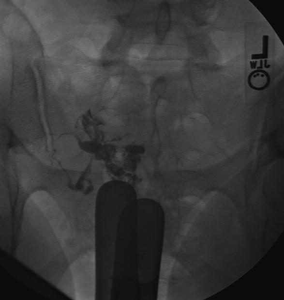
Hysterosalpingography: Asherman's syndrome
-
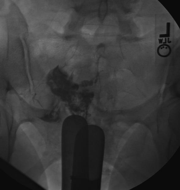
Hysterosalpingography: Asherman's syndrome
-
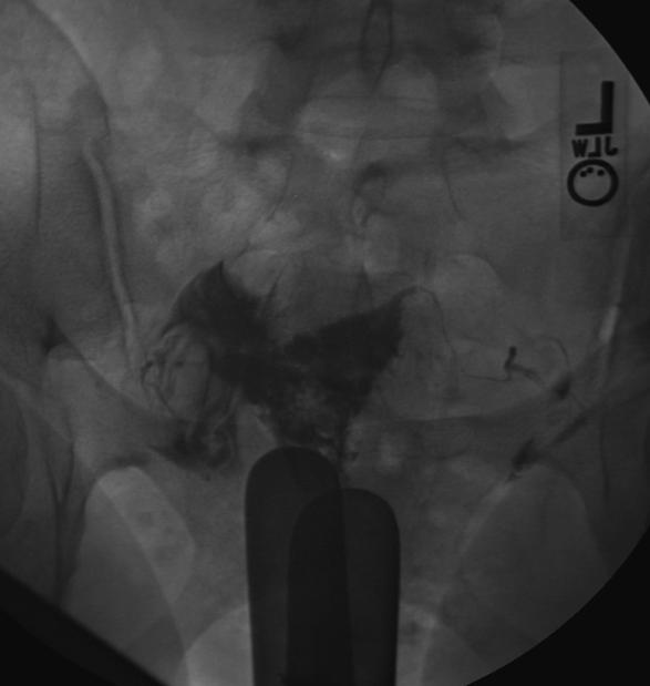
Hysterosalpingography: Asherman's syndrome


References
- ↑ Valle RF, Sciarra JJ. Intrauterine adhesions: hysteroscopic diagnosis, classification, treatment, and reproductive outcome. Am J Obstet Gynecol 1988; 158:1459-1470.