Ascaris infection: Difference between revisions
No edit summary |
No edit summary |
||
| Line 1: | Line 1: | ||
{{CMG}} | |||
{{SI}} | {{SI}} | ||
Revision as of 05:23, 7 January 2009
Editor-In-Chief: C. Michael Gibson, M.S., M.D. [1]
|
WikiDoc Resources for Ascaris infection |
|
Articles |
|---|
|
Most recent articles on Ascaris infection Most cited articles on Ascaris infection |
|
Media |
|
Powerpoint slides on Ascaris infection |
|
Evidence Based Medicine |
|
Cochrane Collaboration on Ascaris infection |
|
Clinical Trials |
|
Ongoing Trials on Ascaris infection at Clinical Trials.gov Trial results on Ascaris infection Clinical Trials on Ascaris infection at Google
|
|
Guidelines / Policies / Govt |
|
US National Guidelines Clearinghouse on Ascaris infection NICE Guidance on Ascaris infection
|
|
Books |
|
News |
|
Commentary |
|
Definitions |
|
Patient Resources / Community |
|
Patient resources on Ascaris infection Discussion groups on Ascaris infection Patient Handouts on Ascaris infection Directions to Hospitals Treating Ascaris infection Risk calculators and risk factors for Ascaris infection
|
|
Healthcare Provider Resources |
|
Causes & Risk Factors for Ascaris infection |
|
Continuing Medical Education (CME) |
|
International |
|
|
|
Business |
|
Experimental / Informatics |
Please Take Over This Page and Apply to be Editor-In-Chief for this topic: There can be one or more than one Editor-In-Chief. You may also apply to be an Associate Editor-In-Chief of one of the subtopics below. Please mail us [2] to indicate your interest in serving either as an Editor-In-Chief of the entire topic or as an Associate Editor-In-Chief for a subtopic. Please be sure to attach your CV and or biographical sketch.
Overview
Ascaris is a worm that lives in the small intestine. Infection with Ascaris is called ascariasis (ass-kuh-rye-uh-sis). Adult female worms can grow over 12 inches in length, adult males are smaller.
Related Key Words and Synonyms:
Intestinal roundworm infection, Ascariasis.
Epidemiology and Demographics
Ascariasis is the most common human worm infection. Infection occurs worldwide and is most common in tropical and subtropical areas where sanitation and hygiene are poor. Children are infected more often than adults. In the United States, infection is rare, but most common in rural areas of the southeast.
How can I get ascariasis?
You or your children can become infected after touching your mouth with your hands that have become contaminated with eggs from soil or other contaminated surfaces or by ingesting contaminated food or water.
Pathophysiology & Etiology
Etiologic agent:
Ascaris lumbricoides is the largest nematode (roundworm) parasitizing the human intestine. (Adult females: 20 to 35 cm; adult male: 15 to 30 cm.)
Life cycle:

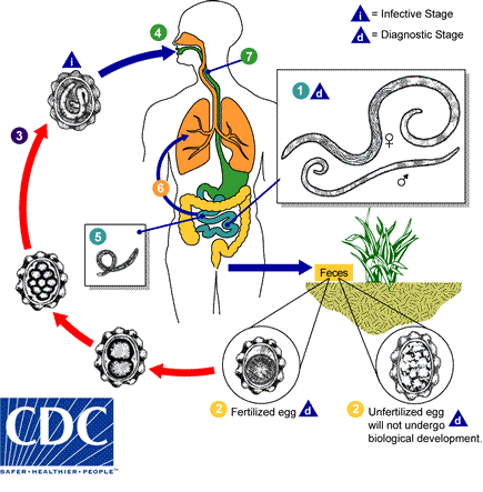
Adult worms 1 live in the lumen of the small intestine. A female may produce approximately 200,000 eggs per day, which are passed with the feces 2. Unfertilized eggs may be ingested but are not infective. Fertile eggs embryonate and become infective after 18 days to several weeks 3, depending on the environmental conditions (optimum: moist, warm, shaded soil). After infective eggs are swallowed 4, the larvae hatch 5 , invade the intestinal mucosa, and are carried via the portal, then systemic circulation to the lungs 6. The larvae mature further in the lungs (10 to 14 days), penetrate the alveolar walls, ascend the bronchial tree to the throat, and are swallowed 7. Upon reaching the small intestine, they develop into adult worms 1. Between 2 and 3 months are required from ingestion of the infective eggs to oviposition by the adult female. Adult worms can live 1 to 2 years.
Diagnosis
Your health care provider will ask you to provide stool samples for testing. Some people notice infection when a worm is passed in their stool or is coughed up. If this happens, bring in the worm specimen to your health care provider for diagnosis. There is no blood test used to diagnose an Ascaris infection.
History and Symptoms
Most people have no symptoms that are noticeable, but infection may cause slower growth and slower weight gain. If you are heavily infected, you may have abdominal pain. Sometimes, while the immature worms migrate through the lungs, you may cough and have difficulty breathing. If you have a very heavy worm infection, your intestines may become blocked.
Laboratory Findings
Microscopic identification of eggs in the stool is the most common method for diagnosing intestinal ascariasis. The recommended procedure is as follows:
- Collect a stool specimen.
- Fix the specimen in 10% formalin.
- Concentrate using the formalin–ethyl acetate sedimentation technique.
- Examine a wet mount of the sediment.
Where concentration procedures are not available, a direct wet mount examination of the specimen is adequate for detecting moderate to heavy infections. For quantitative assessments of infection, various methods such as the Kato-Katz can be used. Larvae can be identified in sputum or gastric aspirate during the pulmonary migration phase (examine formalin-fixed organisms for morphology). Adult worms are occasionally passed in the stool or through the mouth or nose and are recognizable by their macroscopic characteristics.
- Microscopy findings:
Below are several Ascaris eggs seen in wet mounts. Diagnostic characteristics:
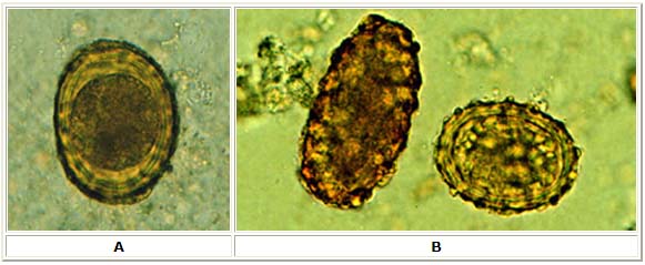
Fertilized eggs (A, B on the right, D, F, G, H) are rounded, have a thick shell, with an external mammillated layer that is often stained brown by bile. In some cases, the outer layer is absent (decorticated eggs: E, F on the right, G). Size: approximately 60 µm in diameter when spherical, and up to 75 µm when ovoid.
Unfertilized eggs (B on the left, C, E) are elongated and larger (up to 90 µm in length); their shell is thinner; and their mammillated layer is more variable, either with large protuberances (C) or practically none (E); these eggs contain mainly a mass of refractile granules.

A: Fertilized Ascaris egg, still at the unicellular stage. Eggs are normally at this stage when passed in the stool. Complete development of the larva requires 18 days under favorable conditions.
B: Unfertilized and fertilized eggs (left and right, respectively).

C: Unfertilized egg. Prominent mammillations of outer layer.
D: Fertilized egg. The embryo can be distinguished inside the egg.
E: Unfertilized egg with no outer mammillated layer (decorticated).

F: Three fertilized eggs (one decorticated, on the right) of Ascaris lumbricoides.

G, H: Two fertilized eggs from the same patient, where embryos have begun to develop (this happens when the stool sample is not processed for several days without refrigeration). The embryos in early stage of division (4 to 6 cells) can be clearly seen. Note that the egg in G has a very thin mammillated outer layer.

I: Egg containing a larva, which will be infective if ingested.
J: Larva hatching from an egg.
- Macroscopic findings:

K: An adult Ascaris worm. Diagnostic characteristics: tapered ends; length 15 to 35 cm (the females tend to be the larger ones). This worm is a female, as evidenced by the size and genital girdle (the dark circular groove at bottom area of image).
Treatment
In the United States, Ascaris infections are generally treated for 1-3 days with medication prescribed by your health care provider. The drugs are effective and appear to have few side effects. Your health care provider will likely request additional stool exams 1 to 2 weeks after therapy; if the infection is still present, treatment will be repeated.
I am pregnant and have just been diagnosed with ascariasis. Can I be treated?
Infection with Ascaris worms is generally light and is not considered an emergency. Unless your infection is heavy, and your health may be at risk, treatment is generally postponed until after delivery of the baby.
Pharmacotherapy
The drugs of choice for treatment of ascariasis are albendazole with mebendazole, ivermectin, and nitazoxanide as alternatives. In the United States, ascariasis is generally treated for 1-3 days with medication prescribed by a health care provider. The drugs are effective and appear to have few side effects.
Acknowledgements
The content on this page was first contributed by: C. Michael Gibson, M.S., M.D.