Aortic arch anomalies
| Aortic arch anomalies |
| Cardiology Network |
 Discuss Aortic arch anomalies further in the WikiDoc Cardiology Network |
| Adult Congenital |
|---|
| Biomarkers |
| Cardiac Rehabilitation |
| Congestive Heart Failure |
| CT Angiography |
| Echocardiography |
| Electrophysiology |
| Cardiology General |
| Genetics |
| Health Economics |
| Hypertension |
| Interventional Cardiology |
| MRI |
| Nuclear Cardiology |
| Peripheral Arterial Disease |
| Prevention |
| Public Policy |
| Pulmonary Embolism |
| Stable Angina |
| Valvular Heart Disease |
| Vascular Medicine |
Editor-In-Chief: C. Michael Gibson, M.S., M.D. [1]
Associate Editors-In-Chief: Cafer Zorkun, M.D., Ph.D. [2]; Keri Shafer, M.D. [3]
Please Join in Editing This Page and Apply to be an Editor-In-Chief for this topic: There can be one or more than one Editor-In-Chief. You may also apply to be an Associate Editor-In-Chief of one of the subtopics below. Please mail us [4] to indicate your interest in serving either as an Editor-In-Chief of the entire topic or as an Associate Editor-In-Chief for a subtopic. Please be sure to attach your CV and or biographical sketch.
Overview
Synonyms: vascular ring, aortic arch anomaly, double arch, isolated vascular ring, congenital aortic defect, double aortic arch, bovine arch
Pathophysiology & Etiology
Vascular rings encircle the trachea and esophagus, which results in variable degrees of compression of both structures. Compression of the trachea causes upper airway obstruction that impairs airflow. The extent of airway compression is variable. Double aortic arch is more often associated airway compression and is also associated with more severe airway compression than other forms of vascular ring.
Variations
There are three common variations to the branching pattern: [1]
- Normal (as below) - seen in ~ 70% of patients.
- Bovine arch - common origin of brachiocephalic and left common carotid artery - seen in approximately 15% of patients (more common in blacks)
- Left common carotid has its origin from the brachiocephalic artery proper, rather than from a common trunk - - seen in approximately 10% of patients (also more common in blacks)
There may be additional branches that arise directly from the arch. These include:
- Thyroidea ima artery, usually between the brachiocephalic and left common carotid
- Left vertebral artery, usually between the left common carotid and the left subclavian arteries.
- Rarely the right subclavian and right common carotid arise independently.
Double Aortic Arch
Double aortic arch has been classified as type 1 (both arches patent) and type 2 (one arch atretic). The atretic arch is almost invariably the left. Type 2 double arches may be subclassified according to the position of the atresia. Subtype 1 the atretic segment is between ductus arteriosus and descending aorta; subtype 2 it is between left subclavian artery and ductus arteriosus; subtype 3 it is between left common carotid artery and left subclavian artery; and subtype 4 the atresia is between ascending aorta and left common carotid artery. In subtype 3 it is common for the left subclavian artery to arise from a diverticulum. The diverticulum has the same embryologic origin as that originally described by Burckhard Kommerell, and may reasonably be referred to as a Kommerell’s diverticulum.
Double aortic arch is an important vascular ring anomaly, as the arches and atretic ligamentous cords surround and potentially compress the trachea and oesophagus. A clue as to the presence of double arch on barium swallow is the presence of focal oesophageal narrowing in both the AP and transverse directions.
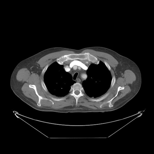
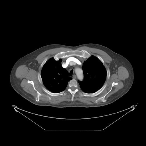
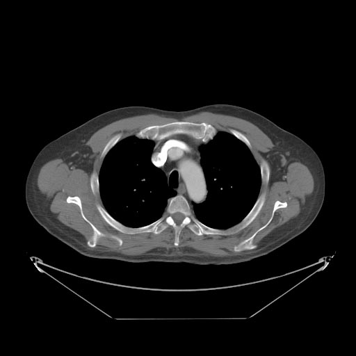
Images shown below are courtesy of Drs Ihor Kociumbas and Laughlin Dawes
-
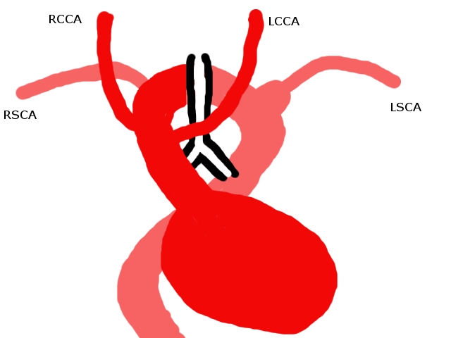
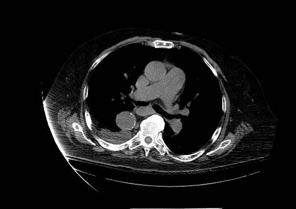
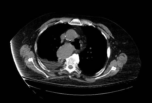
A CT chest was performed in the arterial phase. A diagram of the findings is seen. The left common carotid artery arose from the ascending aorta. The aortic arch was to the right of the trachea, and passed posterior to it. The right common carotid and right subclavian arteries arose from the arch in that order. From the posterior arch, the left subclavian artery arose from a large diverticulum (this corresponds to the left “aortic knuckle” seen on the PA film). The descending aorta then crossed to the right again before passing through the diaphragm in the midline. This appearance may be due to right aortic arch with aberrent left subclavian artery, or to double aortic arch with atresia of the left arch, subtype 3. This case is more likely to be the former, as there was no narrowing of the trachea, and subtype 3 double aortic arch is rare. The anatomic difference between these two anomalies is persistence or not of an atretic segment of the left arch.
-
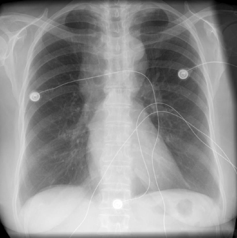
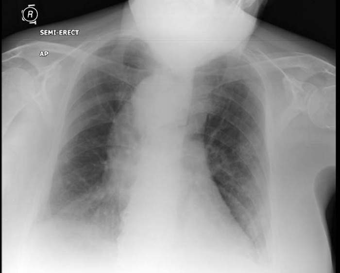
A female patient presented with noisy breathing. The PA chest x-ray shows an abnormal superior mediastinum and cardiac contour. There is a curved bulge to the right of trachea and above the right main bronchus. There is the suggestion of a rounded density to the right of the trachea at T4 level. There is what looks like a left aortic knuckle, but it is somewhat superiorly positioned. There is an abnormal sharp angle in the left mediastinal contour, in the region of the main pulmonary artery. The descending aorta shadow is absent on the left below this level, but there is a double shadow on the right.


Bovine Arch
The bovine arch, refers a common branching patten variant of the aortic arch, where the brachiocephalic artery (aka innominate) shares a common origin with the left common carotid artery.
In actuality it is a misnomer as the true bovine arch - i.e that found in cattle - comprises a common single brachiocephalic trunk which trifurcates into bilateral subclavian arteries and a single bicarotid trunk. This variation is very rare in humans.
(Images shown below are courtesy of radiopaedia.org)
-
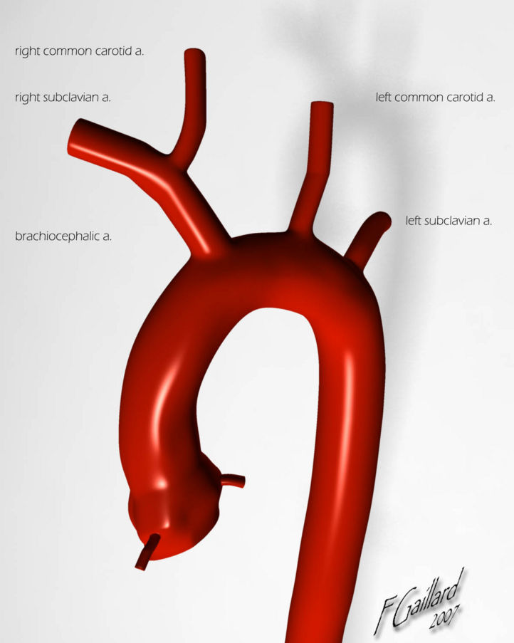
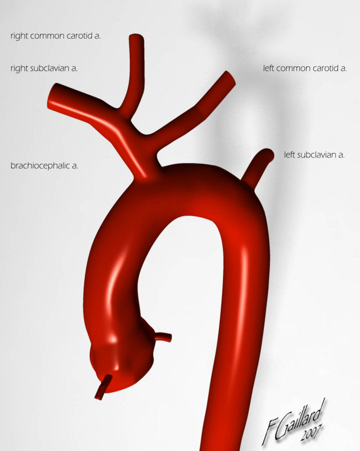
Normal anatomy
-
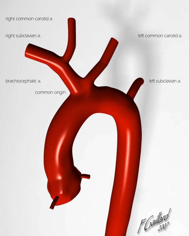
Aortic variations: Bovine arch


-
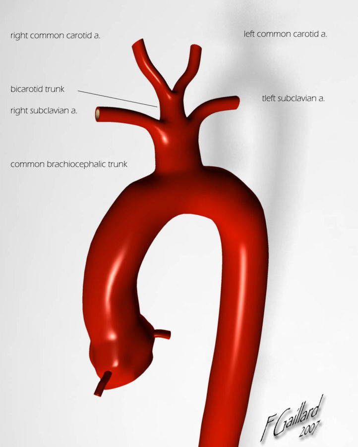
Aortic variations: True Bovine arch
-
Aortic variations: Left common carotid has its origin from brachiocephalic trunk


Genetics
Double aortic arch is associated with a chromosome band 22q11 deletion in approximately 20% of patients. Double aortic arch may be associated with band 22q11 deletion, which has various other possible manifestations. These include, but are not limited to, palatal abnormalities, laryngotracheal anomalies, speech and learning delay, characteristic facial features, hypocalcemia, abnormalities of T-cell–mediated immune function, and neurologic defects.
Natural History and Associated Anomalies
Aortic arch anomolies may be associated with the following:
- Association with VACTERL: (vertebral, anal, cardiac, tracheal, esophageal, renal, and limb) or CHARGE (posterior coloboma, heart defect, choanal atresia, retardation, genital, and ear) associations.
- Esophageal atresia
- Congenital laryngeal web
Diagnosis
Symptoms
In addition to dyspnea, patients may also experience dysphagia due to esophageal compression. Early in life, this may manifest itself as nausea and vomiting and feeding intolerance. Later in life dysphagia is the predominant symptom. Esophageal dysfunction and difficulty swallowing may in turn lead to aspiration. Patients may have both respiratory or esophageal symptoms, but the respiratory symptoms are usually more pervasive in younger patients.
Magnetic Resonance Angiography (MRA)
-
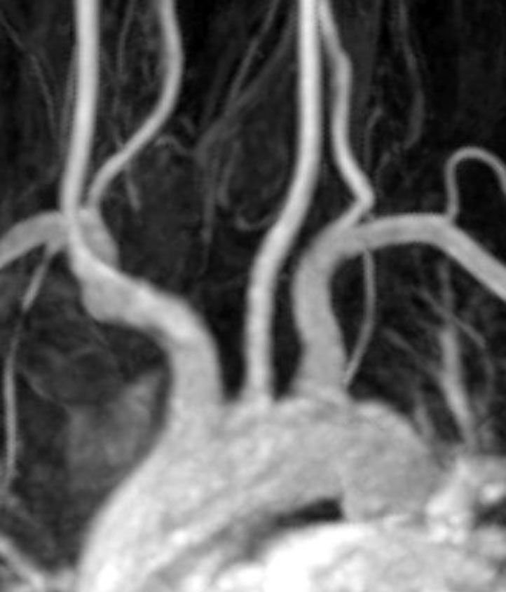
Normal aortic arch (image courtesy of radiopaedia.org)

Multi Slice CT
Images shown below are courtesy of RadsWiki
-
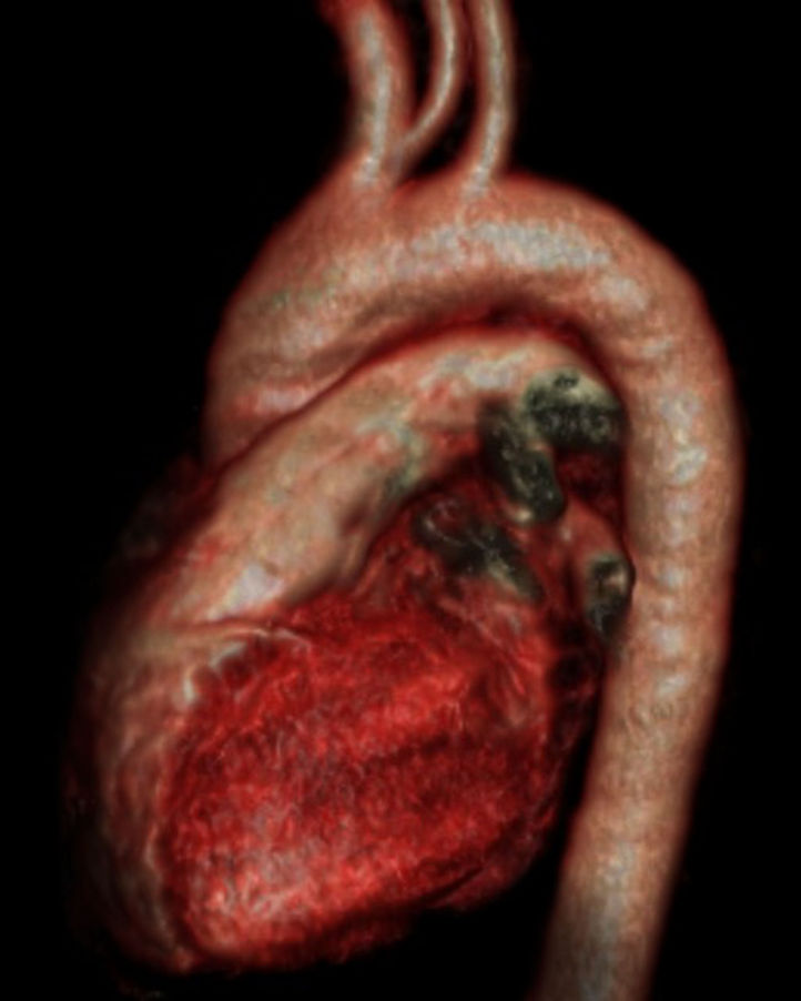
Bovine arch (image courtesy of radiopaedia.org)
-

MSCT: Bovine arch


-

MSCT: Bovine arch
-
MSCT: Bovine arch


-
MSCT: Bovine arch
-
MSCT: Bovine arch


Aortography
Images shown below are courtesy of RadsWiki
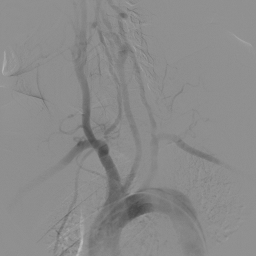
Still aortographic images:
-
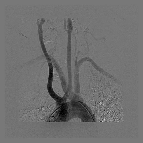
Subclavian Bovine Carotid
-
Bovine Aortic Arch


Right Aortic Arch
Images shown below are courtesy of RadsWiki
-
Right Aortic Arch
-
Right Aortic Arch
-
Right Aortic Arch


Interrupted Aortic Arch
Aortic arch interruption is a rare congenital anomaly and commonly causes death in the neonatal period. Composed of triad of:
- Interrupted aortic arch
- VSD
- PDA (Pulmonary artery supplies lower part of the body).
Locations:
- Type A: distal to left SCA (42%).
- Type B: between left CCA and SCA (53%) associated with DiGeorge syndrome.
- Type C: between innominate and left CCA.
Diagnosis
-
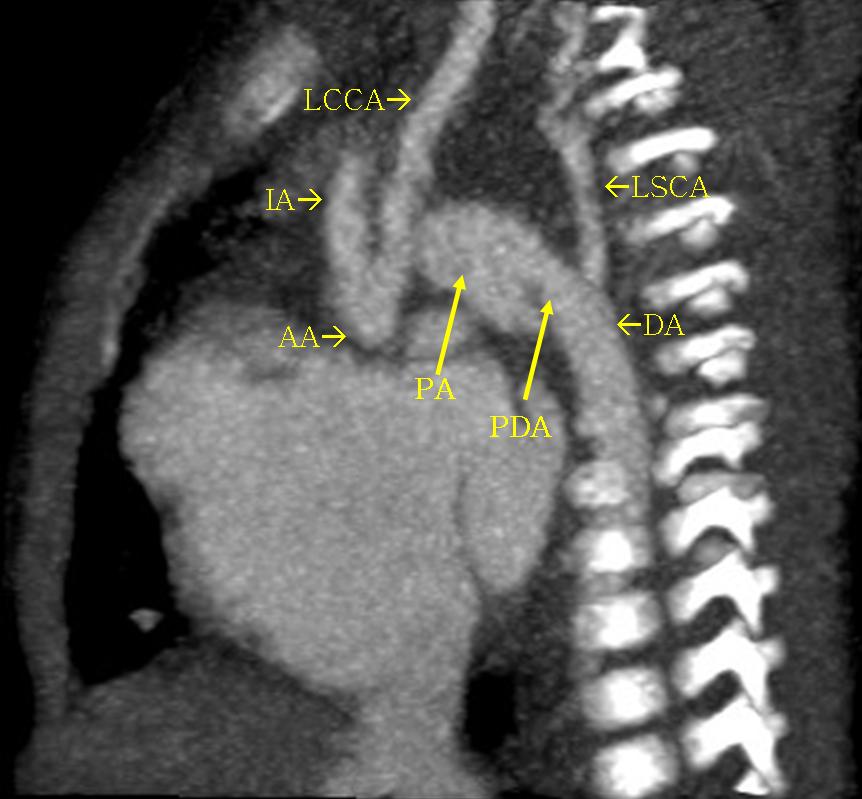
20-days old male presented with heart failure. The arch is interrupted between left CCA and SCA. The pulmonary artery is markedly dilated and connected to descending aorta via large PDA giving the appearance of a low aortic arch. The PA is much larger than ascending aorta. Other associated anomalies in this case included VSD and large sinus venosus ASD.(Image courtesy of Dr Ahmed Haroun)

Prognosis:
76% mortality in 1st month.
References
- ↑ K.F. Layton "Bovine Aortic Arch Variant in Humans: Clarification of a Common Misnomer" AJNR 27: August 2006, 1541-42
Additional Reading
- Moss and Adams' Heart Disease in Infants, Children, and Adolescents Hugh D. Allen, Arthur J. Moss, David J. Driscoll, Forrest H. Adams, Timothy F. Feltes, Robert E. Shaddy, 2007 ISBN 0781786843
- Hurst's the Heart, Fuster V, 12th ed. 2008, ISBN 978-0-07-149928-6
- Willerson JT, Cardiovascular Medicine, 3rd ed., 2007, ISBN 978-1-84628-188-4
External Links
Acknowledgements
The content on this page was first contributed by: C. Michael Gibson, M.S., M.D.
List of contributors: