Anterior jugular vein
| Cardiology Network |
 Discuss Anterior jugular vein further in the WikiDoc Cardiology Network |
| Adult Congenital |
|---|
| Biomarkers |
| Cardiac Rehabilitation |
| Congestive Heart Failure |
| CT Angiography |
| Echocardiography |
| Electrophysiology |
| Cardiology General |
| Genetics |
| Health Economics |
| Hypertension |
| Interventional Cardiology |
| MRI |
| Nuclear Cardiology |
| Peripheral Arterial Disease |
| Prevention |
| Public Policy |
| Pulmonary Embolism |
| Stable Angina |
| Valvular Heart Disease |
| Vascular Medicine |
Editor-In-Chief: C. Michael Gibson, M.S., M.D. [1]
The anterior jugular vein begins near the hyoid bone by the confluence of several superficial veins from the submaxillary region.
It descends between the median line and the anterior border of the Sternocleidomastoideus, and, at the lower part of the neck, passes beneath that muscle to open into the termination of the external jugular, or, in some instances, into the subclavian vein.
It varies considerably in size, bearing usually an inverse proportion to the external jugular; most frequently there are two anterior jugulars, a right and left; but sometimes only one.
Its tributaries are some laryngeal veins, and occasionally a small thyroid vein.
Just above the sternum the two anterior jugular veins communicate by a transverse trunk, the venous jugular arch, which receive tributaries from the inferior thyroid veins; each also communicates with the internal jugular.
There are no valves in this vein.
Additional images
-
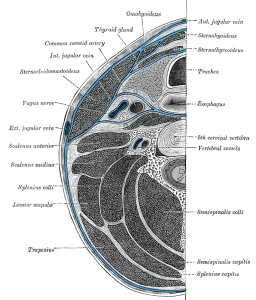
Section of the neck at about the level of the sixth cervical vertebra.
