Allergy chest x ray

Jump to navigation Jump to search
The printable version is no longer supported and may have rendering errors. Please update your browser bookmarks and please use the default browser print function instead.

Allergy Microchapters

Home

Patient Information

Overview

Historical Perspective

Pathophysiology

Causes

Differentiating Allergies from other Diseases

Epidemiology and Demographics

Risk Factors

Screening

Natural History, Complications and Prognosis

Diagnosis

History and Symptoms

Physical Examination

Laboratory Findings

Other Diagnostic Studies

Treatment

Medical Therapy

Primary Prevention

Secondary Prevention

Cost-Effectiveness of Therapy

Future or Investigational Therapies

Case Studies

Case #1

Allergy chest x ray On the Web

Most recent articles

Most cited articles

Review articles

CME Programs

Powerpoint slides

Images

American Roentgen Ray Society Images of Allergy chest x ray

All Images
X-rays
Echo & Ultrasound
CT Images
MRI

Ongoing Trials at Clinical Trials.gov

US National Guidelines Clearinghouse

NICE Guidance

FDA on Allergy chest x ray

CDC on Allergy chest x ray

Allergy chest x ray in the news

Blogs on Allergy chest x ray

Directions to Hospitals Treating Allergies

Risk calculators and risk factors for Allergy chest x ray

Allergic bronchopulmonary aspergillosis
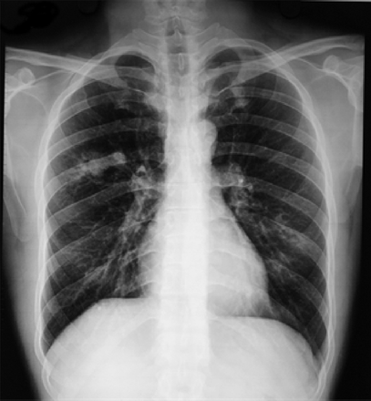
Chest radiograph showing a non-homogeneous opacity in the right mid zone with perihilar patchy infiltrates in the left mid and lower zones. Transient pulmonary infiltrates or fleeting shadows that are characteristic of allergic bronchopulmonary aspergillosis are visible[1].

Hypersensitivity pneumonitis

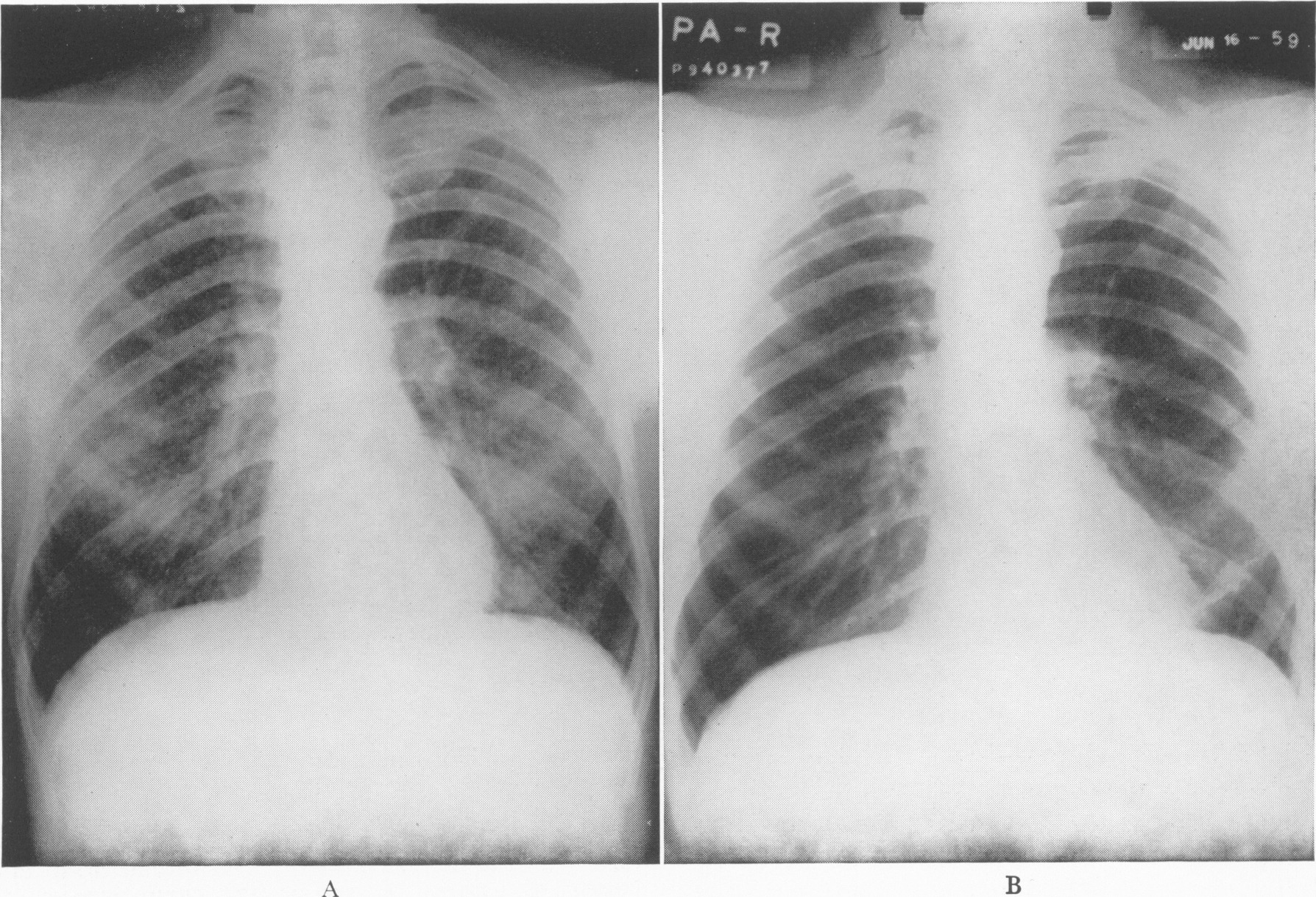
Roentgenograms of the Chest in Case 1 Taken on February 18, Three Weeks after the Onset of Symptoms (A), Showing a Diffuse Reticulonodular Infiltrate, and in June (B), Demonstrating Complete Clearing of the Infiltrate[2].

Eosinophilic Pneumonia

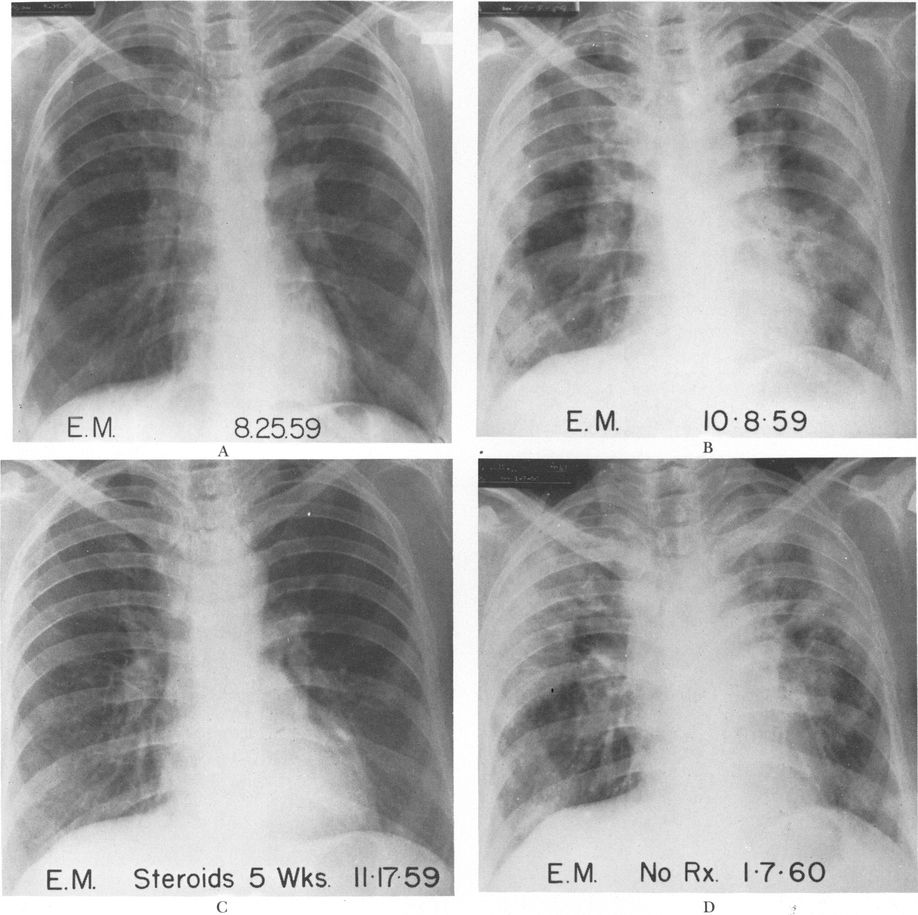
On this admission she was acutely ill, with dyspnea at rest. Occasional inspiratory rales were present over both apexes. No wheezes were heard. The chest roentgenogram showed peculiar bilateral densities, which appeared peripherally and suggested small pockets of pleural fluid (Fig. 7A)[3]


References

  1. Shah A, Panjabi C (2014). "Allergic aspergillosis of the respiratory tract". Eur Respir Rev. 23 (131): 8–29. doi:10.1183/09059180.00007413. PMC 9487274 Check |pmc= value (help). PMID 24591658.
  2. BALDUS WP, PETER JB (1960). "Farmer's lung: report of two cases". N Engl J Med. 262: 700–5. doi:10.1056/NEJM196004072621403. PMID 13796151.
  3. Carrington CB, Addington WW, Goff AM, Madoff IM, Marks A, Schwaber JR; et al. (1969). "Chronic eosinophilic pneumonia". N Engl J Med. 280 (15): 787–98. doi:10.1056/NEJM196904102801501. PMID 5773637.


Template:WikiDoc Sources