Adenoma pathophysiology
Editor-In-Chief: C. Michael Gibson, M.S., M.D. [1]; Associate Editor(s)-in-Chief: Shivali Marketkar, M.B.B.S. [2]
|
Adenoma Microchapters |
|
Diagnosis |
|---|
|
Treatment |
|
Case Studies |
|
Adenoma pathophysiology On the Web |
|
American Roentgen Ray Society Images of Adenoma pathophysiology |
|
Risk calculators and risk factors for Adenoma pathophysiology |
Pathophysiology
Adenoma is a benign epithelial tumor arising in epithelium of mucosa (stomach, small intestine and bowel), glands (endocrine and exocrine) and ducts. In hollow organs (digestive tract) the adenoma grows upwards into the lumen - adenomatous polyp (or polypoid adenoma).
Depending on the type of the insertion base, adenoma may be pedunculated lobular head with a long slender stalk, covered by normal mucosa or sessile (broad base).
The adenomatous proliferation is characterized by different degrees of cell dysplasia (atypia or loss of normal differentiation of epithelium) irregular cells with hyperchromatic nuclei, (pseudo)stratified nuclei, nucleolus, decreased mucosecretion and mitosis. The architecture may be tubular, villous or tubulo-villous. Basement membrane and muscularis mucosae are intact.
Locations
Colon
Adenomas of the colon are quite prevalent. They are found commonly at colonoscopy. They are removed because of their tendency to become malignant and lead to colon cancer.
Renal
This is a tumor which is most often small and asymptomatic and its derived from renal tubules. It may be a precursor lesion to renal carcinoma.
Adrenal
Adrenal adenomas are common, 1 in 10 people have them malignant and asymptomatic. They are often found on of the abdomen, usually not as the focus of investigation; they are usually incidental findings. About one in 10,000 is malignant. Thus, a biopsy is rarely called for, especially if the lesion is homogeneous and smaller than 3 centimeters. Follow-up images in three to six months can confirm the stability of the growth.
While some adrenal adenomas do not secrete hormones at all , often some secrete cortisol causing Cushing's syndrome, aldosterone causing Conn's syndrome or androgens causing hyperandrogenism.
Thyroid
About one in 10 people are found to have solitary thyroid nodules. Investigation is required because a small percentage of these are malignant. Biopsy usually confirms the growth to be an adenoma, but sometimes, excision at surgery is required, especially when the cells found at biopsy are of the follicular type.
Pituitary
Pituitary adenomas are commonly seen in 10% of the neurological patients. A lot of them remain undiagnosed. Treatment is usually surgical, to which patients generally respond well. The most common subtype, prolactinoma, is seen more often in women, and is frequently diagnosed during pregnancy as the hormone progesterone increases its growth. Medical therapy bromocriptine generally suppresses prolactinomas; progesterone antagonist therapy has not proven to be successful.
Liver
Hepatocellular adenoma, Hepatic adenomas are a rare benign tumour of the liver, which may present with hepatomegaly or other symptoms.
Breast
Breast adenomas are called fibroadenomas. They are often very small and difficult to detect. Often there are no symptoms. Treatments can include a needle biopsy, and/or removal.
Appendix
Adenomas can also appear in the appendix. The condition is extremely rare and most physicians will never encounter an actual case, but they do happen. The most common version is called cystadenoma. They are usually discovered in the course of examination of the tissue following an appendectomy. If the appendix has ruptured and a tumor is present this presents challenges, especially if malignant cells have formed and thus spread to the abdomen. In 1995 Former Vice President Dan Quayle was found to have cystadenoma in the appendix.
Gross Pathology
-
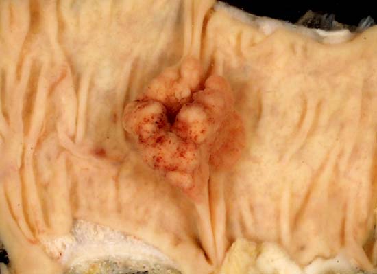
Adenomatous polyp of the colon. Courtesy of Ed Uthman, MD
-
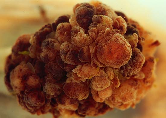
Tubulovillous polyp of the colon. Courtesy of Ed Uthman, MD
-
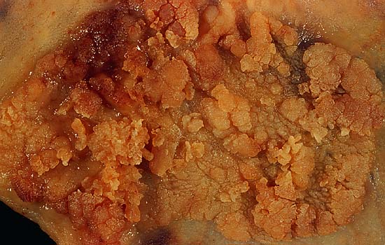
Villous adenoma of the colon. Courtesy of Ed Uthman, MD


Microscopic Pathology
Shown below is a micrograph of a colorectal tubular adenoma with high grade dysplasia.(H&E stain)
The lesional tissue, i.e. dysplastic epithelium, is seen on the left of the image and characterized by:
- Nuclear hyperchromatism (i.e. dark purple nuclei) - that extends to the surface,
- Nuclear crowding (i.e. nuclei are bunched-up),
- Elliptical/cigar-shaped nuclei and loss of goblet cells/reduction in the number of goblet cells.
Normal colonic type epithelium is seen on the right of the image and characterized by small round nuclei and abundant goblet cells.
Video
Shown below is a video describing microscopic features of a tubular adenoma. {{#ev:youtube|XGyjOw6jePY}}