Acoustic neuroma pathophysiology: Difference between revisions
No edit summary |
|||
| Line 5: | Line 5: | ||
==Overview== | ==Overview== | ||
Acoustic neuroma arises from [[Schwann cells]], which are the cells | Acoustic neuroma arises from [[Schwann cells]], which are the [[Cell (biology)|cells]] involved in the conduction of [[Nervous system|nervous]] impulses along [[axons]], [[nerve]] development and [[Nerve regeneration|regeneration]]. On [[microscopic]] [[histopathological]] analysis, acoustic neuroma may display two types of growth patterns: Antoni type A and Antoni type B. Antoni type A growth pattern is composed of elongated [[Cell (biology)|cells]] with [[Cytoplasm|cytoplasmic]] processes arranged in [[Fascicle|fascicles]], little [[stromal]] [[matrix]] and verocay bodies. Antoni type B growth pattern is composed of loose meshwork of [[Cell (biology)|cells]], less dense [[Cell (biology)|cellular]] [[matrix]], microcysts and myxoid change. | ||
==Pathophysiology== | ==Pathophysiology== | ||
Revision as of 19:34, 19 April 2019
|
Acoustic neuroma Microchapters | |
|
Diagnosis | |
|---|---|
|
Treatment | |
|
Case Studies | |
|
Acoustic neuroma pathophysiology On the Web | |
|
American Roentgen Ray Society Images of Acoustic neuroma pathophysiology | |
|
Risk calculators and risk factors for Acoustic neuroma pathophysiology | |
Editor-In-Chief: C. Michael Gibson, M.S., M.D. [1]Associate Editor(s)-in-Chief: Simrat Sarai, M.D. [2] Mohsen Basiri M.D.
Overview
Acoustic neuroma arises from Schwann cells, which are the cells involved in the conduction of nervous impulses along axons, nerve development and regeneration. On microscopic histopathological analysis, acoustic neuroma may display two types of growth patterns: Antoni type A and Antoni type B. Antoni type A growth pattern is composed of elongated cells with cytoplasmic processes arranged in fascicles, little stromal matrix and verocay bodies. Antoni type B growth pattern is composed of loose meshwork of cells, less dense cellular matrix, microcysts and myxoid change.
Pathophysiology
- Acoustic neuromas are benign tumors (WHO grade 1), which usually arise from the intracanalicular segment of the vestibular portion of the vestibulocochlear nerve (CN VIII), near the transition point between glial and Schwann cells (Obersteiner-Redlich zone).
- An acoustic neuroma arises from a type of cell known as the Schwann cell.
- These cells form an insulating layer over all nerves of the peripheral nervous system (i.e., nerves outside of the central nervous system) including the eighth cranial nerve.
- The eighth cranial nerve is separated into two branches:
- The cochlear branch, which transmits sound to the brain
- The vestibular branch, which transmits balance information to the brain
- Most acoustic neuromas occur on the vestibular portion of the eighth cranial nerve.
- As these tumors are made up of Schwann cells, and usually occur on the vestibular portion of the eighth cranial nerve, many physicians prefer the use of the term vestibular schwannoma. However, the term acoustic neuroma is still used more often in the medical literature.[1]
- Acoustic neuromas are well circumscribed encapsulated masses, which unlike neuromas, arise from but are separate from nerve fibers, which they usually splay and displace rather than incorporated.dsfsdfsdf
Genetic
- One the most knowable causes of acoustic neuroma is Neurofibromatosis type 2 (NF2).
- Neurofibromatosis Type 2 is an autosomal dominant disease caused by loss of function mutations.
- Approximately 50% of reported NF2 cases represent new mutations for which no other affected family member can be identified.
- NF2 gene is on chromosome 22q12.2 that encodes a 595–amino acid protein named “moesin- ezrin-radixin–like protein,” otherwise known as “merlin” or “schwannomin.”[2]
- Merlin protein linked with other proteins in cell and are involved in linking cytoskeletal components with the plasma membrane and are located in actin-rich surface projections such as microvilli, membrane surfaces, and cell contact regions.
- Dephosphorylated Merlin proteins are active and roll in the normal cell growth, phosphorylated Merlin ( synthesized due to a mutation in NF2 gene that causes to produce a truncated Merlin protein) inactivated and can not play normal roll in cell growth and causes increased cell growth.[3][4]
Associated Conditions
- Childhood exposure to radiation of the head and neck may be associated with acoustic neuroma.[5][6]
- Exposure to high-dose ionizing radiation is the only definite environmental risk factor associated with an increased risk of developing an acoustic neuroma.[7][8]
- A concomitant history of having had a parathyroid adenoma may have an increased risk of developing vestibular schwannoma.[9]
Microscopic Pathology
- In 1920, Nils Ragnar Euge`ne Antoni (1887–1968), a Swedish neurologist and researcher described 2 distinct patterns of cellular architecture in the peripheral nerve sheath tumors, based his observations on analysis of 30 cases and described a “fibrillary, intensely polar, elongated appearing tissue type” which he called “tissue type A.” [3]
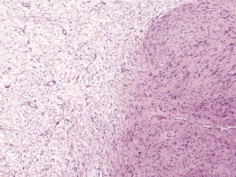
- These highly cellular regions were eventually referred to as Antoni A regions by later authors. [3]
- Antoni also described seemingly distinct loose microcystic tissue adjacent to the Antoni A regions, and these came to be known as Antoni B regions.[3]
They can display two types of growth patterns:[3]
- Antoni A
- Elongated cells with cytoplasmic processes arranged in fascicles
- Little stromal matrix
- Verocay bodies: nuclear free zones of processes lying between regions of nuclear palisading
- Antoni B
- Loose meshwork of cells
- Less densely cellular
- Microcysts and myxoid change
- Photomicrograph of Antoni A tissue and Antoni B tissue within a schwannoma. The highly cellular Antoni A region on the right of the field is contrasted with the loosely organized hypocellular Antoni B region on left of the field (hematoxylin-eosin, original magnification 400).

References
- ↑ Acoustic Schwannoma. Radiopedia(2015) http://radiopaedia.org/articles/acoustic-schwannoma Accessed on October 2 2015
- ↑ Michael E. Sughrue, Andrea H. Yeung, Martin J. Rutkowski, Steven W. Cheung & Andrew T. Parsa (2011). "Molecular biology of familial and sporadic vestibular schwannomas: implications for novel therapeutics". Journal of neurosurgery. 114 (2): 359–366. doi:10.3171/2009.10.JNS091135. PMID 19943731. Unknown parameter
|month=ignored (help) - ↑ 3.0 3.1 3.2 3.3 3.4 Wippold II, F.J (2007). "Neuropathology for the Neuroradiologist: Antoni A and Antoni B Tissue Patterns". AJNR Am J Neuroradiol.
- ↑ SUGHRUE, MICHAEL E. (2011). "Molecular biology of familial and sporadic vestibular schwannomas: implications for novel therapeutics". J Neurosurg. 114.
- ↑ Arthur B. Schneider, Elaine Ron, Jay Lubin, Marilyn Stovall, Eileen Shore-Freedman, Jocelyn Tolentino & Barbara J. Collins (2008). "Acoustic neuromas following childhood radiation treatment for benign conditions of the head and neck". Neuro-oncology. 10 (1): 73–78. doi:10.1215/15228517-2007-047. PMID 18079359. Unknown parameter
|month=ignored (help) - ↑ E. Shore-Freedman, C. Abrahams, W. Recant & A. B. Schneider (1983). "Neurilemomas and salivary gland tumors of the head and neck following childhood irradiation". Cancer. 51 (12): 2159–2163. PMID 6850504. Unknown parameter
|month=ignored (help) - ↑ Oyebode Taiwo, Deron Galusha, Baylah Tessier-Sherman, Sharon Kirsche, Linda Cantley, Martin D. Slade, Mark R. Cullen & A. Michael Donoghue (2014). "Acoustic neuroma: potential risk factors and audiometric surveillance in the aluminium industry". Occupational and environmental medicine. 71 (9): 624–628. doi:10.1136/oemed-2014-102094. PMID 25015928. Unknown parameter
|month=ignored (help) - ↑ Mantao Chen, Zuoxu Fan, Xiujue Zheng, Fei Cao & Liang Wang (2016). "Risk Factors of Acoustic Neuroma: Systematic Review and Meta-Analysis". Yonsei medical journal. 57 (3): 776–783. doi:10.3349/ymj.2016.57.3.776. PMID 26996581. Unknown parameter
|month=ignored (help) - ↑ L. Magnus Backlund, Dan Grander, Lena Brandt, Per Hall & Anders Ekbom (2005). "Parathyroid adenoma and primary CNS tumors". International journal of cancer. 113 (6): 866–869. doi:10.1002/ijc.20743. PMID 15515018. Unknown parameter
|month=ignored (help)