Ascariasis laboratory tests
|
Ascariasis Microchapters |
|
Diagnosis |
|---|
|
Treatment |
|
Case Studies |
|
Ascariasis laboratory tests On the Web |
|
American Roentgen Ray Society Images of Ascariasis laboratory tests |
|
Risk calculators and risk factors for Ascariasis laboratory tests |
Editor-In-Chief: C. Michael Gibson, M.S., M.D. [1]; Associate Editor-In-Chief: Imtiaz Ahmed Wani, M.B.B.S
Diagnostic Findings
Microscopic identification of eggs in the stool is the most common method for diagnosing intestinal ascariasis. The recommended procedure is as follows:
- Collect a stool specimen.
- Fix the specimen in 10% formalin.
- Concentrate using the formalin–ethyl acetate sedimentation technique.
- Examine a wet mount of the sediment.
Where concentration procedures are not available, a direct wet mount examination of the specimen is adequate for detecting moderate to heavy infections. For quantitative assessments of infection, various methods such as the Kato-Katz can be used. Larvae can be identified in sputum or gastric aspirate during the pulmonary migration phase (examine formalin-fixed organisms for morphology). Adult worms are occasionally passed in the stool or through the mouth or nose and are recognizable by their macroscopic characteristics.
Macroscopic Findings
(Images courtesy of Dr. Imtiaz Ahmed Wani)
-

Enterotomy for worm removal
-
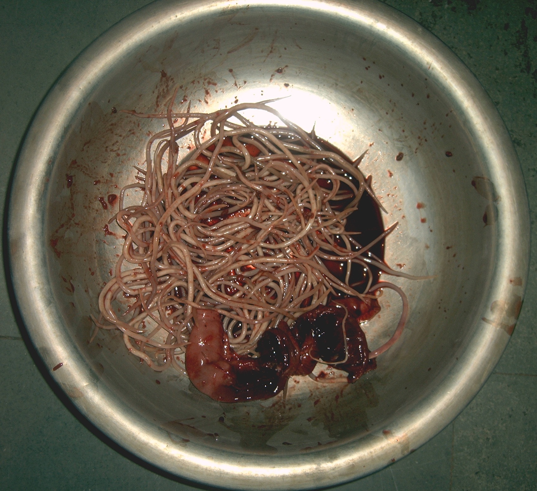
Ascaris lumbricoides caused gangrene of ileum (shown worms removed from a child)


Microscopic Findings
Below are several Ascaris eggs seen in wet mounts. Diagnostic characteristics:
Fertilized eggs (A, B on the right, D, F, G, H) are rounded, have a thick shell, with an external mammillated layer that is often stained brown by bile. In some cases, the outer layer is absent (decorticated eggs:E, F on the right, G). Size: approximately 60 µm in diameter when spherical, and up to 75 µm when ovoid.
Unfertilized eggs (B on the left, C, E) are elongated and larger (up to 90 µm in length); their shell is thinner; and their mammillated layer is more variable, either with large protuberances (C) or practically none (E); these eggs contain mainly a mass of refractile granules.

A: Fertilized Ascaris egg, still at the unicellular stage. Eggs are normally at this stage when passed in the stool. Complete development of the larva requires 18 days under favorable conditions.
B: Unfertilized and fertilized eggs (left and right, respectively).

C: Unfertilized egg. Prominent mammillations of outer layer.
D: Fertilized egg. The embryo can be distinguished inside the egg.
E: Unfertilized egg with no outer mammillated layer (decorticated).

F: Three fertilized eggs (one decorticated, on the right) of Ascaris lumbricoides.

G, H: Two fertilized eggs from the same patient, where embryos have begun to develop (this happens when the stool sample is not processed for several days without refrigeration). The embryos in early stage of division (4 to 6 cells) can be clearly seen. Note that the egg in G has a very thin mammillated outer layer.

I: Egg containing a larva, which will be infective if ingested.
J: Larva hatching from an egg.
References
de:Spulwurm hu:Orsóférgek io:Askaridiko id:Askariasis it:Ascaridiasi nl:Spoelworm ps:اسکاريس لومبريکويډېس sk:Hlísta detská