Basal cell carcinoma pathophysiology
|
Basal cell carcinoma Microchapters |
|
Diagnosis |
|---|
|
Case Studies |
|
Basal cell carcinoma pathophysiology On the Web |
|
American Roentgen Ray Society Images of Basal cell carcinoma pathophysiology |
|
Risk calculators and risk factors for Basal cell carcinoma pathophysiology |
Editor-In-Chief: C. Michael Gibson, M.S., M.D. [1];Associate Editor(s)-in-Chief: Shivali Marketkar, M.B.B.S. [2]
Overview
Basal cell carcinomas develop in the basal cell layer of the skin. Cumulative DNA damage caused by chronic sunlight exposure results in DNA mutations that predispose to the development of basal cell carcinoma. On gross pathology, basal cell carcinoma lesions may demonstrate pearly pink nodules with telangiectasias, rolled borders, and central crusting with or without an ulcerating lesion (rodent ulcer). On microscopic analysis, peripheral palisading nuclei are characteristic.
Pathophysiology
Genetics
- A number of aberrations involving the sonic hedgehog signaling pathway(SHH) are noted[1][2]
- The majority of mutations in sporadic basal cell carcinoma and basal cell nevus syndrome(BCNS) patients occur in PTCH1 gene, a protein that inhibits Smoothened gene(SMO)
- The second most common mutation in sporadic basal cell carcinoma and basal cell nevus syndrome(BCNS) patients are gain-of-function mutations of the Smoothened gene(SMO)
- Loss of PTCH1 results in the failure of Smoothened inhibition, subsecuently leading to increases in GLI1 levels, changes in transcription, and subsequent tumorigenesis
Enviromental Exposure
- Basal cell carcinomas develop in the basal cell layer of the skin
- Cumulative DNA damage caused by chronic sunlight exposure results in DNA mutations that predispose to the development of basal cell carcinoma
- While DNA repair eliminates most UV-induced damage, not all cross-links are excised, which eventually results in mutations
- Apart from the mutagenesis, sunlight depresses the local immune system, possibly decreasing immune surveillance for new tumor cells
Gross Pathology

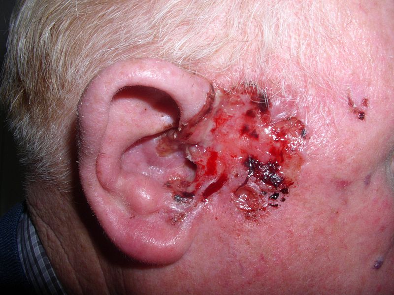
The image above depicts basal cell carcinoma in a 75 year old male. The following features are characteristic on gross pathology:
- Pearly pink nodules
- Telangiectasias
- Rolled borders
- Central crusting
- Ulcerating lesion (rodent ulcer)
Microscopic Pathology
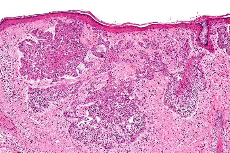
Shown below is a classic micrograph of basal cell carcinoma(H&E stain).The following features are characteristic:
- Peripheral palisading nuclei
- Myxoid stroma
- Artefactual clefting

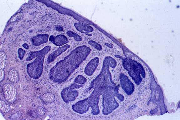
Shown below is the image of nodular variant of Basal cell carcinoma

Video
{{#ev:youtube|JnJXrFnvOKs}}
References
- ↑ Mohan SV, Chang AL (2014). "Advanced Basal Cell Carcinoma: Epidemiology and Therapeutic Innovations". Curr Dermatol Rep. 3: 40–45. doi:10.1007/s13671-014-0069-y. PMC 3931971. PMID 24587976.
- ↑ Pellegrini C, Maturo MG, Di Nardo L, Ciciarelli V, Gutiérrez García-Rodrigo C, Fargnoli MC (November 2017). "Understanding the Molecular Genetics of Basal Cell Carcinoma". Int J Mol Sci. 18 (11). doi:10.3390/ijms18112485. PMC 5713451. PMID 29165358.