Rhinitis pathophysiology
|
Rhinitis Microchapters |
|
Diagnosis |
|---|
|
Treatment |
|
Case Studies |
|
Rhinitis pathophysiology On the Web |
|
American Roentgen Ray Society Images of Rhinitis pathophysiology |
|
Risk calculators and risk factors for Rhinitis pathophysiology |
Editor-In-Chief: C. Michael Gibson, M.S., M.D. [1] Associate Editor(s)-in-Chief: Fatimo Biobaku M.B.B.S [2]
Overview
Pathophysiology
Clinically relevant anatomy and physiology of the nose[1][2]

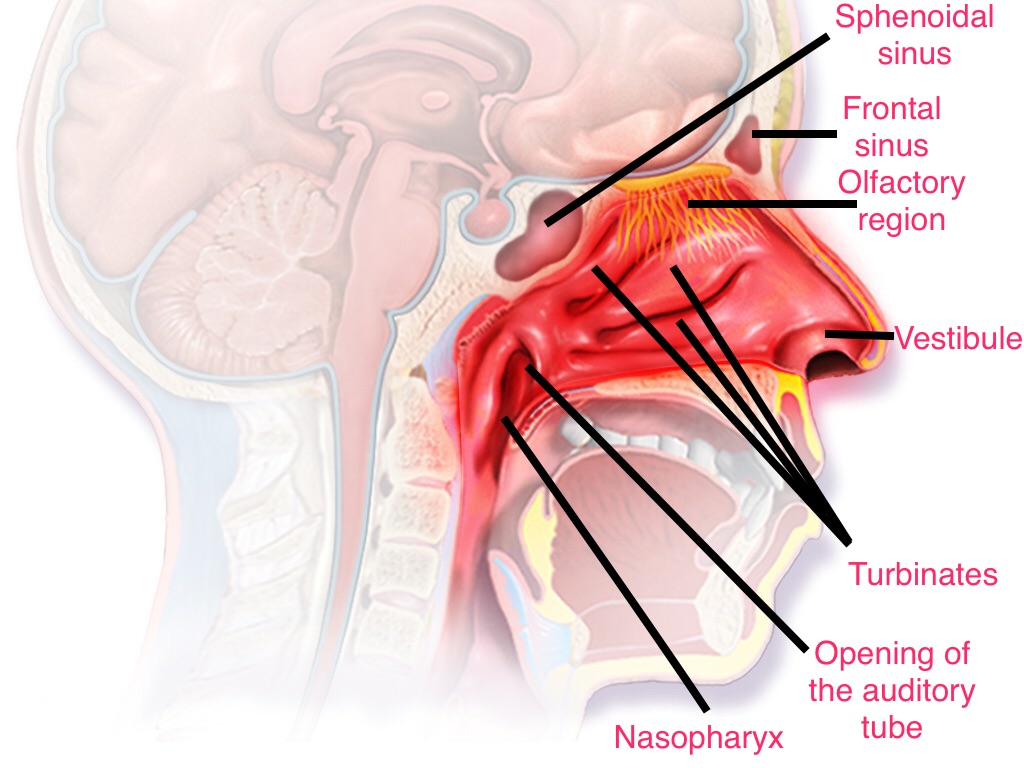
The human nose- It is both a respiratory and an olfactory organ. The nose is a highly vascular organ, the nasal blood vessels receive parasympathetic innervation and dense sympathetic innervation. Parasympathetic nerve stimulation promotes secretion from nasal airway glands and nasal congestion while sympathetic nerve stimulation cause a reduction in nasal blood flow, and significant nasal decongestion. The nasal cavity is divided into right and left halves by the nasal septum. The nasal cavity extends from the vestibule to the nasopharynx, and it is generally divided into three parts namely:
- The vestibule- the area which surrounds the external opening to the nasal cavity.
- The olfactory region- located at the apex of the nasal cavity, it is lined by olfactory cells.
- The respiratory region- this is the largest part of the nasal cavity. The turbinates/conchae project from the lateral wall of the nasal cavity, and they promote air filtration, humidification and temperature regulation.The respiratory region is lined by pseudostratified columnar epithelial cells(about 80% of these cells are ciliated). Interspersed within the epithelium are mucus-secreting goblet cells which are necessary for the maintenance of mucociliary clearance. Factors such as dryness and temperature significantly affect the ciliary function of epithelial cells. Ciliary action stops after 8-10mins at 50% relative humidity of inspired air, and after 3-5mins at 30% relative humidity of inspired air. Ciliary activity ceases at temperatures between 7-12”C. Significant impairment of ciliary function can also occur due to factors such as environmental exposure to large amounts of wood dust and chromium vapors,tobacco smoke, inhaled gases, locally applied drugs, infection. Ciliary structure changes has been noted in patients with longstanding allergic rhinitis.
The paranasal sinuses- The paranasal sinuses drain into the nasal cavity. The nose and sinuses are contiguous structures, and they share vascular, neuronal and interconnecting anatomic pathways.
Pathophisiology of Allergic Rhinitis[3][4]
Overview
Allergic rhinitis is a multifactorial disease, its development is influenced by an interplay of genetic and environmental factors. Aeroallergens in the nasal tissue undergo antigen processing, eliciting allergen-specific allergic responses and also promoting the development of allergic airway disease. Proteins, glycoproteins and rarely, glycans in indoor and outdoor inhalant allergens such as dust mite fecal particles, cockroach residues, animal danders, molds, and pollens are common aeroallergens causing allergic rhinitis. It is usually an IgE mediated disease with varying degrees of nasal inflammation.
Pathophysiology
The inherent enzymatic proteolytic activity of aeroallergens promote their access to antigen presenting cells by cleaving tight junctions in the airway epithelium and also via activation of receptors on epithelial cells. These activated epithelial cells produce proinflammatory mediators (such as cytokines, chemokines, thymic stromal lymphopoietin) which interact with subepithelial and interepithelial dendritic cells, skewing T-cell development and adaptive allergic sensitization. Antigen presenting cells (dendritic cells expressing CD1a and CD11c and macrophages) process nasal allergens in the epithelial mucosa of the nose. These antigen presenting cells then present the allergenic peptides by MHC class II molecules to T-cell receptors on resting CD4+ T lymphocytes in regional lymph nodes. Costimulatory signals result in the proliferation of allergen-stimulated Tcells into TH2-biased cells which release interleukins(IL-3, IL-4, IL-5, IL-13) and other cytokines. These cytokines lead to a cascade of events which promote B-cell isotype switching with subsequent local and systemic allergen-specific IgE antibody production by plasma cells. Allergen-specific IgE antibodies then become fixed to the high affinity receptor for IgE on the membranes of mast cells and basophils. These aggregation of receptor-bound IgE molecules, on exposure to specific allergen, results in the production of mediators (histamine, leukotrienes and others) that produce the allergic response, resulting in itching, sneezing, rhinorrhoea and blockage in the nose. Mediators and cytokines released during the early response also act on postcapillary endothelial cells, promoting the expression of adhesion molecules(such as intercellular adhesion molecule 1, E-selectin, and vascular cell adhesion molecule 1). These adhesion molecules promote the adherence of circulating leukocytes, such as eosinophils, to endothelial cells. Factors with chemoattractant properties( such as IL-5 for eosinophils) also promote the infiltration of the superficial lamina propria of the mucosa with many eosinophils, some neutrophils and basophils, and eventually CD4+ (TH2) lymphocytes and macrophages. These cells become activated and release more mediators, which in turn activate many of the proinflammatory reactions seen in the immediate response.
The role of IgE-mediated reaction in rhinitis and asthma have been further confirmed by the effect of an anti-IgE monoclonal antibody in these diseases. Studies of cells infiltrating the nasal mucosa during the pollen season show increment in the amount of various inflammatory cells which correlates with the severity of symptoms and nasal nonspecific hyperactivity. Eosinophils are almost always found in the mucosa between nondesquamated epithelial cells, in the submucosa and in nasal secretions. Degranulated mast cells are present in increased numbers in the epithelium and the submucosa. CD4+ T-cells are increased in number during the pollen season and there is an increase in Langerhan-like cells (CD1+) during the pollen season in allergic patients.
Pathophysiology of Nonallergic Rhinitis and other Rhinitis Syndromes
overview
Nonallergic rhinitis is a heterogenous group of conditions which is poorly defined and understood.[5]
References
- ↑ Jones N (2001). "The nose and paranasal sinuses physiology and anatomy". Adv Drug Deliv Rev. 51 (1–3): 5–19. PMID 11516776.
- ↑ Watelet JB, Van Cauwenberge P (1999). "Applied anatomy and physiology of the nose and paranasal sinuses". Allergy. 54 Suppl 57: 14–25. PMID 10565476.
- ↑ Bousquet J, Khaltaev N, Cruz AA, Denburg J, Fokkens WJ, Togias A; et al. (2008). "Allergic Rhinitis and its Impact on Asthma (ARIA) 2008 update (in collaboration with the World Health Organization, GA(2)LEN and AllerGen)". Allergy. 63 Suppl 86: 8–160. doi:10.1111/j.1398-9995.2007.01620.x. PMID 18331513 PMID: 18331513 Check
|pmid=value (help). - ↑ Dykewicz MS, Hamilos DL (2010). "Rhinitis and sinusitis". J Allergy Clin Immunol. 125 (2 Suppl 2): S103–15. doi:10.1016/j.jaci.2009.12.989. PMID 20176255.
- ↑ Sin B, Togias A (2011). "Pathophysiology of allergic and nonallergic rhinitis". Proc Am Thorac Soc. 8 (1): 106–14. doi:10.1513/pats.201008-057RN. PMID 21364228.