Pulmonary amyloidosis: Difference between revisions
No edit summary |
No edit summary |
||
| Line 1: | Line 1: | ||
__NOTOC__ | __NOTOC__ | ||
{{ | {{Pulmonary amyloidosis}} | ||
{{CMG}}; '''Associate Editor-In-Chief:''' {{CZ}} | {{CMG}}; '''Associate Editor-In-Chief:''' {{CZ}} | ||
Revision as of 02:45, 25 September 2012
|
Pulmonary amyloidosis Microchapters |
|
Diagnosis |
|---|
|
Treatment |
|
Case Studies |
|
Pulmonary amyloidosis On the Web |
|
American Roentgen Ray Society Images of Pulmonary amyloidosis |
Editor-In-Chief: C. Michael Gibson, M.S., M.D. [1]; Associate Editor-In-Chief: Cafer Zorkun, M.D., Ph.D. [2]
Overview
- Amyloidosis limited to the respiratory tract is uncommon.
- Pulmonary amyloidosis occurs in three histopathologic types
- Focal deposits in the tracheobronchial tree
- Diffuse parenchymal opacities
- Single or multiple pulmonary nodules.
- Nodular pulmonary amyloidosis is usually found incidentally on chest nadiognaphs in asymptomatic, older adults.
- The deposits originate in the muscular walls of small blood vessels, and as they enlarge, they spread into the interstitial tissues.
Diagnosis
Chest x-ray
- Nodular pulmonary amyloidosis
- Nodules of pulmonary amyloidosis are usually 1-4 cm in size
- They may enlarge, calcify, ossify, or cavitate.
Images shown below are courtesy of RadsWiki and copylefted


CT
- Nodular pulmonary amyloidosis
- Nodules of pulmonary amyloidosis are usually 1-4 cm in size
- They may enlarge, calcify, ossify, or cavitate.
Images shown below are courtesy of RadsWiki and copylefted


Histopathological Images
-
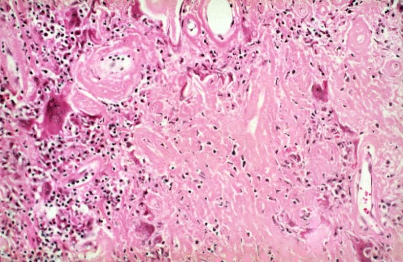
NODULAR PARENCHYMAL AMYLOIDOSIS: Lower Respiratory Tract. The amyloid consists of solid masses and bands of amorphous, eosinophilic, extracellular material. A multinucleated giant cell reaction is present, a typical finding in pulmonary amyloidosis.
-
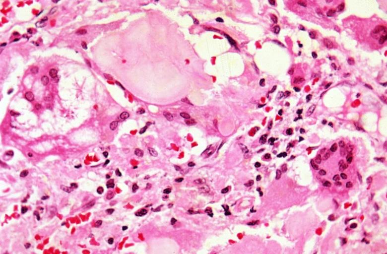
PULMONARY AMYLOIDOSIS. Lower Respiratory Tract. Multinucleated giant cells are closely applied to the periphery of the islands of amyloid. There are also numbers of lymphocytes and plasma cells present.

