Breast cancer classification: Difference between revisions
No edit summary |
|||
| Line 4: | Line 4: | ||
==Overview== | ==Overview== | ||
==Classification Based on Histopathology== | |||
===Malignant Tumors=== | |||
= | {| style="border: 0px; font-size: 90%; margin: 3px; width: 800px" | ||
* | |valign=top| | ||
* | |+ | ||
*[[ | ! style="background: #4479BA; width: 150px;" | {{fontcolor|#FFF|Type}} | ||
* | ! style="background: #4479BA; width: 300px;" | {{fontcolor|#FFF|Subtype}} | ||
|- | |||
| style="padding: 5px 5px; background: #DCDCDC; font-weight: bold" | | |||
'''Adenocarcinoma'''<ref name = class> Breast Neoplasm. Radiopedia. (2015) http://radiopaedia.org/articles/breast-neoplasms Accessed on January 16, 2016</ref> | |||
|- | |||
| style="padding: 5px 5px; background: #DCDCDC; font-weight: bold" | | |||
'''Ductal''' | |||
| style="padding: 5px 5px; background: #F5F5F5;" | | |||
*Ductal carcinoma in situ (DCIS) | |||
:*comedo type: ~60% of DCIS | |||
:*non comedo type: ~40% of DCIS | |||
::*papillary | |||
::*micropapillary | |||
::*cribriform | |||
::*solid | |||
:*intracystic papillary carcinoma in situ | |||
*'''invasive ductal carcinoma''' | |||
:*invasive ductal carcinoma not otherwise specified (NOS): ~65% | |||
:*tubular carcinoma of breast: ~7-8% | |||
::*tubulolobular carcinoma of breast | |||
:*medullary carcinoma of breast (MCB): ~2% | |||
:*mucinous (colloid) carcinoma: ~2% | |||
:*malignant papillary lesions of the breast | |||
::*papillary carcinoma of breast: 1-2% 1 | |||
|- | |||
| style="padding: 5px 5px; background: #DCDCDC; font-weight: bold" | | |||
'''Lobular''' | |||
| style="padding: 5px 5px; background: #F5F5F5;" | | |||
*lobular carcinoma in situ (LCIS) | |||
*invasive lobular carcinoma: ~10% | |||
|- | |||
| style="padding: 5px 5px; background: #DCDCDC; font-weight: bold" | | |||
'''Other malignant breast tumors''' | |||
| style="padding: 5px 5px; background: #F5F5F5;" | | |||
*inflammatory carcinoma of breast: 1-4% | |||
*Paget disease of the nipple | |||
*triple negative and basal-like breast cancers | |||
*metaplastic carcinoma of the breast | |||
*adenoid cystic carcinoma of the breast: <0.4% | |||
*apocrine carcinoma of the breast | |||
|- | |||
| style="padding: 5px 5px; background: #DCDCDC; font-weight: bold" | | |||
'''Sarcoma''' | |||
| style="padding: 5px 5px; background: #F5F5F5;" | | |||
*angiosarcoma of the breast | |||
*fibrosarcoma of breast | |||
*extra-skeletal osteosarcoma of breast | |||
*malignant phyllodes tumor | |||
*angiosarcoma | |||
*rhabdomyosarcoma | |||
|- | |||
| style="padding: 5px 5px; background: #DCDCDC; font-weight: bold" | | |||
'''Lymphoma''' | |||
| style="padding: 5px 5px; background: #F5F5F5;" | | |||
*Non-hodgkin lymphoma | |||
|- | |||
| style="padding: 5px 5px; background: #DCDCDC; font-weight: bold" | | |||
'''Metastases to the breast''' | |||
| style="padding: 5px 5px; background: #F5F5F5;" | | |||
The most common extra-mammary cancers that metastasise to breast are: | |||
*[[lymphoma]]/[[leukaemia]]: most common extra mammary source | |||
*[[melanoma]] | |||
*[[sarcomas]] | |||
*[[prostate cancer]]: considered on the most frequent primary sites in men 4 | |||
*[[lung cancer]] | |||
*[[gastric cancer]] | |||
*[[ovarian cancer]] | |||
*[[renal cell cancer]] | |||
|- | |||
|} | |||
===Benign Tumors=== | |||
*Phyllodes tumor<ref name = ben> Breast Neoplasms. Radiopaedia (2015) http://radiopaedia.org/articles/breast-neoplasms Accessed on january 16, 2016 </ref> | |||
*mammary fibromatosis: 0.2% of all breast tumours 5 | |||
*benign papillary lesions of the breast | |||
:*papilloma | |||
::*Intraductal papilloma | |||
::*solitary papilloma of breast | |||
::*central solitary papilloma of breast | |||
::*peripheral solitary papilloma of breast | |||
::*multiple papillomata of breast | |||
:::*juvenile papillomatosis of breast | |||
*granular cell tumour of the breast | |||
==Staging== | ==Staging== | ||
The breast cancer stage is based on the results of testing that is done on the tumor and lymph nodes removed during surgery and other tests.<ref name = stage> Breast Cancer. National Cancer Institute (2016) http://www.cancer.gov/types/breast/patient/breast-treatment-pdq#link/_148 Accessed on January 16, 2016 </ref> | |||
The breast cancer stage is based on the results of testing that is done on the tumor and lymph nodes removed during surgery and other tests. | |||
===Stage 0=== | ===Stage 0=== | ||
There are 3 types of breast carcinoma in situ: | There are 3 types of breast carcinoma in situ: | ||
| Line 27: | Line 102: | ||
*Paget disease of the nipple is a condition in which abnormal cells are found in the nipple only. | *Paget disease of the nipple is a condition in which abnormal cells are found in the nipple only. | ||
===Stage 1=== | ===Stage 1=== | ||
In stage I, cancer has formed. Stage I is divided into stages IA and IB. | In stage I, cancer has formed. Stage I is divided into stages IA and IB. | ||
*In stage IA, the tumor is 2 centimeters or smaller. Cancer has not spread outside the breast. | *In stage IA, the tumor is 2 centimeters or smaller. Cancer has not spread outside the breast. | ||
*In stage IB, small clusters of breast cancer cells (larger than 0.2 millimeter but not larger than 2 millimeters) are found in the lymph nodes and either: | *In stage IB, small clusters of breast cancer cells (larger than 0.2 millimeter but not larger than 2 millimeters) are found in the lymph nodes and either: | ||
:*no tumor is found in the breast; or | :*no tumor is found in the breast; or | ||
:*the tumor is 2 centimeters or smaller. | :*the tumor is 2 centimeters or smaller. | ||
===Stage II=== | ===Stage II=== | ||
Stage II is divided into stages IIA and IIB. | Stage II is divided into stages IIA and IIB. | ||
| Line 96: | Line 170: | ||
[[Metastasis|'''M'''etastases]] - There are two metastatic classification values (M0 or M1) which depend on the presence or absence of breast cancer cells in locations other than the breast and lymph nodes (so-called distant metastases, e.g. to bone, brain, lung). | [[Metastasis|'''M'''etastases]] - There are two metastatic classification values (M0 or M1) which depend on the presence or absence of breast cancer cells in locations other than the breast and lymph nodes (so-called distant metastases, e.g. to bone, brain, lung). | ||
==Gallery== | ==Gallery== | ||
Revision as of 00:33, 17 January 2016
|
Breast Cancer Microchapters |
|
Diagnosis |
|---|
|
Treatment |
|
Case Studies |
|
Breast cancer classification On the Web |
|
American Roentgen Ray Society Images of Breast cancer classification |
|
Risk calculators and risk factors for Breast cancer classification |
Editor-In-Chief: C. Michael Gibson, M.S., M.D. [1], Assistant Editor(s)-In-Chief: Jack Khouri, Mirdula Sharma, MBBS [2]
Overview
Classification Based on Histopathology
Malignant Tumors
| Type | Subtype |
|---|---|
|
Adenocarcinoma[1] | |
|
Ductal |
|
|
Lobular |
|
|
Other malignant breast tumors |
|
|
Sarcoma |
|
|
Lymphoma |
|
|
Metastases to the breast |
The most common extra-mammary cancers that metastasise to breast are:
|
Benign Tumors
- Phyllodes tumor[2]
- mammary fibromatosis: 0.2% of all breast tumours 5
- benign papillary lesions of the breast
- papilloma
- Intraductal papilloma
- solitary papilloma of breast
- central solitary papilloma of breast
- peripheral solitary papilloma of breast
- multiple papillomata of breast
- juvenile papillomatosis of breast
- granular cell tumour of the breast
Staging
The breast cancer stage is based on the results of testing that is done on the tumor and lymph nodes removed during surgery and other tests.[3]
Stage 0
There are 3 types of breast carcinoma in situ:
- Ductal carcinoma in situ (DCIS) is a noninvasive condition in which abnormal cells are found in the lining of a breast duct. The abnormal cells have not spread outside the duct to other tissues in the breast. In some cases, DCIS may become invasive cancer and spread to other tissues. At this time, there is no way to know which lesions could become invasive.
- Lobular carcinoma in situ (LCIS) is a condition in which abnormal cells are found in the lobules of the breast. This condition seldom becomes invasive cancer.
- Paget disease of the nipple is a condition in which abnormal cells are found in the nipple only.
Stage 1
In stage I, cancer has formed. Stage I is divided into stages IA and IB.
- In stage IA, the tumor is 2 centimeters or smaller. Cancer has not spread outside the breast.
- In stage IB, small clusters of breast cancer cells (larger than 0.2 millimeter but not larger than 2 millimeters) are found in the lymph nodes and either:
- no tumor is found in the breast; or
- the tumor is 2 centimeters or smaller.
Stage II
Stage II is divided into stages IIA and IIB.
- In stage IIA:
- no tumor is found in the breast or the tumor is 2 centimeters or smaller. Cancer (larger than 2 millimeters) is found in 1 to 3 axillary lymph nodes or in the lymph nodes near the breastbone (found during a sentinel lymph node biopsy); or
- the tumor is larger than 2 centimeters but not larger than 5 centimeters. Cancer has not spread to the lymph nodes.
- In stage IIB, the tumor is:
- larger than 2 centimeters but not larger than 5 centimeters. Small clusters of breast cancer cells (larger than 0.2 millimeter but not larger than 2 millimeters) are found in the lymph nodes; or
- larger than 2 centimeters but not larger than 5 centimeters. Cancer has spread to 1 to 3 axillary lymph nodes or to the lymph nodes near the breastbone (found during a sentinel lymph node biopsy); or
- larger than 5 centimeters. Cancer has not spread to the lymph nodes.
Stage III
Stage III is divided into IIIA, IIIB, and IIIC.
- Stage IIIA
- no tumor is found in the breast or the tumor may be any size. Cancer is found in 4 to 9 axillary lymph nodes or in the lymph nodes near the breastbone (found during imaging tests or a physical exam); or
- the tumor is larger than 5 centimeters. Small clusters of breast cancer cells (larger than 0.2 millimeter but not larger than 2 millimeters) are found in the lymph nodes; or
- the tumor is larger than 5 centimeters. Cancer has spread to 1 to 3 axillary lymph nodes or to the lymph nodes near the breastbone (found during a sentinel lymph node biopsy).
- Stage IIIB
- the tumor may be any size and cancer has spread to the chest wall and/or to the skin of the breast and caused swelling or an ulcer.
- cancer may have spread to:
- up to 9 axillary lymph nodes; or
- the lymph nodes near the breastbone
- Stage IIIC
- no tumor is found in the breast or the tumor may be any size. Cancer may have spread to the skin of the breast and caused swelling or an ulcer and/or has spread to the chest wall.
- cancer has spread to:
- 10 or more axillary lymph nodes; or
- lymph nodes above or below the collarbone; or
- axillary lymph nodes and lymph nodes near the breastbone
- For treatment, stage IIIC breast cancer is divided into operable and inoperable stage IIIC.
Stage IV
- In stage IV, cancer has spread to other organs of the body, most often the bones, lungs, liver, or brain.
Inflammatory Breast Cancer
- In inflammatory breast cancer, cancer has spread to the skin of the breast and the breast looks red and swollen and feels warm. The redness and warmth occur because the cancer cells block the lymph vessels in the skin. The skin of the breast may also show the dimpled appearance called peau d’orange (like the skin of an orange). There may not be any lumps in the breast that can be felt.
- Inflammatory breast cancer may be stage IIIB, stage IIIC, or stage IV.
Tumor - There are five tumor classification values (Tis, T1, T2, T3 or T4) which depend on the presence or absence of invasive cancer, the dimensions of the invasive cancer, and the presence or absence of invasion outside of the breast (e.g. to the skin of the breast, to the muscle, or to the rib cage underneath):
- Tx - Primary tumor cannot be assessed
- T0 - No evidence of primary tumor
- Tis - Carcinoma in situ
- Tis(DCIS) - Intraductal Carcinoma in situ
- Tis(LCIS) - Lobular Carcinoma in situ
- Tis(Paget's) - Paget's disease of the nipple with no tumor
- T1 - Tumor 2cm or less in its greatest dimension
- T1mic - Microinvasion 0.1cm or less in greatest dimension
- T1a - Tumor more than 0.1cm but not more than 0.5cm in its greatest dimension
- T1b - Tumor more than 0.5cm but not more than 1.0cm in its greatest dimension
- T1c - Tumor more than 1.0cm but not more than 2.0cm in its greatest dimension
- T2 - Tumor more than 2.0cm but not more than 5.0cm in its greatest dimension
- T3 - Tumor more than 5cm in its greatest dimension
- T4 - Tumor of any size with direct extension to (a) chest wall or (b) skin as described below:
- T4a - Extension to chest wall
- T4b - Edema (including peau d'orange) or ulceration of the breast skin, or satellite skin nodules confined to the same breast
- T4c - Both T4a and T4b
- T4d - Inflammatory breast cancer
Lymph Node - There are four lymph node classification values (N0, N1, N2 or N3) which depend on the number, size, and location of breast cancer cell deposits in lymph nodes.
- Nx - regional lymph nodes cannot be assessed, perhaps due to previous removal
- N0 - no regional lymph node metastasis
- N1 - metastasis to movable regional axillary lymph nodes on the same side as the affected breast
- N2 - metastasis to fixed regional axillary lymph nodes, or metastasis to the internal mammary lymph nodes, on the same side as the affected breast
- N3 - metastasis to supraclavicular lymph nodes or infraclavicular lymph nodes or metastasis to the internal mammary lymph nodes with metastasis to the axillary lymph nodes
Metastases - There are two metastatic classification values (M0 or M1) which depend on the presence or absence of breast cancer cells in locations other than the breast and lymph nodes (so-called distant metastases, e.g. to bone, brain, lung).
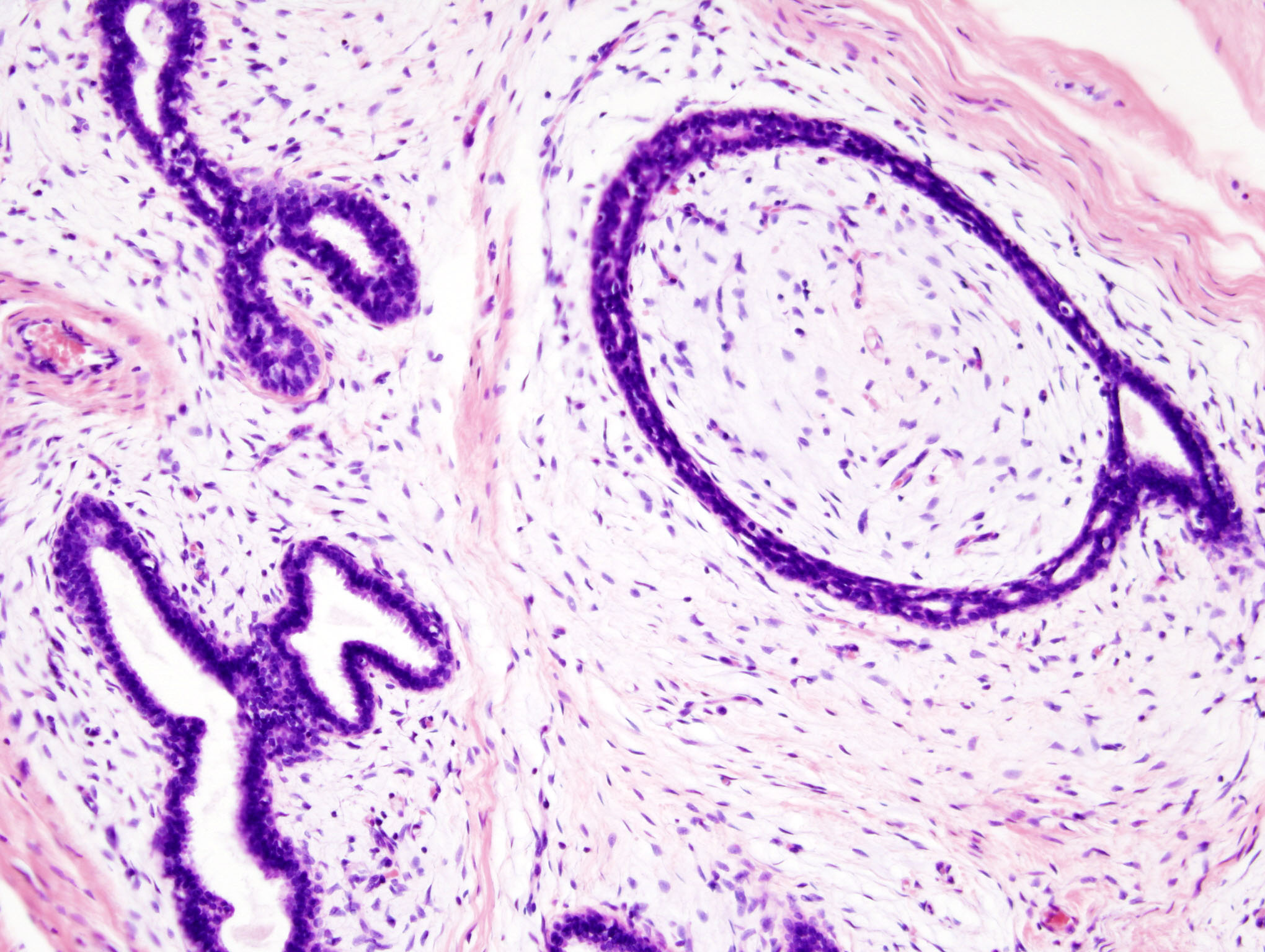
Gallery
-
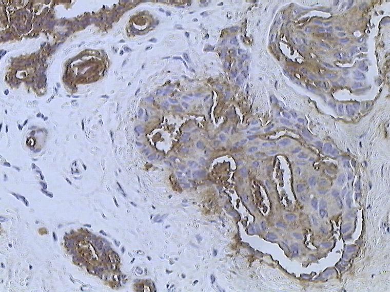
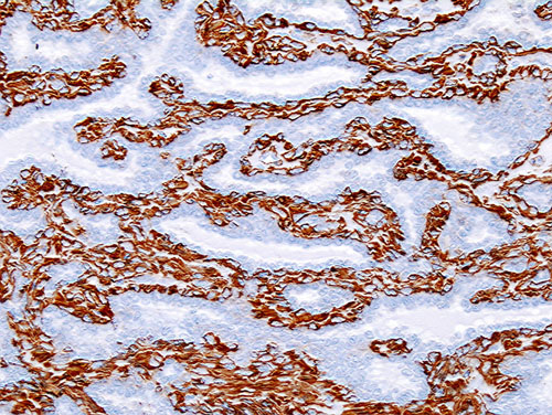
Infiltrating ductal carcinoma of the Breast assayed with anti Mucin 1 antibody
-
Breast cancer (Infiltrating ductal carcinoma of the breast) assayed with anti HER-2 (ErbB2) antibody
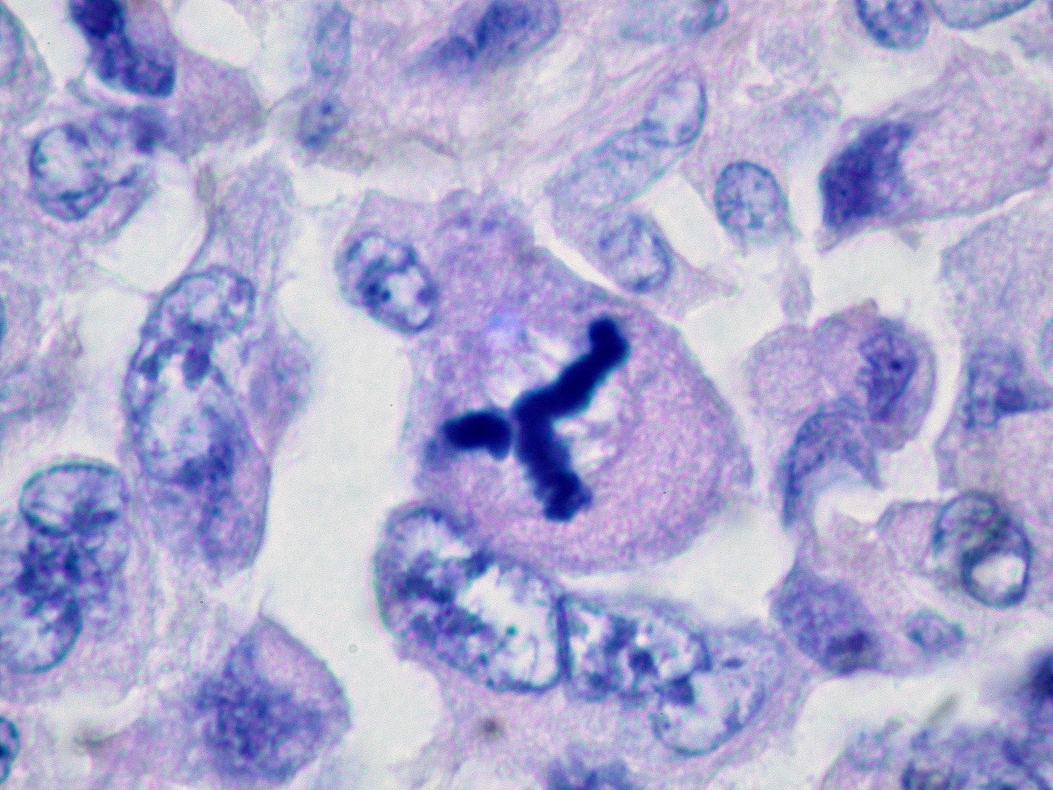
-
Invasive lobular carcinoma of the breast demonstrating a predominantly lobular growth pattern
-
Metaplastic (sarcomatoid) carcinoma of the breast.
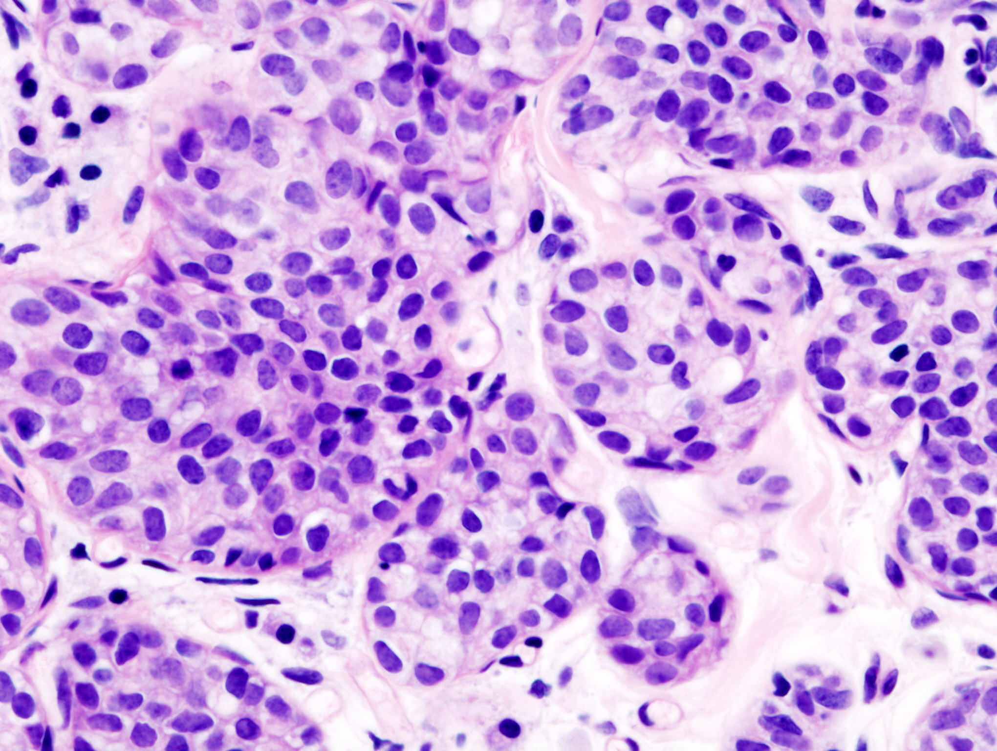
-
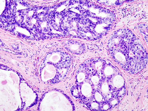
Histopathologic image from ductal cell carcinoma in situ (DCIS) of breast. Hematoxylin-eosin stain.
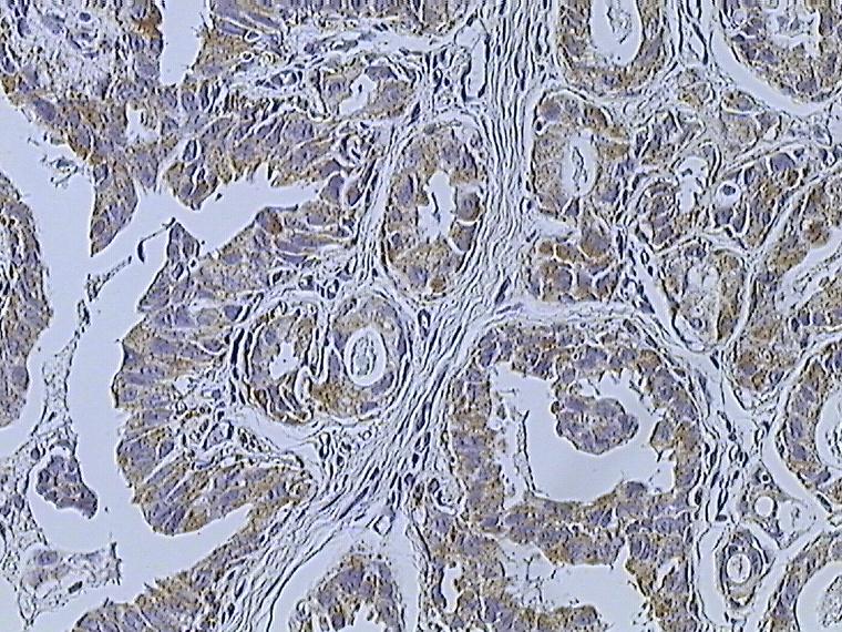
-
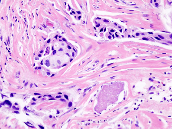
Histopathology of invasive ductal carcinoma of the breast representing a scirrhous growth. Core needle biopsy. Hematoxylin and eosin stain.
-
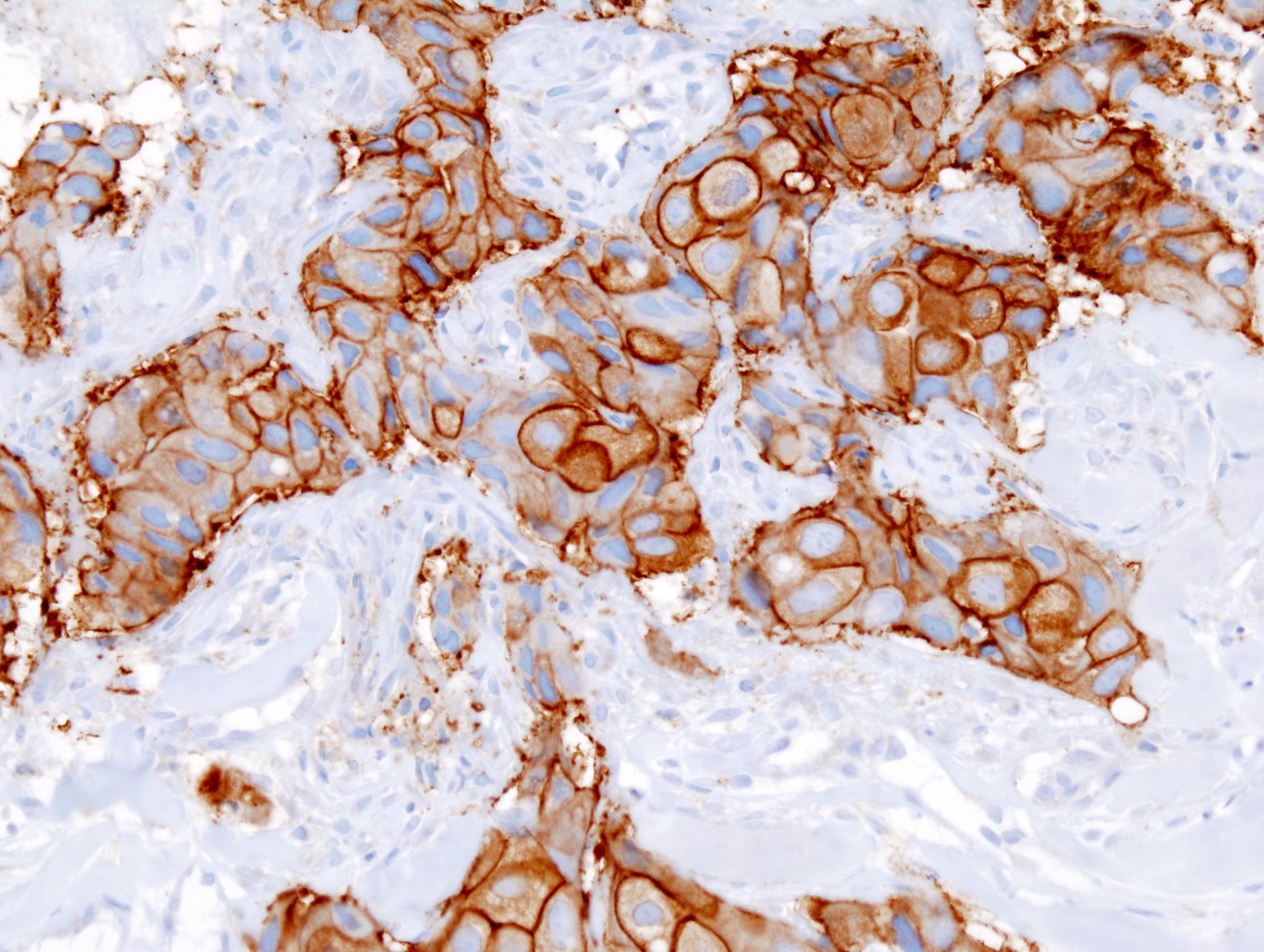
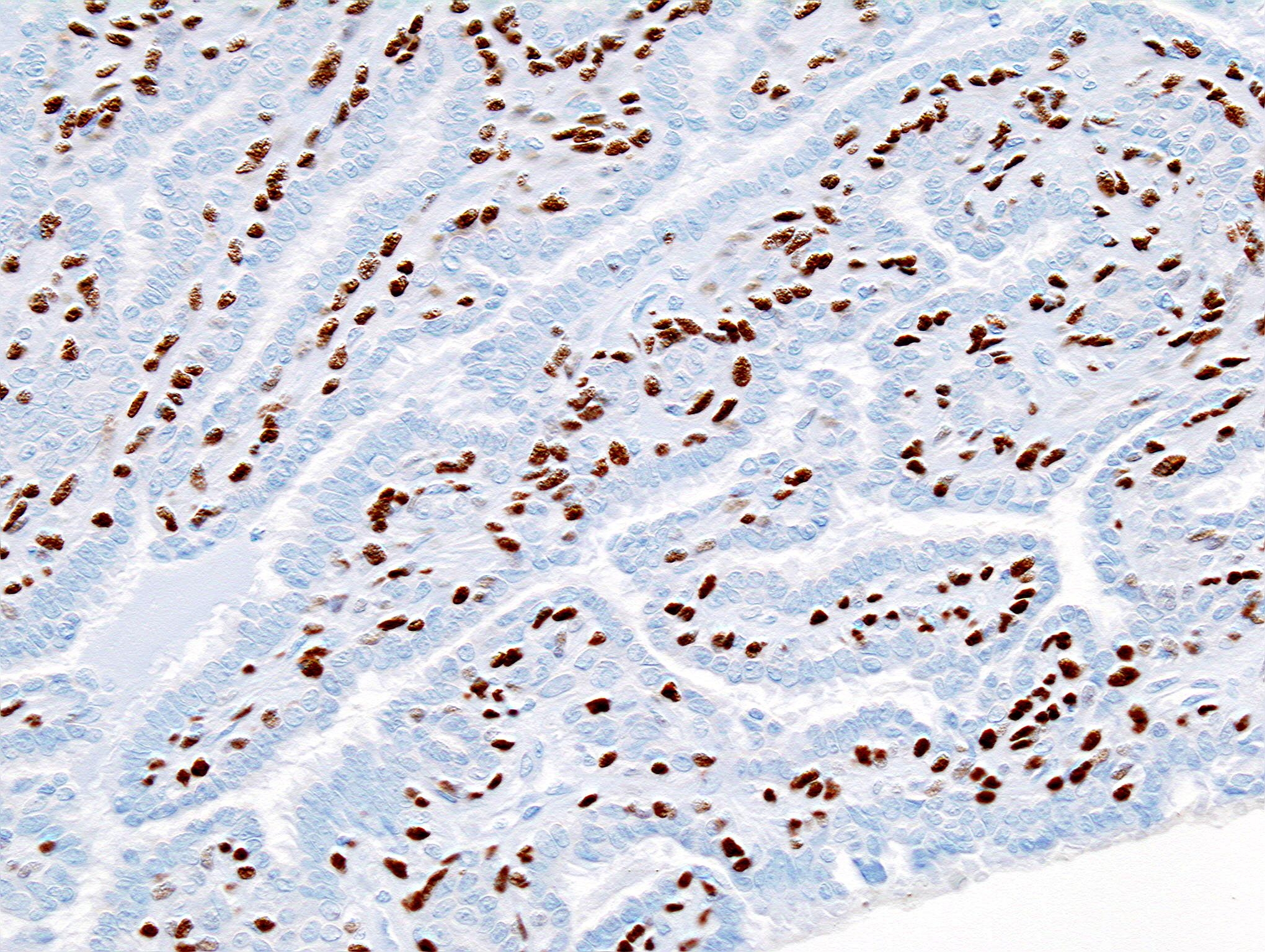
Histopathology of invasive ductal carcinoma of the breast representing a scirrhous growth. Core needle biopsy. HER-2/neu oncoprotein expression by Ventana immunostaining system.
-

Breast cancer (Infiltrating ductal carcinoma of the breast) dyed with H&E
-
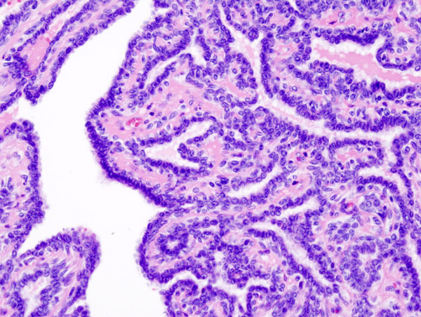
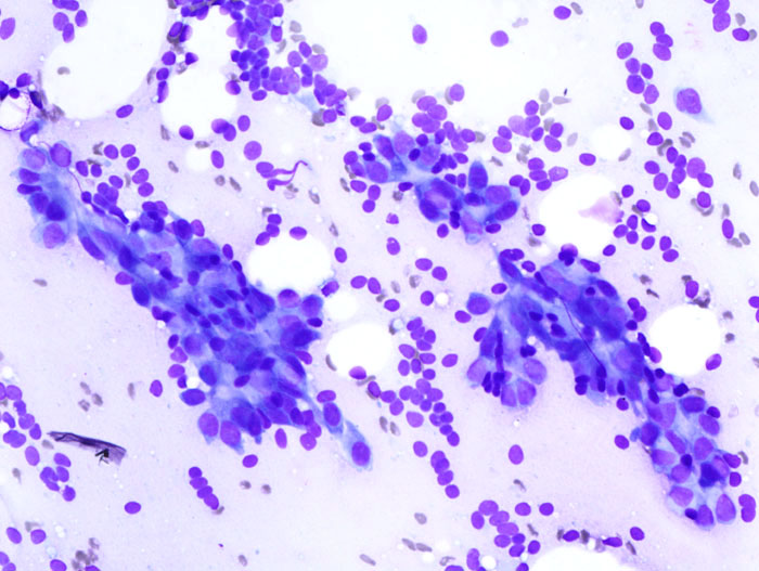
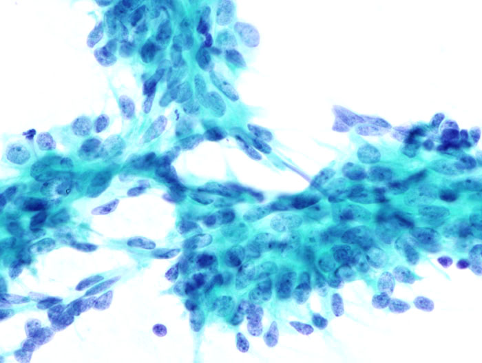
Histopathology of intraductal papilloma of the breast by excisional biopsy. Hematoxylin and eosin stain.
-
Histopathology of intraductal papilloma of the breast by excisional biopsy. Immunostaining for alpha-smooth muscle actin.
-
Histopathology of intraductal papilloma of the breast by excisional biopsy. Immunostaining for p63 protein.
-
Breast fibroadenoma
-
Breast fibroadenoma
-
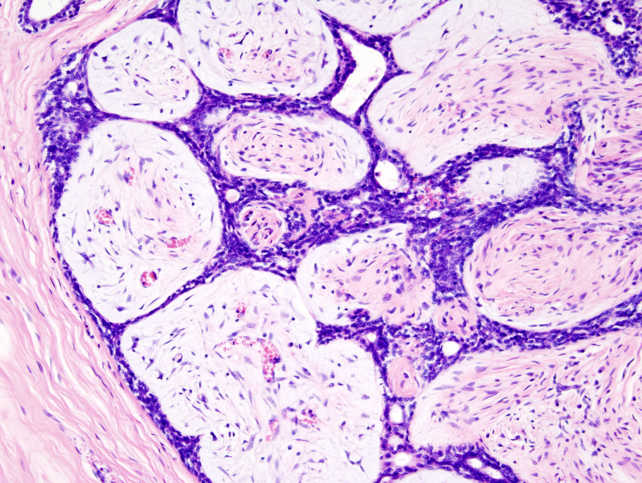
Histopathologic image of breast fibroadenoma. Core needle biopsy. Hematoxylin & eosin stain.
-
Histopathologic image of breast fibroadenoma. Core needle biopsy. Hematoxylin & eosin stain.

.jpg)
.jpg)

.jpg)
.jpg)
_HER2_expression.JPG)

.jpg)
_smooth_muscle_actin.JPG)
_p63.JPG)
_DG_stain.jpg)
_PAP_stain.jpg)
.jpg)
.jpg)
References
- ↑ Breast Neoplasm. Radiopedia. (2015) http://radiopaedia.org/articles/breast-neoplasms Accessed on January 16, 2016
- ↑ Breast Neoplasms. Radiopaedia (2015) http://radiopaedia.org/articles/breast-neoplasms Accessed on january 16, 2016
- ↑ Breast Cancer. National Cancer Institute (2016) http://www.cancer.gov/types/breast/patient/breast-treatment-pdq#link/_148 Accessed on January 16, 2016