Pancreatic cancer CT: Difference between revisions
Jump to navigation
Jump to search
No edit summary |
No edit summary |
||
| Line 3: | Line 3: | ||
{{CMG}}; '''Associate Editor-In-Chief:''' {{CZ}} | {{CMG}}; '''Associate Editor-In-Chief:''' {{CZ}} | ||
==CT== | ==CT== | ||
===Pancreatic Adenocarcinoma=== | ===Pancreatic Adenocarcinoma=== | ||
| Line 21: | Line 19: | ||
[[Category:Oncology]] | [[Category:Oncology]] | ||
[[Category:Mature chapter]] | [[Category:Mature chapter]] | ||
[[Category:Primary care]] | [[Category:Primary care]] | ||
[[Category:Needs overview]] | [[Category:Needs overview]] | ||
Revision as of 15:32, 17 August 2015
|
Pancreatic cancer Microchapters |
|
Diagnosis |
|---|
|
Treatment |
|
Case Studies |
|
Pancreatic cancer CT On the Web |
|
American Roentgen Ray Society Images of Pancreatic cancer CT |
Editor-In-Chief: C. Michael Gibson, M.S., M.D. [1]; Associate Editor-In-Chief: Cafer Zorkun, M.D., Ph.D. [2]
CT
Pancreatic Adenocarcinoma
-
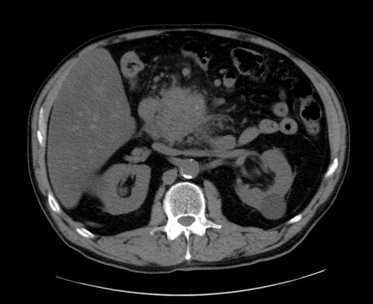
Pancreatic adenocarcinoma
-
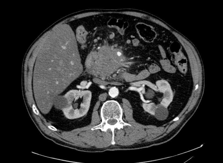
Pancreatic adenocarcinoma
-
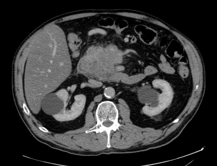
Pancreatic adenocarcinoma


