Hydronephrosis echocardiography or ultrasound: Difference between revisions
m (Anmol Pitliya moved page Hydronephrosis ultrasound to Hydronephrosis echocardiography or ultrasound without leaving a redirect) |
Usama Talib (talk | contribs) No edit summary |
||
| Line 2: | Line 2: | ||
{{Hydronephrosis}} | {{Hydronephrosis}} | ||
{{CMG}}; {{AE}} | |||
==Overview== | |||
There are no echocardiography/ultrasound findings associated with [disease name]. | |||
OR | |||
Echocardiography/ultrasound may be helpful in the diagnosis of [disease name]. Findings on an echocardiography/ultrasound suggestive of/diagnostic of [disease name] include [finding 1], [finding 2], and [finding 3]. | |||
OR | |||
There are no echocardiography/ultrasound findings associated with [disease name]. However, an echocardiography/ultrasound may be helpful in the diagnosis of complications of [disease name], which include [complication 1], [complication 2], and [complication 3]. | |||
==Echocardiography/Ultrasound== | |||
*There are no echocardiography/ultrasound findings associated with [disease name]. | |||
OR | |||
*Echocardiography/ultrasound may be helpful in the diagnosis of [disease name]. Findings on an echocardiography/ultrasound suggestive of/diagnostic of [disease name] include: | |||
**[Finding 1] | |||
**[Finding 2] | |||
**[Finding 3] | |||
OR | |||
*There are no echocardiography/ultrasound findings associated with [disease name]. However, an echocardiography/ultrasound may be helpful in the diagnosis of complications of [disease name], which include: | |||
**[Complication 1] | |||
**[Complication 2] | |||
**[Complication 3] | |||
Ultrasound allows for visualization of the ureters and kidneys and can be used to assess the presence of hydronephrosis and/or hydroureter. | Ultrasound allows for visualization of the ureters and kidneys and can be used to assess the presence of hydronephrosis and/or hydroureter. | ||
Revision as of 15:16, 5 June 2018
|
Hydronephrosis Microchapters |
|
Diagnosis |
|---|
|
Treatment |
|
Case Studies |
|
Hydronephrosis echocardiography or ultrasound On the Web |
|
American Roentgen Ray Society Images of Hydronephrosis echocardiography or ultrasound |
|
Risk calculators and risk factors for Hydronephrosis echocardiography or ultrasound |
Editor-In-Chief: C. Michael Gibson, M.S., M.D. [1]; Associate Editor(s)-in-Chief:
Overview
There are no echocardiography/ultrasound findings associated with [disease name].
OR
Echocardiography/ultrasound may be helpful in the diagnosis of [disease name]. Findings on an echocardiography/ultrasound suggestive of/diagnostic of [disease name] include [finding 1], [finding 2], and [finding 3].
OR
There are no echocardiography/ultrasound findings associated with [disease name]. However, an echocardiography/ultrasound may be helpful in the diagnosis of complications of [disease name], which include [complication 1], [complication 2], and [complication 3].
Echocardiography/Ultrasound
- There are no echocardiography/ultrasound findings associated with [disease name].
OR
- Echocardiography/ultrasound may be helpful in the diagnosis of [disease name]. Findings on an echocardiography/ultrasound suggestive of/diagnostic of [disease name] include:
- [Finding 1]
- [Finding 2]
- [Finding 3]
OR
- There are no echocardiography/ultrasound findings associated with [disease name]. However, an echocardiography/ultrasound may be helpful in the diagnosis of complications of [disease name], which include:
- [Complication 1]
- [Complication 2]
- [Complication 3]
Ultrasound allows for visualization of the ureters and kidneys and can be used to assess the presence of hydronephrosis and/or hydroureter.
-
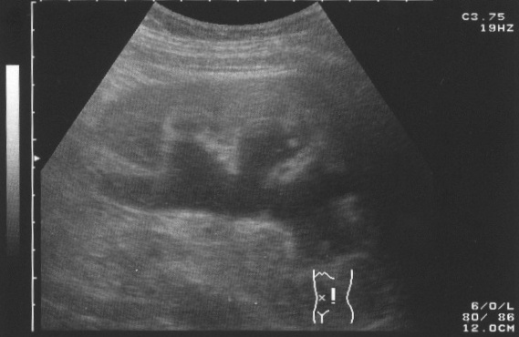
Ultrasound picture of hydronephrosis caused by a left ureteral stone

Prenatal diagnosis is possible.[1]
References
- ↑ Estrada CR (2008). "Prenatal hydronephrosis: early evaluation". Curr Opin Urol. 18 (4): 401–3. doi:10.1097/MOU.0b013e328302edfe. PMID 18520762. Unknown parameter
|month=ignored (help)