|
|
| (88 intermediate revisions by 2 users not shown) |
| Line 1: |
Line 1: |
| | ==Physical examination== |
| | ==References== |
| | {{reflist|2}} |
| | |
| | {{WH}} |
| | {{WS}} |
| | |
| | ==References== |
| | {{Reflist|2}} |
| | |
| | |
| ===Pathophysiology prev=== | | ===Pathophysiology prev=== |
| <div style="-webkit-user-select: none;"> | | <div style="-webkit-user-select: none;"> |
| Line 9: |
Line 20: |
| {{Cirrhosis}} | | {{Cirrhosis}} |
| {{CMG}} {{AE}} | | {{CMG}} {{AE}} |
| ==Table==
| |
| <div style="width: 85%;"><small>
| |
| {| class="wikitable"
| |
| ! rowspan="3" |Disease
| |
| ! rowspan="3" |Cause
| |
| ! colspan="9" |Symptoms
| |
| !Diagnosis
| |
| ! rowspan="3" |Other findings
| |
| |-
| |
| ! colspan="3" |Pain
| |
| ! rowspan="2" |Nausea
| |
| &
| |
|
| |
|
| Vomiting
| | |
| ! rowspan="2" |Heartburn
| | ===Pathophysiology prev=== |
| ! rowspan="2" |Belching or
| | <div style="-webkit-user-select: none;"> |
| Bloating
| | {| class="infobox" style="position: fixed; top: 65%; right: 10px; margin: 0 0 0 0; border: 0; float: right;" |
| ! rowspan="2" |Weight loss
| |
| ! rowspan="2" |Loss of
| |
| Appetite
| |
| ! rowspan="2" |Stools
| |
| ! rowspan="2" |Endoscopy findings
| |
| |- | | |- |
| !Location
| | | {{#ev:youtube|https://https://www.youtube.com/watch?v=5szNmKtyBW4|350}} |
| !Aggravating Factors
| |
| !Alleviating Factors
| |
| |- | | |- |
| !Gastric outlet obstruction
| | |} |
| | | | __NOTOC__ |
| *[[Peptic ulcer|PUD]]: 5% cases (most commonly affecting [[pylorus]] and initial part of the [[duodenum]])
| | {{Cirrhosis}} |
| *[[Polyps|Gastric polyps]]<ref name="pmid7129059">{{cite journal |vauthors=Miner PB, Harri JE, McPhee MS |title=Intermittent gastric outlet obstruction from a pedunculated gastric polyp |journal=Gastrointest. Endosc. |volume=28 |issue=3 |pages=219–20 |year=1982 |pmid=7129059 |doi= |url=}}</ref><ref name="pmid12831404">{{cite journal |vauthors=Gencosmanoglu R, Sen-Oran E, Kurtkaya-Yapicier O, Tozun N |title=Antral hyperplastic polyp causing intermittent gastric outlet obstruction: case report |journal=BMC Gastroenterol |volume=3 |issue= |pages=16 |year=2003 |pmid=12831404 |pmc=166166 |doi=10.1186/1471-230X-3-16 |url=}}</ref>
| | {{CMG}} {{AE}} |
| *[[Caustic|Caustic ingestion]]<ref name="pmid2753330">{{cite journal |vauthors=Zargar SA, Kochhar R, Nagi B, Mehta S, Mehta SK |title=Ingestion of corrosive acids. Spectrum of injury to upper gastrointestinal tract and natural history |journal=Gastroenterology |volume=97 |issue=3 |pages=702–7 |year=1989 |pmid=2753330 |doi= |url=}}</ref>
| | |
| *[[Stenosis|Duodenal stricture]] <ref name="pmid2000520">{{cite journal |vauthors=Taylor SM, Adams DB, Anderson MC |title=Duodenal stricture: a complication of chronic fibrocalcific pancreatitis |journal=South. Med. J. |volume=84 |issue=3 |pages=338–41 |year=1991 |pmid=2000520 |doi= |url=}}</ref>
| | == History and Symptoms == |
| *Systemic [[amyloidosis]] of the [[gastrointestinal tract]] <ref name="pmid8331978">{{cite journal |vauthors=Menke DM, Kyle RA, Fleming CR, Wolfe JT, Kurtin PJ, Oldenburg WA |title=Symptomatic gastric amyloidosis in patients with primary systemic amyloidosis |journal=Mayo Clin. Proc. |volume=68 |issue=8 |pages=763–7 |year=1993 |pmid=8331978 |doi= |url=}}</ref><ref name="pmid9891699">{{cite journal |vauthors=Friedman S, Janowitz HD |title=Systemic amyloidosis and the gastrointestinal tract |journal=Gastroenterol. Clin. North Am. |volume=27 |issue=3 |pages=595–614, vi |year=1998 |pmid=9891699 |doi= |url=}}</ref>
| | |
| *Eosinophillic [[gastroenteritis]] <ref name="pmid10660821">{{cite journal |vauthors=Khan S, Orenstein SR |title=Eosinophilic gastroenteritis masquerading as pyloric stenosis |journal=Clin Pediatr (Phila) |volume=39 |issue=1 |pages=55–7 |year=2000 |pmid=10660821 |doi=10.1177/000992280003900109 |url=}}</ref><ref name="pmid11400803">{{cite journal |vauthors=Chaudhary R, Shrivastava RK, Mukhopadhyay HG, Diwan RN, Das AK |title=Eosinophilic gastritis--an unusual cause of gastric outlet obstruction |journal=Indian J Gastroenterol |volume=20 |issue=3 |pages=110 |year=2001 |pmid=11400803 |doi= |url=}}</ref><ref name="pmid17614041">{{cite journal |vauthors=Tursi A, Rella G, Inchingolo CD, Maiorano M |title=Gastric outlet obstruction due to gastroduodenal eosinophilic gastroenteritis |journal=Endoscopy |volume=39 Suppl 1 |issue= |pages=E184 |year=2007 |pmid=17614041 |doi=10.1055/s-2006-945125 |url=}}</ref><ref name="pmid14669340">{{cite journal |vauthors=Chen MJ, Chu CH, Lin SC, Shih SC, Wang TE |title=Eosinophilic gastroenteritis: clinical experience with 15 patients |journal=World J. Gastroenterol. |volume=9 |issue=12 |pages=2813–6 |year=2003 |pmid=14669340 |pmc=4612059 |doi= |url=}}</ref><ref name="pmid8420276">{{cite journal |vauthors=Lee CM, Changchien CS, Chen PC, Lin DY, Sheen IS, Wang CS, Tai DI, Sheen-Chen SM, Chen WJ, Wu CS |title=Eosinophilic gastroenteritis: 10 years experience |journal=Am. J. Gastroenterol. |volume=88 |issue=1 |pages=70–4 |year=1993 |pmid=8420276 |doi= |url=}}</ref> | | * History should include: |
| *[[Obstruction]] by [[Gallstone disease|gallstones]] (Bouveret syndrome) | | ** Appearance of bowel movements |
| *Complication of [[acute pancreatitis]]: [[pancreatic pseudocyst]] formation<ref name="pmid6732492">{{cite journal |vauthors=Aranha GV, Prinz RA, Greenlee HB, Freeark RJ |title=Gastric outlet and duodenal obstruction from inflammatory pancreatic disease |journal=Arch Surg |volume=119 |issue=7 |pages=833–5 |year=1984 |pmid=6732492 |doi= |url=}}</ref><ref name="pmid4811173">{{cite journal |vauthors=Agrawal NM, Gyr N, McDowell W, Font RG |title=Intestinal obstruction due to acute pancreatitis. Case report and review of literature |journal=Am J Dig Dis |volume=19 |issue=2 |pages=179–85 |year=1974 |pmid=4811173 |doi= |url=}}</ref> | | ** Travel history |
| *[[Chronic pancreatitis]] <ref name="pmid2658160">{{cite journal |vauthors=Bradley EL |title=Complications of chronic pancreatitis |journal=Surg. Clin. North Am. |volume=69 |issue=3 |pages=481–97 |year=1989 |pmid=2658160 |doi= |url=}}</ref><ref name="pmid19629001">{{cite journal |vauthors=Levenick JM, Gordon SR, Sutton JE, Suriawinata A, Gardner TB |title=A comprehensive, case-based review of groove pancreatitis |journal=Pancreas |volume=38 |issue=6 |pages=e169–75 |year=2009 |pmid=19629001 |doi=10.1097/MPA.0b013e3181ac73f1 |url=}}</ref> | | ** Associated symptoms |
| *[[Sarcoidosis]] of the [[Gastrointestinal tract|GIT]] <ref name="pmid2180656">{{cite journal |vauthors=Stampfl DA, Grimm IS, Barbot DJ, Rosato FE, Gordon SJ |title=Sarcoidosis causing duodenal obstruction. Case report and review of gastrointestinal manifestations |journal=Dig. Dis. Sci. |volume=35 |issue=4 |pages=526–32 |year=1990 |pmid=2180656 |doi= |url=}}</ref><ref name="pmid807981">{{cite journal |vauthors=Johnson FE, Humbert JR, Kuzela DC, Todd JK, Lilly JR |title=Gastric outlet obstruction due to X-linked chronic granulomatous disease |journal=Surgery |volume=78 |issue=2 |pages=217–23 |year=1975 |pmid=807981 |doi= |url=}}</ref><ref name="pmid6623357">{{cite journal |vauthors=Mulholland MW, Delaney JP, Simmons RL |title=Gastrointestinal complications of chronic granulomatous disease: surgical implications |journal=Surgery |volume=94 |issue=4 |pages=569–75 |year=1983 |pmid=6623357 |doi= |url=}}</ref><ref name="pmid16970572">{{cite journal |vauthors=Huang A, Abbasakoor F, Vaizey CJ |title=Gastrointestinal manifestations of chronic granulomatous disease |journal=Colorectal Dis |volume=8 |issue=8 |pages=637–44 |year=2006 |pmid=16970572 |doi=10.1111/j.1463-1318.2006.01030.x |url=}}</ref> | | ** Immune status |
| *[[Bezoar|Bezoars]]<ref name="pmid9291515">{{cite journal |vauthors=Bakken DA, Abramo TJ |title=Gastric lactobezoar: a rare cause of gastric outlet obstruction |journal=Pediatr Emerg Care |volume=13 |issue=4 |pages=264–7 |year=1997 |pmid=9291515 |doi= |url=}}</ref><ref name="pmid10328129">{{cite journal |vauthors=De Backer A, Van Nooten V, Vandenplas Y |title=Huge gastric trichobezoar in a 10-year-old girl: case report with emphasis on endoscopy in diagnosis and therapy |journal=J. Pediatr. Gastroenterol. Nutr. |volume=28 |issue=5 |pages=513–5 |year=1999 |pmid=10328129 |doi= |url=}}</ref><ref name="pmid9663194">{{cite journal |vauthors=Phillips MR, Zaheer S, Drugas GT |title=Gastric trichobezoar: case report and literature review |journal=Mayo Clin. Proc. |volume=73 |issue=7 |pages=653–6 |year=1998 |pmid=9663194 |doi=10.1016/S0025-6196(11)64889-1 |url=}}</ref><ref name="pmid14738689">{{cite journal |vauthors=White NB, Gibbs KE, Goodwin A, Teixeira J |title=Gastric bezoar complicating laparoscopic adjustable gastric banding, and review of literature |journal=Obes Surg |volume=13 |issue=6 |pages=948–50 |year=2003 |pmid=14738689 |doi=10.1381/096089203322618849 |url=}}</ref><ref name="pmid16448609">{{cite journal |vauthors=Zapata R, Castillo F, Córdova A |title=[Gastric food bezoar as a complication of bariatric surgery. Case report and review of the literature] |language=Spanish; Castilian |journal=Gastroenterol Hepatol |volume=29 |issue=2 |pages=77–80 |year=2006 |pmid=16448609 |doi= |url=}}</ref> | | ** Woodland exposure |
| *[[Crohn's disease]] involving the [[duodenum]] <ref name="pmid2919581">{{cite journal |vauthors=Nugent FW, Roy MA |title=Duodenal Crohn's disease: an analysis of 89 cases |journal=Am. J. Gastroenterol. |volume=84 |issue=3 |pages=249–54 |year=1989 |pmid=2919581 |doi= |url=}}</ref><ref name="pmid16278730">{{cite journal |vauthors=Kefalas CH |title=Gastroduodenal Crohn's disease |journal=Proc (Bayl Univ Med Cent) |volume=16 |issue=2 |pages=147–51 |year=2003 |pmid=16278730 |pmc=1201000 |doi= |url=}}</ref><ref name="pmid9360875">{{cite journal |vauthors=Matsui T, Hatakeyama S, Ikeda K, Yao T, Takenaka K, Sakurai T |title=Long-term outcome of endoscopic balloon dilation in obstructive gastroduodenal Crohn's disease |journal=Endoscopy |volume=29 |issue=7 |pages=640–5 |year=1997 |pmid=9360875 |doi=10.1055/s-2007-1004271 |url=}}</ref><ref name="pmid6106466">{{cite journal |vauthors=Fitzgibbons TJ, Green G, Silberman H, Eliasoph J, Halls JM, Yellin AE |title=Management of Crohn's disease involving the duodenum, including duodenal cutaneous fistula |journal=Arch Surg |volume=115 |issue=9 |pages=1022–8 |year=1980 |pmid=6106466 |doi= |url=}}</ref> | | ==References== |
| *[[Stomach|Gastro]]-[[Duodenum|duodenal]] [[tuberculosis]]<ref name="pmid12703983">{{cite journal |vauthors=Amarapurkar DN, Patel ND, Amarapurkar AD |title=Primary gastric tuberculosis--report of 5 cases |journal=BMC Gastroenterol |volume=3 |issue= |pages=6 |year=2003 |pmid=12703983 |pmc=155648 |doi= |url=}}</ref><ref name="pmid15540690">{{cite journal |vauthors=Rao YG, Pande GK, Sahni P, Chattopadhyay TK |title=Gastroduodenal tuberculosis management guidelines, based on a large experience and a review of the literature |journal=Can J Surg |volume=47 |issue=5 |pages=364–8 |year=2004 |pmid=15540690 |pmc=3211943 |doi= |url=}}</ref><ref name="pmid16217956">{{cite journal |vauthors=Padussis J, Loffredo B, McAneny D |title=Minimally invasive management of obstructive gastroduodenal tuberculosis |journal=Am Surg |volume=71 |issue=8 |pages=698–700 |year=2005 |pmid=16217956 |doi= |url=}}</ref><ref name="pmid8677960">{{cite journal |vauthors=Di Placido R, Pietroletti R, Leardi S, Simi M |title=Primary gastroduodenal tuberculous infection presenting as pyloric outlet obstruction |journal=Am. J. Gastroenterol. |volume=91 |issue=4 |pages=807–8 |year=1996 |pmid=8677960 |doi= |url=}}</ref><ref name="pmid3605037">{{cite journal |vauthors=Subei I, Attar B, Schmitt G, Levendoglu H |title=Primary gastric tuberculosis: a case report and literature review |journal=Am. J. Gastroenterol. |volume=82 |issue=8 |pages=769–72 |year=1987 |pmid=3605037 |doi= |url=}}</ref>
| | {{reflist|2}} |
| * Pyloric stenosis
| | |
| * Ingestion of [[corrosive|corrosives]]
| | {{WH}} |
| |
| | {{WS}} |
| *Early stages:<ref name="pmid7129059">{{cite journal |vauthors=Miner PB, Harri JE, McPhee MS |title=Intermittent gastric outlet obstruction from a pedunculated gastric polyp |journal=Gastrointest. Endosc. |volume=28 |issue=3 |pages=219–20 |year=1982 |pmid=7129059 |doi= |url=}}</ref><ref name="pmid7771437">{{cite journal |vauthors=Urayama S, Kozarek R, Ball T, Brandabur J, Traverso L, Ryan J, Wechter D |title=Presentation and treatment of annular pancreas in an adult population |journal=Am. J. Gastroenterol. |volume=90 |issue=6 |pages=995–9 |year=1995 |pmid=7771437 |doi= |url=}}</ref>
| | |
| * [[Nausea and vomiting|Nausea]] | | ==Other Imaging Findings== |
| * [[Nausea and vomiting|Vomiting]]: characteristic feature
| | * [[Endoscopy]] |
| ** Intermittent
| | * [[Barium enema]] |
| ** Occurs one hour after [[ingestion]] | | * [[Colonoscopy]] |
| ** Non [[Bile|bilious]] | | * [[Sigmoidoscopy]] |
| ** Contains undigested particles of food
| | |
| ** Patient has intolerance to solids, followed by liquids
| | ==Other diagnostic studies== |
| ** [[Dehydration]]
| | == Other Diagnostic Studies == |
| ** [[Electrolyte disturbance|Electrolyte abnormalities]]
| | |
| Late stages:<ref name="pmid2207566">{{cite journal |vauthors=Johnson CD, Ellis H |title=Gastric outlet obstruction now predicts malignancy |journal=Br J Surg |volume=77 |issue=9 |pages=1023–4 |year=1990 |pmid=2207566 |doi= |url=}}</ref><ref name="pmid7572891">{{cite journal |vauthors=Shone DN, Nikoomanesh P, Smith-Meek MM, Bender JS |title=Malignancy is the most common cause of gastric outlet obstruction in the era of H2 blockers |journal=Am. J. Gastroenterol. |volume=90 |issue=10 |pages=1769–70 |year=1995 |pmid=7572891 |doi= |url=}}</ref><ref name="pmid16817848">{{cite journal |vauthors=Cappell MS, Davis M |title=Characterization of Bouveret's syndrome: a comprehensive review of 128 cases |journal=Am. J. Gastroenterol. |volume=101 |issue=9 |pages=2139–46 |year=2006 |pmid=16817848 |doi=10.1111/j.1572-0241.2006.00645.x |url=}}</ref><ref name="pmid717362">{{cite journal |vauthors=Dubois A, Price SF, Castell DO |title=Gastric retention in peptic ulcer disease. A reappraisal |journal=Am J Dig Dis |volume=23 |issue=11 |pages=993–7 |year=1978 |pmid=717362 |doi= |url=}}</ref>
| | * Breath hydrogen test |
| * [[Weight loss]]
| | |
| * [[Malnutrition]]: more pronounced in patients with [[Cancer|malignancy]] | | * [[HIV test]]ing for those patients suspected of having HIV |
| * [[Abdominal distension]]
| | |
| * Features of incomplete [[obstruction]] | | == |
| * [[Stomach|Gastric]] retention: presenting as early [[satiety]]
| |
| * [[Bloating]]
| |
| * Fullness of [[epigastrium]]
| |
| * [[Aspiration pneumonia]]: due to [[Dilation|dilatation]] of [[stomach]], loss of [[contractility]] and accumulation of undigested food contents
| |
| |Food
| |
| |-
| |
| |✔
| |
| |✔
| |
| |✔
| |
| |may be present in case of GOO due to malignancy
| |
| |✔
| |
| |[[Melena|Black stools]] in case of GOO due to PUD
| |
| |
| |
| *Helps in the determination of site of [[obstruction]]
| |
| *Helps in the visualization of the [[Stomach|gastric]] silhouette:
| |
| *Helps note the following:
| |
| **[[Stomach|Gastric]] [[dilation]]
| |
| **Narrowing of the [[pylorus]]
| |
| **Presence of [[Ulcer|ulcers]]
| |
| **[[Tumor|Tumors]]
| |
| **Differentiation of GOO from [[gastroparesis]] where gastric [[dilation]] is not associated with the narrowing of the [[pylorus]]
| |
| |<nowiki>-</nowiki>
| |
| |-
| |
| ![[Acute gastritis]]
| |
| |
| |
| * ''[[H. pylori]]''
| |
| * [[NSAIDS]]
| |
| * [[Corticosteroids]]
| |
| * [[Alcohol]]
| |
| * Spicy food
| |
| * Viral infections
| |
| * [[Crohn's disease]]
| |
| * [[Autoimmune diseases]]
| |
| * Bile reflux
| |
| * [[Cocaine]] use
| |
| * Breathing machine or ventilator
| |
| * Ingestion of [[corrosive|corrosives]]
| |
| |
| |
| * [[Epigastric pain]]
| |
| |Food
| |
| |[[Antacids]]
| |
| |✔
| |
| |✔
| |
| |✔
| |
| |<nowiki>-</nowiki>
| |
| |✔
| |
| |[[Melena|Black stools]]
| |
| |
| |
| * Pangastritis or antral [[gastritis]]
| |
| * [[Gastric erosion|Erosive]] (Superficial, deep, hemorrhagic)
| |
| * Nonerosive (''[[H. pylori]]'')
| |
| |<nowiki>-</nowiki>
| |
| |-
| |
| ![[Gastritis|Chronic gastritis]]
| |
| |
| |
| * ''[[H. pylori]]''
| |
| * [[Alcohol]]
| |
| * Medications
| |
| * [[Autoimmune diseases]]
| |
| * Chronic stress
| |
| |
| |
| * [[Epigastric pain]]
| |
| |Food
| |
| |[[Antacids]]
| |
| |✔
| |
| |✔
| |
| |✔
| |
| |✔
| |
| |✔
| |
| |<nowiki>-</nowiki>
| |
| |''[[H. pylori]] [[gastritis]]''
| |
| * [[Atrophy]]
| |
| * Intestinal [[metaplasia]]
| |
| Lymphocytic gastritis
| |
| * Enlarged folds
| |
| * Aphthoid erosions
| |
| |<nowiki>-</nowiki>
| |
| |-
| |
| ![[Atrophic gastritis]]
| |
| |
| |
| * ''[[H. pylori]]''
| |
| * [[Autoimmune disease]]
| |
| |[[Epigastric pain]]
| |
| |<nowiki>-</nowiki>
| |
| |<nowiki>-</nowiki>
| |
| |✔
| |
| |<nowiki>-</nowiki>
| |
| |
| |
| |✔
| |
| |✔
| |
| |<nowiki>-</nowiki>
| |
| |''[[H. pylori]]''
| |
| * Mucosal [[atrophy]]
| |
| [[Autoimmune]]
| |
| * Mucosal [[atrophy]]
| |
| |
| |
| * [[Iron deficiency anemia]]
| |
| Autoimmune gastritis diagnosis include:
| |
| * Antiparietal and anti-IF antibodies
| |
| * [[Achlorhydria]] and hypergastrinemia
| |
| * Low serum [[vitamin B12|cobalamine]]
| |
| |-
| |
| ![[Crohn's disease]]
| |
| |
| |
| * [[Autoimmune disease]]
| |
| |
| |
| * [[Abdominal pain]]
| |
| |<nowiki>-</nowiki>
| |
| |<nowiki>-</nowiki>
| |
| |<nowiki>-</nowiki>
| |
| |<nowiki>-</nowiki>
| |
| |<nowiki>-</nowiki>
| |
| |✔
| |
| |✔
| |
| |
| |
| * Chronic [[diarrhea]] often bloody with [[pus]] or [[mucus]]
| |
| * [[Rectal bleeding]]
| |
| |
| |
| * Mucosal nodularity with cobblestoning
| |
| * Multiple [[aphthous ulcers]]
| |
| * Linier or serpiginous ulcerations
| |
| * Thickened antral folds
| |
| * Antral narrowing
| |
| * Hypoperistalsis
| |
| * Duodenal strictures
| |
| |
| |
| * [[Fever]]
| |
| * [[Fatigue]]
| |
| * [[Anemia]] ([[pernicious anemia]])
| |
| |-
| |
| ![[GERD]]
| |
| |
| |
| * Lower esophageal sphincter abnormalities
| |
|
| |
|
| * [[Hiatal hernia]]
| | ==Overview== |
| * Abnormal esophageal contractions
| |
| * Prolonged emptying of [[stomach]]
| |
| * [[Gastrinomas]]
| |
| |
| |
| * [[Epigastric pain]]
| |
| |
| |
| * Spicy food
| |
| * Tight fitting clothing
| |
| |
| |
| * [[Antacids]]
| |
| * Head elevation during sleep
| |
| |✔
| |
|
| |
|
| (Suspect delayed gastric emptying)
| | ==References== |
| |✔ | | {{reflist|2}} |
| |<nowiki>-</nowiki>
| |
| |<nowiki>-</nowiki>
| |
| |<nowiki>-</nowiki>
| |
| |<nowiki>-</nowiki>
| |
| |
| |
| * [[Esophagitis]]
| |
| * Barrette esophagus
| |
| * [[Strictures]]
| |
| |Other symptoms:
| |
| * [[Dysphagia]]
| |
| * [[Regurgitation]]
| |
| * [[Cough|Nocturnal cough]]
| |
| * [[Hoarseness]]
| |
| Complications
| |
| * [[Esophagitis]]
| |
| * [[Strictures]]
| |
| * Barrette esophagus
| |
| |-
| |
| ![[Peptic ulcer disease]]
| |
| |
| |
| * ''[[H. pylori]]''
| |
| * [[Smoking]]
| |
| * [[Alcohol]]
| |
| * [[Radiation therapy]]
| |
| * Medications
| |
| * Zollinger-ellison syndrome
| |
| |
| |
| * [[Epigastric pain]] sometimes extending to back
| |
| * [[Right upper quadrant pain]]
| |
| |
| |
| '''[[Duodenal ulcer]]'''
| |
| *Pain aggravates with empty stomach
| |
| '''[[Gastric ulcer]]'''
| |
| *Pain aggravates with food
| |
| |
| |
| * [[Antacids]]
| |
|
| |
|
| * [[Duodenal ulcer]]
| | {{WH}} |
| :*Pain alleviates with food
| | {{WS}} |
| |✔
| |
| |✔
| |
| |<nowiki>-</nowiki>
| |
| |<nowiki>-</nowiki>
| |
| |<nowiki>-</nowiki>
| |
| |
| |
| * [[Melena|Black stools]]
| |
| |'''Gastric ulcers'''
| |
| * Discrete mucosal lesions with a punched-out smooth ulcer base with whitish fibrinoid base
| |
| * Most [[ulcers]] are at the junction of [[fundus]] and antrum
| |
| * 0.5-2.5cm
| |
| '''Duodenal ulcers'''
| |
| * Well-demarcated break in the [[mucosa]] that may extend into the [[muscularis propria]] of the [[duodenum]]
| |
| * Found in the first part of [[duodenum]]
| |
| * <1cm
| |
| |'''Other diagnostic tests'''
| |
| * Serum [[gastrin]] levels
| |
| * [[Secretin]] stimulation test
| |
| * [[Biopsy]]
| |
| |-
| |
| ![[Gastrinoma]]
| |
| |
| |
| * Associated with [[MEN type 1]]
| |
| |
| |
| * [[Abdominal pain]]
| |
| |<nowiki>-</nowiki>
| |
| |<nowiki>-</nowiki>
| |
| |✔
| |
|
| |
|
| (suspect [[gastric outlet obstruction]])
| | ===Pathophysiology prev=== |
| |✔
| | <div style="-webkit-user-select: none;"> |
| |<nowiki>-</nowiki>
| | {| class="infobox" style="position: fixed; top: 65%; right: 10px; margin: 0 0 0 0; border: 0; float: right;" |
| |<nowiki>-</nowiki>
| |
| |<nowiki>-</nowiki>
| |
| | | |
| * [[Melena|Black stools]]
| |
| |Useful in collecting the tissue for [[biopsy]]
| |
| |
| |
| * May present with symptoms of [[GERD]] or [[peptic ulcer disease]]
| |
| * Associated with [[MEN type 1]]
| |
| '''Diagnostic tests'''
| |
| * Serum [[gastrin]] levels
| |
| * [[Somatostatin]] receptor [[scintigraphy]]
| |
| * [[CT]] and [[MRI]]
| |
| |- | | |- |
| ![[Gastric Cancer|Gastric Adenocarcinoma]]
| | | {{#ev:youtube|https://https://www.youtube.com/watch?v=5szNmKtyBW4|350}} |
| | | |
| * ''[[H. pylori]]'' infection
| |
| * Smoked and salted food
| |
| |
| |
| * [[Abdominal pain]]
| |
| |<nowiki>-</nowiki>
| |
| |<nowiki>-</nowiki>
| |
| |✔
| |
| |✔
| |
| |✔
| |
| |✔
| |
| |✔
| |
| |
| |
| * [[Melena|Black stools]], or blood in stools
| |
| |'''Esophagogastroduodenoscopy'''
| |
| * Multiple biopsies are taken to establish the diagnosis
| |
| |'''Other symptoms''' | |
| * [[Dysphagia]]
| |
| * Early [[satiety]]
| |
| * Frequent [[burping]]
| |
| |- | | |- |
| ![[Gastric lymphoma|Primary gastric lymphoma]]
| |
| |
| |
| * ''[[H. pylori]]'' infection
| |
| |
| |
| * [[Abdominal pain]]
| |
| * [[Chest pain]]
| |
| |<nowiki>-</nowiki>
| |
| |<nowiki>-</nowiki>
| |
| |<nowiki>-</nowiki>
| |
| |<nowiki>-</nowiki>
| |
| |<nowiki>-</nowiki>
| |
| |✔
| |
| |<nowiki>-</nowiki>
| |
| |<nowiki>-</nowiki>
| |
| |Useful in collecting the tissue for [[biopsy]]
| |
| |'''Other symptoms'''
| |
| * Painless swollen [[lymph nodes]] in neck and armpit
| |
| * Night sweats
| |
| * [[Fatigue]]
| |
| * [[Fever]]
| |
| * [[Cough]] or trouble breathing
| |
| |} | | |} |
| | __NOTOC__ |
| | {{Cirrhosis}} |
| | {{CMG}} {{AE}} |
|
| |
|
| ==Video codes== | | ==Video codes== |
| Line 354: |
Line 87: |
| {{#ev:youtube|4uSSvD1BAHg}} | | {{#ev:youtube|4uSSvD1BAHg}} |
| {{#ev:youtube|PQXb5D-5UZw}} | | {{#ev:youtube|PQXb5D-5UZw}} |
| | {{#ev:youtube|UVJYQlUm2A8}} |
|
| |
|
| ===Video in table=== | | ===Video in table=== |
| Line 385: |
Line 119: |
| ===Image and text to the right=== | | ===Image and text to the right=== |
|
| |
|
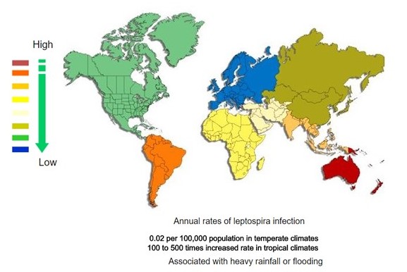
| <figure-inline><figure-inline><figure-inline><figure-inline><figure-inline><figure-inline><figure-inline><figure-inline><figure-inline><figure-inline><figure-inline><figure-inline><figure-inline><figure-inline><figure-inline><figure-inline><figure-inline><figure-inline><figure-inline><figure-inline><figure-inline>[[File:Global distribution of leptospirosis.jpg|577x577px]]</figure-inline></figure-inline></figure-inline></figure-inline></figure-inline></figure-inline></figure-inline></figure-inline></figure-inline></figure-inline></figure-inline></figure-inline></figure-inline></figure-inline></figure-inline></figure-inline></figure-inline></figure-inline></figure-inline></figure-inline></figure-inline> Recent out break of leptospirosis is reported in Bronx, New York and found 3 cases in the months January and February, 2017. | | <figure-inline><figure-inline><figure-inline><figure-inline><figure-inline><figure-inline><figure-inline><figure-inline><figure-inline><figure-inline><figure-inline><figure-inline><figure-inline><figure-inline><figure-inline><figure-inline><figure-inline><figure-inline><figure-inline><figure-inline><figure-inline><figure-inline><figure-inline><figure-inline><figure-inline><figure-inline><figure-inline>[[File:Global distribution of leptospirosis.jpg|577x577px]]</figure-inline></figure-inline></figure-inline></figure-inline></figure-inline></figure-inline></figure-inline></figure-inline></figure-inline></figure-inline></figure-inline></figure-inline></figure-inline></figure-inline></figure-inline></figure-inline></figure-inline></figure-inline></figure-inline></figure-inline></figure-inline></figure-inline></figure-inline></figure-inline></figure-inline></figure-inline></figure-inline> Recent out break of leptospirosis is reported in Bronx, New York and found 3 cases in the months January and February, 2017. |
|
| |
|
| ===Gallery=== | | ===Gallery=== |
| Line 400: |
Line 134: |
| ==References== | | ==References== |
| {{Reflist|2}} | | {{Reflist|2}} |
|
| |
| [[Category:Gastroenterology]]
| |
| [[Category:Hepatology]]
| |
| [[Category:Disease]]
| |
|
| |
| {{WS}} | | {{WS}} |
| {{WH}} | | {{WH}} |
| Line 411: |
Line 140: |
| REFERENCES | | REFERENCES |
| <references /> | | <references /> |
| | |
| | [[Category:Gastroenterology]] |
| | [[Category:Needs overview]] |
| | [[Category:Hepatology]] |
| | [[Category:Disease]] |


 </figure-inline></figure-inline></figure-inline></figure-inline></figure-inline></figure-inline></figure-inline></figure-inline></figure-inline></figure-inline></figure-inline></figure-inline></figure-inline></figure-inline></figure-inline></figure-inline></figure-inline></figure-inline></figure-inline></figure-inline></figure-inline></figure-inline></figure-inline></figure-inline></figure-inline></figure-inline></figure-inline> Recent out break of leptospirosis is reported in Bronx, New York and found 3 cases in the months January and February, 2017.
![Histopathology of a pancreatic endocrine tumor (insulinoma). Source:https://librepathology.org/wiki/Neuroendocrine_tumour_of_the_pancreas[1]](/images/2/2f/Pancreatic_insulinoma_histology_2.JPG)
![Histopathology of a pancreatic endocrine tumor (insulinoma). Chromogranin A immunostain. Source:https://librepathology.org/wiki/Neuroendocrine_tumour_of_the_pancreas[1]](/images/a/a3/Pancreatic_insulinoma_histopathology_3.JPG)
![Histopathology of a pancreatic endocrine tumor (insulinoma). Insulin immunostain. Source:https://librepathology.org/wiki/Neuroendocrine_tumour_of_the_pancreas[1]](/images/d/d5/Pancreatic_insulinoma_histology_4.JPG)