Coccidioidomycosis pathophysiology: Difference between revisions
No edit summary |
m (Bot: Removing from Primary care) |
||
| (35 intermediate revisions by 6 users not shown) | |||
| Line 1: | Line 1: | ||
__NOTOC__ | __NOTOC__ | ||
{{Coccidioidomycosis}} | {{Coccidioidomycosis}} | ||
{{CMG}}; {{AE}}; {{VB}} | {{CMG}}; {{AE}}; {{VB}}; {{ADG}} | ||
==Overview== | |||
[[Coccidioidomycosis]] is a [[fungal]] [[infection]], that is acquired through [[inhalation]] of the [[spores]] that is present in the environment. Following transmission, [[coccidioidomycosis]] are deposited into termina[[Bronchioles|l bronchioles]] and enlarge, become rounded and develop internal septations to form what are known as the spherules. It then disseminates to the [[lymphatics]] and [[blood]] stream to gain access to any organ of the body.<ref name="pmid26739609">{{cite journal |vauthors=Stockamp NW, Thompson GR |title=Coccidioidomycosis |journal=Infect. Dis. Clin. North Am. |volume=30 |issue=1 |pages=229–46 |year=2016 |pmid=26739609 |doi=10.1016/j.idc.2015.10.008 |url=}}</ref><ref name="pmid26398540">{{cite journal |vauthors=Twarog M, Thompson GR |title=Coccidioidomycosis: Recent Updates |journal=Semin Respir Crit Care Med |volume=36 |issue=5 |pages=746–55 |year=2015 |pmid=26398540 |doi=10.1055/s-0035-1562900 |url=}}</ref><ref name="pmid25577855">{{cite journal |vauthors=DiCaudo DJ |title=Coccidioidomycosis |journal=Semin Cutan Med Surg |volume=33 |issue=3 |pages=140–5 |year=2014 |pmid=25577855 |doi= |url=}}</ref><ref name="pmid24575994">{{cite journal |vauthors=Malo J, Luraschi-Monjagatta C, Wolk DM, Thompson R, Hage CA, Knox KS |title=Update on the diagnosis of pulmonary coccidioidomycosis |journal=Ann Am Thorac Soc |volume=11 |issue=2 |pages=243–53 |year=2014 |pmid=24575994 |doi=10.1513/AnnalsATS.201308-286FR |url=}}</ref> | |||
== | ==Pathogenesis== | ||
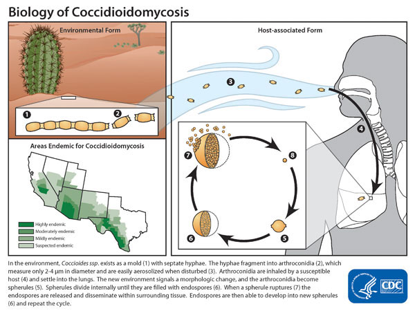
The pathogenesis of coccidioidomycosis can be described in following steps.<ref name="pmid26739609">{{cite journal |vauthors=Stockamp NW, Thompson GR |title=Coccidioidomycosis |journal=Infect. Dis. Clin. North Am. |volume=30 |issue=1 |pages=229–46 |year=2016 |pmid=26739609 |doi=10.1016/j.idc.2015.10.008 |url=}}</ref><ref name="pmid26398540">{{cite journal |vauthors=Twarog M, Thompson GR |title=Coccidioidomycosis: Recent Updates |journal=Semin Respir Crit Care Med |volume=36 |issue=5 |pages=746–55 |year=2015 |pmid=26398540 |doi=10.1055/s-0035-1562900 |url=}}</ref><ref name="pmid25577855">{{cite journal |vauthors=DiCaudo DJ |title=Coccidioidomycosis |journal=Semin Cutan Med Surg |volume=33 |issue=3 |pages=140–5 |year=2014 |pmid=25577855 |doi= |url=}}</ref><ref name="pmid24575994">{{cite journal |vauthors=Malo J, Luraschi-Monjagatta C, Wolk DM, Thompson R, Hage CA, Knox KS |title=Update on the diagnosis of pulmonary coccidioidomycosis |journal=Ann Am Thorac Soc |volume=11 |issue=2 |pages=243–53 |year=2014 |pmid=24575994 |doi=10.1513/AnnalsATS.201308-286FR |url=}}</ref> | |||
[[Image:Coccidioidomycosis-lifecycle.jpg|center|thumb|Life cycle and epidemiology - Source: https://www.cdc.gov/]] | |||
===Transmission=== | |||
*Coccidioiodomycosis exist as mycelia in the soil with septations. | |||
*During hot climate or dry environment, they proliferate asexually, transforming into spores, known as [[arthroconidia]]. | |||
*[[Infection]] is caused by [[inhalation]] of these [[arthroconidia]]. | |||
*The disease is not transmitted from person to person. | |||
===Incubation period=== | |||
*Incubation period of coccidioidomycosis varies from one to three weeks. | |||
===Dissemination=== | |||
*Following inhalation, [[arthroconidia]] reach terminal [[bronchioles]]. | |||
*Then they are ingested by [[Macrophages|pulmonary macrophages]]. | |||
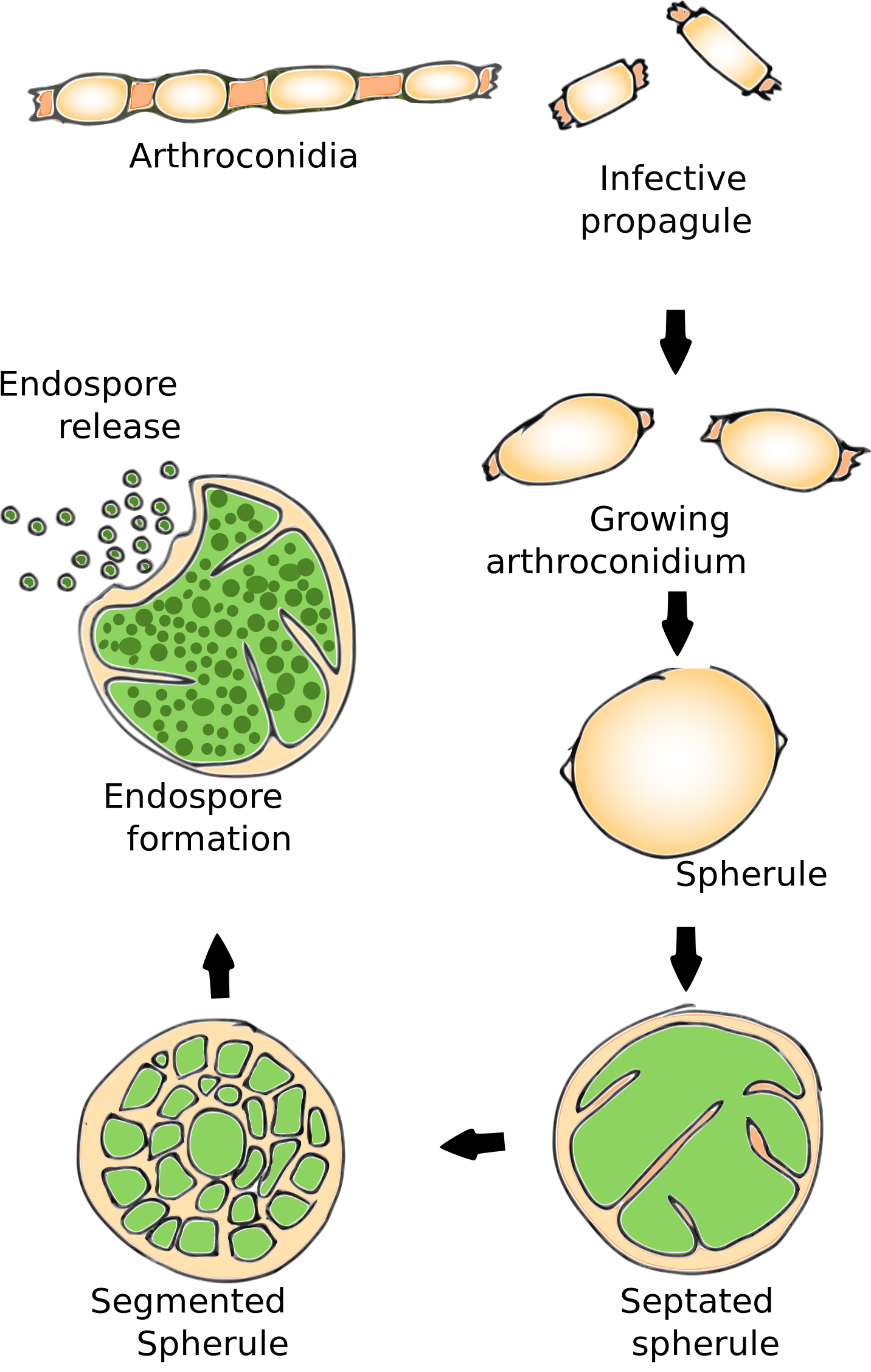
*Inside [[macrophages]] these [[arthroconidia]] enlarge, become rounded and develop internal septations to form what are known as the spherules. | |||
*It then disseminates to the [[lymphatics]] and [[blood]] stream to gain access to any organ of the body. | |||
===Seeding=== | |||
*Spherules contain uni-nuclear cells called as [[endospores]] which may propagate the infection further as they have the capability to develop into spherules. | |||
*This conversion of [[arthroconidia]] into spherules initiates an [[Inflammatory response|inflammatory reaction]] and leads to a chemotaxic response (peptides derived from activation of the [[Complement System|complement pathway]], [[leukotrienes]] ) which attracts [[neutrophils]] and [[eosinophils]] to the site of [[inflammation]]. | |||
*[[Cell-mediated immunity|Cell mediated immunity]] keeps the [[infection]] in check and keeps them limited to the organ of origin by forming [[Granulomas|granulomas.]] | |||
===Immune response=== | |||
Coccidioidomycosis elicits [[Cell-mediated immune response|cell-mediated immune responses]]. | |||
*[[Hypersensitivity|Delayed type hypersensitivity]] to coccidioidal [[antigens]] is common after acute infection has resolved. | |||
*Dissemination usually occurs via the l[[Lymphatic system|ymphatic]]<nowiki/>s and is more common in [[immune]] suppressed in whom the primary [[infection]] is not contained. | |||
[[Image:Life cycle of coccidioides.svg.png| | [[Image:Life cycle of coccidioides.svg.png|center|Life cycle of coccidiodes|788x788px]] | ||
==Genetics== | |||
There is no known genetic association to coccidioidomycosis. | |||
==Microscopic pathology== | |||
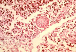
It is a dimorphic fungus and on microscopy, the following can be seen | |||
*Spherule with [[endospores]] | |||
*Rarely as [[hyphae]] in lung biopsy | |||
[[Image:Microscopy valley fever.jpg|center|Histopathological changes in coccidioidomycosis]] | |||
<gallery> | |||
Image: Coccidioidomycosis19.jpeg| Methenamine silver stain reveals reveals spherules of Coccidioides immitis in brain tissue. <SMALL><SMALL>''[http://phil.cdc.gov/phil/home.asp From Public Health Image Library (PHIL).] ''<ref name=PHIL> {{Cite web | title = Public Health Image Library (PHIL) | url = http://phil.cdc.gov/phil/home.asp}}</ref></SMALL></SMALL> | |||
Image: Coccidioidomycosis07.jpeg| PAS-stained photomicrograph of an unknown tissue specimen revealed the presence of three Coccidioides immitis spherules. <SMALL><SMALL>''[http://phil.cdc.gov/phil/home.asp From Public Health Image Library (PHIL).] ''<ref name=PHIL> {{Cite web | title = Public Health Image Library (PHIL) | url = http://phil.cdc.gov/phil/home.asp}}</ref></SMALL></SMALL> | |||
Image: Coccidioidomycosis06.jpeg| PAS-stained photomicrograph of an unknown tissue specimen revealed the presence of three Coccidioides immitis spherules. <SMALL><SMALL>''[http://phil.cdc.gov/phil/home.asp From Public Health Image Library (PHIL).] ''<ref name=PHIL> {{Cite web | title = Public Health Image Library (PHIL) | url = http://phil.cdc.gov/phil/home.asp}}</ref></SMALL></SMALL> | |||
Image: Coccidioidomycosis05.jpeg| Photomicrograph of of pus from a Guinea pig, Cavia porcellus, reveals presence of numbers of Coccidioides sp. fungal sporangia. <SMALL><SMALL>''[http://phil.cdc.gov/phil/home.asp From Public Health Image Library (PHIL).] ''<ref name=PHIL> {{Cite web | title = Public Health Image Library (PHIL) | url = http://phil.cdc.gov/phil/home.asp}}</ref></SMALL></SMALL> | |||
Image: Coccidioidomycosis04.jpeg| Photomicrograph of of pus from a Guinea pig, Cavia porcellus, reveals presence of numbers of Coccidioides sp. fungal sporangia. <SMALL><SMALL>''[http://phil.cdc.gov/phil/home.asp From Public Health Image Library (PHIL).] ''<ref name=PHIL> {{Cite web | title = Public Health Image Library (PHIL) | url = http://phil.cdc.gov/phil/home.asp}}</ref></SMALL></SMALL> | |||
Image: Coccidioidomycosis15.jpeg| Histopathologic characteristics found within a pus specimen, prepared using potassium hudroxide (KOH). Specimen harvested from a skin lesion in a case of cutaneous coccidioidomycosis. <SMALL><SMALL>''[http://phil.cdc.gov/phil/home.asp From Public Health Image Library (PHIL).] ''<ref name=PHIL> {{Cite web | title = Public Health Image Library (PHIL) | url = http://phil.cdc.gov/phil/home.asp}}</ref></SMALL></SMALL> | |||
Image: Coccidioidomycosis14.jpeg| Histopathologic characteristics found within a pus specimen, prepared using periodic acid-Schiff (PAS). Specimen harvested from a skin lesion in a case of cutaneous coccidioidomycosis. <SMALL><SMALL>''[http://phil.cdc.gov/phil/home.asp From Public Health Image Library (PHIL).] ''<ref name=PHIL> {{Cite web | title = Public Health Image Library (PHIL) | url = http://phil.cdc.gov/phil/home.asp}}</ref></SMALL></SMALL> | |||
Image: Coccidioidomycosis12.jpeg| Histopathologic characteristics found within a pus specimen, prepared using periodic acid-Schiff (PAS). Specimen harvested from a skin lesion in a case of cutaneous coccidioidomycosis. <SMALL><SMALL>''[http://phil.cdc.gov/phil/home.asp From Public Health Image Library (PHIL).] ''<ref name=PHIL> {{Cite web | title = Public Health Image Library (PHIL) | url = http://phil.cdc.gov/phil/home.asp}}</ref></SMALL></SMALL> | |||
Image: Coccidioidomycosis25.jpeg| Histopathology of coccidioidomycosis, lung. Methenamine silver stain. <SMALL><SMALL>''[http://phil.cdc.gov/phil/home.asp From Public Health Image Library (PHIL).] ''<ref name=PHIL> {{Cite web | title = Public Health Image Library (PHIL) | url = http://phil.cdc.gov/phil/home.asp}}</ref></SMALL></SMALL> | |||
Image: Coccidioidomycosis24.jpeg| Histopathology of coccidioidomycosis, retroperitoneal area. <SMALL><SMALL>''[http://phil.cdc.gov/phil/home.asp From Public Health Image Library (PHIL).] ''<ref name=PHIL> {{Cite web | title = Public Health Image Library (PHIL) | url = http://phil.cdc.gov/phil/home.asp}}</ref></SMALL></SMALL> | |||
Image: Coccidioidomycosis23.jpeg| Histopathologic changes due to coccidioidomycosis of the lung caused by Coccidioides immitis. <SMALL><SMALL>''[http://phil.cdc.gov/phil/home.asp From Public Health Image Library (PHIL).] ''<ref name=PHIL> {{Cite web | title = Public Health Image Library (PHIL) | url = http://phil.cdc.gov/phil/home.asp}}</ref></SMALL></SMALL> | |||
Image: Coccidioidomycosis22.jpeg| Histopathologic changes in a case of coccidioidomycosis of the lung showing a large fibrocaseous nodule. <SMALL><SMALL>''[http://phil.cdc.gov/phil/home.asp From Public Health Image Library (PHIL).] ''<ref name=PHIL> {{Cite web | title = Public Health Image Library (PHIL) | url = http://phil.cdc.gov/phil/home.asp}}</ref></SMALL></SMALL> | |||
Image: Coccidioidomycosis30.jpeg| Histopathology of coccidioidomycosis of lung. <SMALL><SMALL>''[http://phil.cdc.gov/phil/home.asp From Public Health Image Library (PHIL).] ''<ref name=PHIL> {{Cite web | title = Public Health Image Library (PHIL) | url = http://phil.cdc.gov/phil/home.asp}}</ref></SMALL></SMALL> | |||
</gallery> | |||
{{#ev:youtube|RtpvzCfFwfg}} | {{#ev:youtube|RtpvzCfFwfg}} | ||
==References== | ==References== | ||
{{Reflist|2}} | {{Reflist|2}} | ||
{{WH}} | |||
{{WS}} | |||
[[Category:Pulmonology]] | [[Category:Pulmonology]] | ||
[[Category:Fungal diseases]] | [[Category:Fungal diseases]] | ||
[[Category:Biological weapons]] | [[Category:Biological weapons]] | ||
[[Category:Mature chapter]] | [[Category:Mature chapter]] | ||
[[Category:Disease]] | [[Category:Disease]] | ||
[[Category:Needs overview]] | [[Category:Needs overview]] | ||
[[Category:Emergency medicine]] | |||
[[Category:Up-To-Date]] | |||
[[Category:Infectious disease]] | |||
Latest revision as of 21:00, 29 July 2020
|
Coccidioidomycosis Microchapters |
|
Diagnosis |
|---|
|
Treatment |
|
Case Studies |
|
Coccidioidomycosis pathophysiology On the Web |
|
American Roentgen Ray Society Images of Coccidioidomycosis pathophysiology |
|
Risk calculators and risk factors for Coccidioidomycosis pathophysiology |
Editor-In-Chief: C. Michael Gibson, M.S., M.D. [1]; Associate Editor(s)-in-Chief: ; Vidit Bhargava, M.B.B.S [2]; Aditya Ganti M.B.B.S. [3]
Overview
Coccidioidomycosis is a fungal infection, that is acquired through inhalation of the spores that is present in the environment. Following transmission, coccidioidomycosis are deposited into terminal bronchioles and enlarge, become rounded and develop internal septations to form what are known as the spherules. It then disseminates to the lymphatics and blood stream to gain access to any organ of the body.[1][2][3][4]
Pathogenesis
The pathogenesis of coccidioidomycosis can be described in following steps.[1][2][3][4]

Transmission
- Coccidioiodomycosis exist as mycelia in the soil with septations.
- During hot climate or dry environment, they proliferate asexually, transforming into spores, known as arthroconidia.
- Infection is caused by inhalation of these arthroconidia.
- The disease is not transmitted from person to person.
Incubation period
- Incubation period of coccidioidomycosis varies from one to three weeks.
Dissemination
- Following inhalation, arthroconidia reach terminal bronchioles.
- Then they are ingested by pulmonary macrophages.
- Inside macrophages these arthroconidia enlarge, become rounded and develop internal septations to form what are known as the spherules.
- It then disseminates to the lymphatics and blood stream to gain access to any organ of the body.
Seeding
- Spherules contain uni-nuclear cells called as endospores which may propagate the infection further as they have the capability to develop into spherules.
- This conversion of arthroconidia into spherules initiates an inflammatory reaction and leads to a chemotaxic response (peptides derived from activation of the complement pathway, leukotrienes ) which attracts neutrophils and eosinophils to the site of inflammation.
- Cell mediated immunity keeps the infection in check and keeps them limited to the organ of origin by forming granulomas.
Immune response
Coccidioidomycosis elicits cell-mediated immune responses.
- Delayed type hypersensitivity to coccidioidal antigens is common after acute infection has resolved.
- Dissemination usually occurs via the lymphatics and is more common in immune suppressed in whom the primary infection is not contained.

Genetics
There is no known genetic association to coccidioidomycosis.
Microscopic pathology
It is a dimorphic fungus and on microscopy, the following can be seen
- Spherule with endospores
- Rarely as hyphae in lung biopsy

-
Methenamine silver stain reveals reveals spherules of Coccidioides immitis in brain tissue. From Public Health Image Library (PHIL). [5]
-
PAS-stained photomicrograph of an unknown tissue specimen revealed the presence of three Coccidioides immitis spherules. From Public Health Image Library (PHIL). [5]
-
PAS-stained photomicrograph of an unknown tissue specimen revealed the presence of three Coccidioides immitis spherules. From Public Health Image Library (PHIL). [5]
-
Photomicrograph of of pus from a Guinea pig, Cavia porcellus, reveals presence of numbers of Coccidioides sp. fungal sporangia. From Public Health Image Library (PHIL). [5]
-
Photomicrograph of of pus from a Guinea pig, Cavia porcellus, reveals presence of numbers of Coccidioides sp. fungal sporangia. From Public Health Image Library (PHIL). [5]
-
Histopathologic characteristics found within a pus specimen, prepared using potassium hudroxide (KOH). Specimen harvested from a skin lesion in a case of cutaneous coccidioidomycosis. From Public Health Image Library (PHIL). [5]
-
Histopathologic characteristics found within a pus specimen, prepared using periodic acid-Schiff (PAS). Specimen harvested from a skin lesion in a case of cutaneous coccidioidomycosis. From Public Health Image Library (PHIL). [5]
-
Histopathologic characteristics found within a pus specimen, prepared using periodic acid-Schiff (PAS). Specimen harvested from a skin lesion in a case of cutaneous coccidioidomycosis. From Public Health Image Library (PHIL). [5]
-
Histopathology of coccidioidomycosis, lung. Methenamine silver stain. From Public Health Image Library (PHIL). [5]
-
Histopathology of coccidioidomycosis, retroperitoneal area. From Public Health Image Library (PHIL). [5]
-
Histopathologic changes due to coccidioidomycosis of the lung caused by Coccidioides immitis. From Public Health Image Library (PHIL). [5]
-
Histopathologic changes in a case of coccidioidomycosis of the lung showing a large fibrocaseous nodule. From Public Health Image Library (PHIL). [5]
-
Histopathology of coccidioidomycosis of lung. From Public Health Image Library (PHIL). [5]
{{#ev:youtube|RtpvzCfFwfg}}
References
- ↑ 1.0 1.1 Stockamp NW, Thompson GR (2016). "Coccidioidomycosis". Infect. Dis. Clin. North Am. 30 (1): 229–46. doi:10.1016/j.idc.2015.10.008. PMID 26739609.
- ↑ 2.0 2.1 Twarog M, Thompson GR (2015). "Coccidioidomycosis: Recent Updates". Semin Respir Crit Care Med. 36 (5): 746–55. doi:10.1055/s-0035-1562900. PMID 26398540.
- ↑ 3.0 3.1 DiCaudo DJ (2014). "Coccidioidomycosis". Semin Cutan Med Surg. 33 (3): 140–5. PMID 25577855.
- ↑ 4.0 4.1 Malo J, Luraschi-Monjagatta C, Wolk DM, Thompson R, Hage CA, Knox KS (2014). "Update on the diagnosis of pulmonary coccidioidomycosis". Ann Am Thorac Soc. 11 (2): 243–53. doi:10.1513/AnnalsATS.201308-286FR. PMID 24575994.
- ↑ 5.00 5.01 5.02 5.03 5.04 5.05 5.06 5.07 5.08 5.09 5.10 5.11 5.12 "Public Health Image Library (PHIL)".
![Methenamine silver stain reveals reveals spherules of Coccidioides immitis in brain tissue. From Public Health Image Library (PHIL). [5]](/images/b/b0/Coccidioidomycosis19.jpeg)
![PAS-stained photomicrograph of an unknown tissue specimen revealed the presence of three Coccidioides immitis spherules. From Public Health Image Library (PHIL). [5]](/images/d/d3/Coccidioidomycosis07.jpeg)
![PAS-stained photomicrograph of an unknown tissue specimen revealed the presence of three Coccidioides immitis spherules. From Public Health Image Library (PHIL). [5]](/images/6/6c/Coccidioidomycosis06.jpeg)
![Photomicrograph of of pus from a Guinea pig, Cavia porcellus, reveals presence of numbers of Coccidioides sp. fungal sporangia. From Public Health Image Library (PHIL). [5]](/images/d/de/Coccidioidomycosis05.jpeg)
![Photomicrograph of of pus from a Guinea pig, Cavia porcellus, reveals presence of numbers of Coccidioides sp. fungal sporangia. From Public Health Image Library (PHIL). [5]](/images/6/6a/Coccidioidomycosis04.jpeg)
![Histopathologic characteristics found within a pus specimen, prepared using potassium hudroxide (KOH). Specimen harvested from a skin lesion in a case of cutaneous coccidioidomycosis. From Public Health Image Library (PHIL). [5]](/images/c/ca/Coccidioidomycosis15.jpeg)
![Histopathologic characteristics found within a pus specimen, prepared using periodic acid-Schiff (PAS). Specimen harvested from a skin lesion in a case of cutaneous coccidioidomycosis. From Public Health Image Library (PHIL). [5]](/images/f/fb/Coccidioidomycosis14.jpeg)
![Histopathologic characteristics found within a pus specimen, prepared using periodic acid-Schiff (PAS). Specimen harvested from a skin lesion in a case of cutaneous coccidioidomycosis. From Public Health Image Library (PHIL). [5]](/images/d/df/Coccidioidomycosis12.jpeg)
![Histopathology of coccidioidomycosis, lung. Methenamine silver stain. From Public Health Image Library (PHIL). [5]](/images/9/9e/Coccidioidomycosis25.jpeg)
![Histopathology of coccidioidomycosis, retroperitoneal area. From Public Health Image Library (PHIL). [5]](/images/a/a6/Coccidioidomycosis24.jpeg)
![Histopathologic changes due to coccidioidomycosis of the lung caused by Coccidioides immitis. From Public Health Image Library (PHIL). [5]](/images/6/6d/Coccidioidomycosis23.jpeg)
![Histopathologic changes in a case of coccidioidomycosis of the lung showing a large fibrocaseous nodule. From Public Health Image Library (PHIL). [5]](/images/4/49/Coccidioidomycosis22.jpeg)
![Histopathology of coccidioidomycosis of lung. From Public Health Image Library (PHIL). [5]](/images/4/4d/Coccidioidomycosis30.jpeg)