Chondroma CT: Difference between revisions
Jump to navigation
Jump to search
No edit summary |
No edit summary |
||
| (22 intermediate revisions by 7 users not shown) | |||
| Line 1: | Line 1: | ||
__NOTOC__ | __NOTOC__ | ||
{{Chondroma}} | {{Chondroma}} | ||
{{CMG}}{{AE}} {{STM}} | {{CMG}}; {{AE}} {{Rohan}}, {{F.K}}, {{STM}} | ||
==Overview== | ==Overview== | ||
Findings on CT scan suggestive of [[enchondroma]] include sharply defined scalloped margins, expansion of the overlying [[cortex]] may be present, no [[periosteal reaction]] and soft tissue mass. | |||
==CT Scan== | ==CT Scan== | ||
*Bone CT may be helpful in the diagnosis of | *Bone CT scan may be helpful in the diagnosis of [[enchondroma]]. Findings on CT scan suggestive of [[enchondroma]] include:<ref name="pmid26137286">{{cite journal |vauthors=Nishio J, Arashiro Y, Mori S, Iwasaki H, Naito M |title=Periosteal chondroma of the distal tibia: Computed tomography and magnetic resonance imaging characteristics and correlation with histological findings |journal=Mol Clin Oncol |volume=3 |issue=3 |pages=677–681 |date=May 2015 |pmid=26137286 |pmc=4471519 |doi=10.3892/mco.2015.492 |url=}}</ref><ref name="pmid22983260">{{cite journal |vauthors=Duan F, Qiu S, Jiang J, Chang J, Liu Z, Lv X, Feng X, Xiong W, An J, Chen J, Yang W, Wen C |title=Characteristic CT and MRI findings of intracranial chondroma |journal=Acta Radiol |volume=53 |issue=10 |pages=1146–54 |date=December 2012 |pmid=22983260 |doi=10.1258/ar.2012.120433 |url=}}</ref> | ||
{| align="right" | |||
| | |||
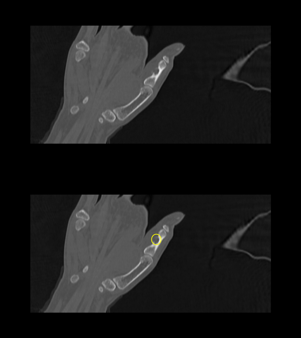
[[File:CT Chondroma.gif|300px|thumb|CT of phalanx showing chondroma.[https://radiopaedia.org/cases/juxtacortical-chondroma?lang=us Source: Case courtesy of Dr Vinay V Belaval, Radiopaedia.org, rID: 63471]]] | |||
* | |} | ||
** | |||
**Sharply defined scalloped margins | |||
< | **Expansion of the overlying cortex may be present, but there should not be cortical breakthrough unless fractured | ||
**No [[periosteal reaction]] | |||
**No [[soft tissue]] mass | |||
<br><br><br><br><br><br><br> | |||
< | |||
==References== | ==References== | ||
{{reflist|2}} | {{reflist|2}} | ||
{{WikiDoc Help Menu}} | {{WikiDoc Help Menu}} | ||
{{WikiDoc Sources}} | {{WikiDoc Sources}} | ||
[[Category:Up-To-Date]] | |||
[[Category:Oncology]] | |||
[[Category:Medicine]] | |||
[[Category:Orthopedics]] | |||
Latest revision as of 18:23, 24 January 2019
|
Chondroma Microchapters |
|
Diagnosis |
|---|
|
Treatment |
|
Chondroma CT On the Web |
|
American Roentgen Ray Society Images of Chondroma CT |
Editor-In-Chief: C. Michael Gibson, M.S., M.D. [1]; Associate Editor(s)-in-Chief: Rohan A. Bhimani, M.B.B.S., D.N.B., M.Ch.[2], Farima Kahe M.D. [3], Soujanya Thummathati, MBBS [4]
Overview
Findings on CT scan suggestive of enchondroma include sharply defined scalloped margins, expansion of the overlying cortex may be present, no periosteal reaction and soft tissue mass.
CT Scan
- Bone CT scan may be helpful in the diagnosis of enchondroma. Findings on CT scan suggestive of enchondroma include:[1][2]
 |
- Sharply defined scalloped margins
- Expansion of the overlying cortex may be present, but there should not be cortical breakthrough unless fractured
- No periosteal reaction
- No soft tissue mass
References
- ↑ Nishio J, Arashiro Y, Mori S, Iwasaki H, Naito M (May 2015). "Periosteal chondroma of the distal tibia: Computed tomography and magnetic resonance imaging characteristics and correlation with histological findings". Mol Clin Oncol. 3 (3): 677–681. doi:10.3892/mco.2015.492. PMC 4471519. PMID 26137286.
- ↑ Duan F, Qiu S, Jiang J, Chang J, Liu Z, Lv X, Feng X, Xiong W, An J, Chen J, Yang W, Wen C (December 2012). "Characteristic CT and MRI findings of intracranial chondroma". Acta Radiol. 53 (10): 1146–54. doi:10.1258/ar.2012.120433. PMID 22983260.